. 2013 Jul 23;8(7):e69327.
doi: 10.1371/journal.pone.0069327. Print 2013.

Dysregulation of mitochondrial quality control processes contribute to sarcopenia in a mouse model of premature aging

Affiliations

Dysregulation of mitochondrial quality control processes contribute to sarcopenia in a mouse model of premature aging

Anna-Maria Joseph et al. PLoS One. .

Abstract

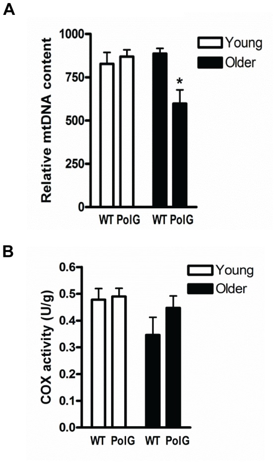
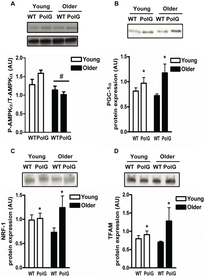
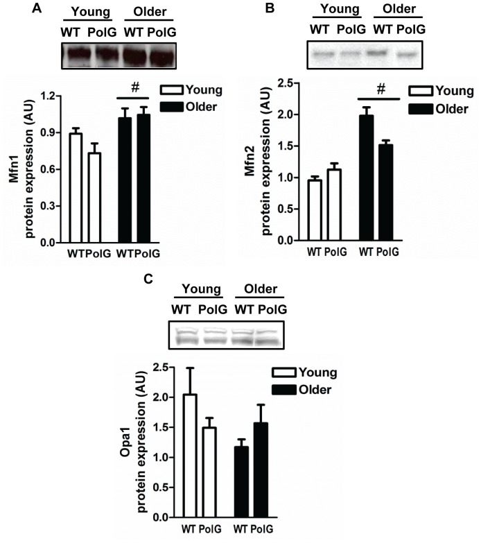
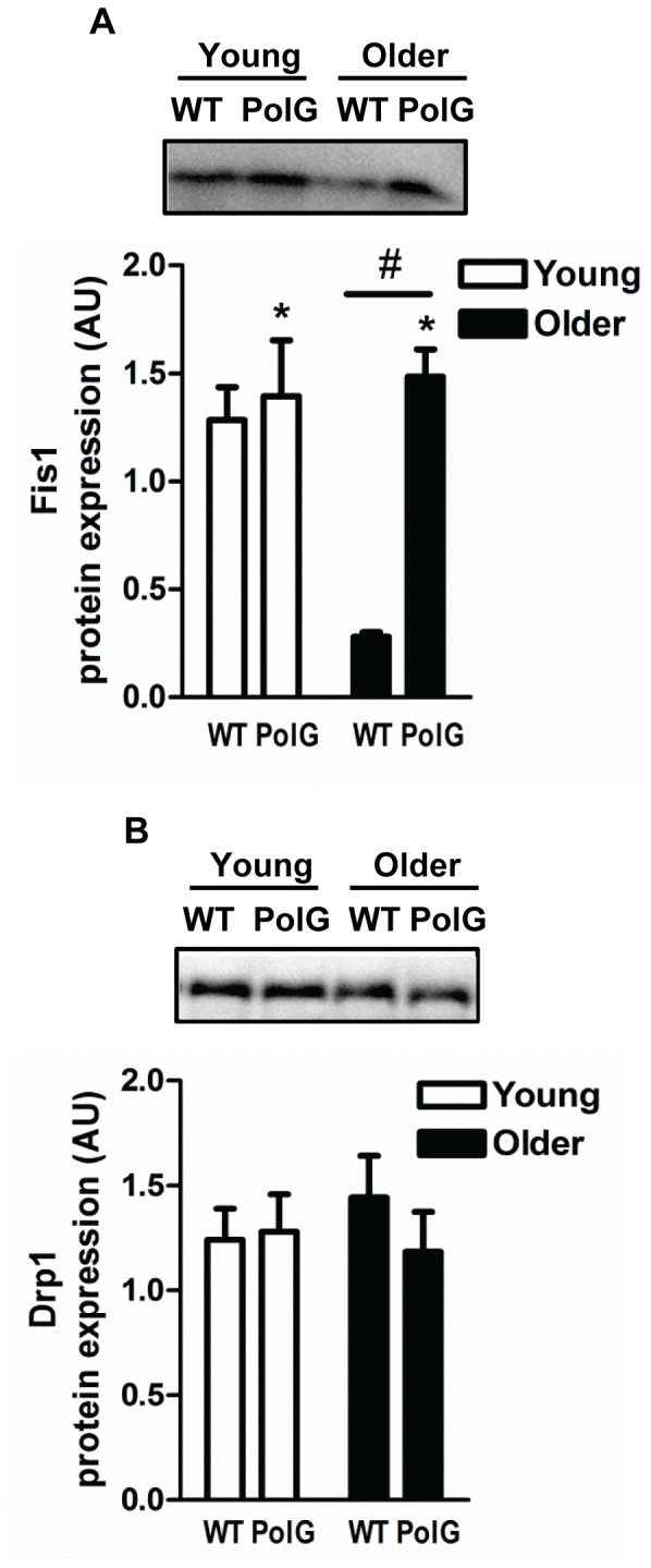
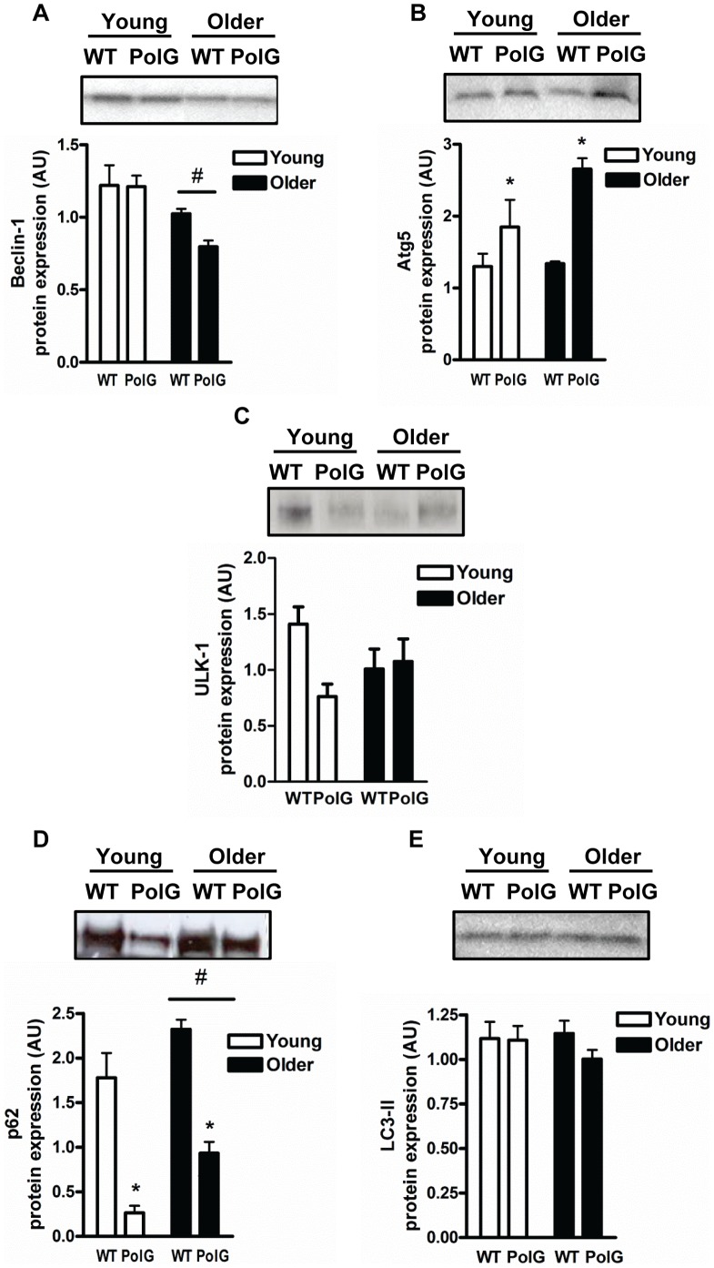
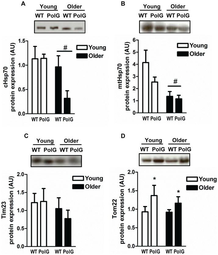
Mitochondrial DNA (mtDNA) mutations lead to decrements in mitochondrial function and accelerated rates of these mutations has been linked to skeletal muscle loss (sarcopenia). The purpose of this study was to investigate the effect of mtDNA mutations on mitochondrial quality control processes in skeletal muscle from animals (young; 3-6 months and older; 8-15 months) expressing a proofreading-deficient version of mtDNA polymerase gamma (PolG). This progeroid aging model exhibits elevated mtDNA mutation rates, mitochondrial dysfunction, and a premature aging phenotype that includes sarcopenia. We found increased expression of the mitochondrial biogenesis regulator peroxisome proliferator-activated receptor gamma coactivator-1α (PGC-1α) and its target proteins, nuclear respiratory factor 1 (NRF-1) and mitochondrial transcription factor A (Tfam) in PolG animals compared to wild-type (WT) (P<0.05). Muscle from older PolG animals displayed higher mitochondrial fission protein 1 (Fis1) concurrent with greater induction of autophagy, as indicated by changes in Atg5 and p62 protein content (P<0.05). Additionally, levels of the Tom22 import protein were higher in PolG animals when compared to WT (P<0.05). In contrast, muscle from normally-aged animals exhibited a distinctly different expression profile compared to PolG animals. Older WT animals appeared to have higher fusion (greater Mfn1/Mfn2, and lower Fis1) and lower autophagy (Beclin-1 and p62) compared to young WT suggesting that autophagy is impaired in aging muscle. In conclusion, muscle from mtDNA mutator mice display higher mitochondrial fission and autophagy levels that likely contribute to the sarcopenic phenotype observed in premature aging and this differs from the response observed in normally-aged muscle.


Conflict of interest statement

Competing Interests: The authors have declared that no competing interests exist.

Figures

Figure 1
Figure 1. Mitochondrial DNA content and enzyme activity in muscle from PolG mice.
(A) Mitochondrial DNA (mtDNA) was measured by Real Time-PCR (qT-PCR) in muscle from young (3–6 mo) and older (8–15 mo) WT and PolG mice. Relative mtDNA content was normalized to β-actin and a graphical representation of the summary data is shown (n = 7–9). (B) Cytochrome c oxidase (COX) activity was expressed as unit per gram of tissue (n = 7–9). Significance was set at P<0.05 and all data are represented as mean ± SE. *P<0.05 vs. age-matched WT.
Figure 2
Figure 2. PolG mice display higher levels of mitochondrial regulatory proteins and transcription factors.
AMPKα activation (A), PGC-1α (B), NRF-1 (C), and Tfam (D) were determined by Western Blotting in muscle from young (3–6 mo) and older (8–15 mo) WT and PolG animals. AMPKα activation is determined by phosphorylated AMPKα over total AMPKα. Representative blots are shown above with a graphical summary of the data below (n = 7–13). Significance was set at P<0.05 and all data are represented as mean ± SE. Data are expressed as arbitrary units (AU). #P<0.05 main effect of age. *P<0.05 main effect of genotype.
Figure 3
Figure 3. Altered mitochondrial fusion proteins in mtDNA mutator mice.
Immunoblotting of mitochondrial fusion proteins Mfn1 (A), Mfn2 (B) and Opa1 (C) in skeletal muscle from young (3–6 mo) and older (8–15 mo) WT and PolG animals. Representative blots are shown above with a graphical summary of the data below (n = 7–13). Two bands were detected for Opa1 representing the long and short isoforms. Both bands were quantified and total Opa1 protein content displayed in the graph. Significance was set at P<0.05 and all data are represented as mean ± SE. Data are expressed as arbitrary units (AU). #P<0.05 main effect of age.
Figure 4
Figure 4. Altered expression of mitochondrial fission proteins in mtDNA mutator mice.
Fis1 (A) and Drp1 (B) protein content was determined in muscle from young (3–6 mo) and older (8–15 mo) WT and PolG animals. A graphical summary along with representative blots are depicted (n = 7–13). Significance was set at P<0.05 and all data are represented as mean ± SE. Data are expressed as arbitrary units (AU). #P<0.05 main effect of age. *P<0.05 main effect of genotype.
Figure 5
Figure 5. Autophagy proteins are upregulated in muscle from PolG animals.
The content of autophagy proteins Beclin-1 (A), Atg5 (B), ULK1 (C), p62 (D), and LC3-II (E) was measured with Western Blotting in skeletal muscle from young (3–6 mo) and older (8–15 mo) WT and PolG animals. Representative blots are shown above with a graphical summary of the data below (n = 7–13). Significance was set at P<0.05 and all data are represented as mean ± SE. Data are expressed as arbitrary units (AU). #P<0.05 main effect of age. *P<0.05 main effect of genotype.
Figure 6
Figure 6. Effect of mtDNA mutations on mitochondrial protein import machinery.
Levels of mitochondrial protein import machinery cytosolic Hsp70 (A), mitochondrial Hsp70 (B), Tim23 (C), and Tom22 (D) were measured in muscle from young (3–6 mo) and older (8–15 mo) WT and PolG animals. Representative blots are shown above with a graphical summary of the data below (n = 7–13). Significance was set at P<0.05 and all data are represented as mean ± SE. Data are expressed as arbitrary units (AU). #P<0.05 main effect of age. *P<0.05 main effect of genotype.

References

    1. Calvani R, Joseph AM, Adhihetty PJ, Miccheli A, Bossola M, et al. (2013) Mitochondrial pathways in sarcopenia of aging and disuse muscle atrophy. Biol Chem 394: 393–414. - PMC - PubMed
    1. Harman D (1972) Free radical theory of aging: dietary implications. Am J Clin Nutr 25: 839–843. - PubMed
    1. Johnson ML, Robinson MM, Nair KS (2013) Skeletal muscle aging and the mitochondrion. Trends Endocrinol Metab. - PMC - PubMed
    1. Larsson NG (2010) Somatic mitochondrial DNA mutations in mammalian aging. Annu Rev Biochem 79: 683–706. - PubMed
    1. Corral-Debrinski M, Horton T, Lott MT, Shoffner JM, Beal MF, et al. (1992) Mitochondrial DNA deletions in human brain: regional variability and increase with advanced age. Nat Genet 2: 324–329. - PubMed

Publication types