. 2013 Jul 23;8(7):e69538.
doi: 10.1371/journal.pone.0069538. Print 2013.

Structure of the SCAN domain of human paternally expressed gene 3 protein

Affiliations

Structure of the SCAN domain of human paternally expressed gene 3 protein

Vadim Rimsa et al. PLoS One. .

Abstract

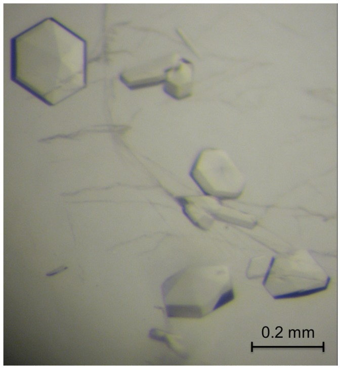
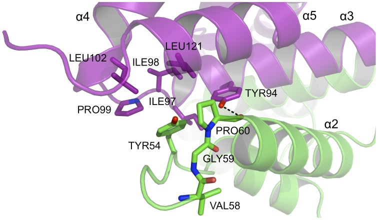
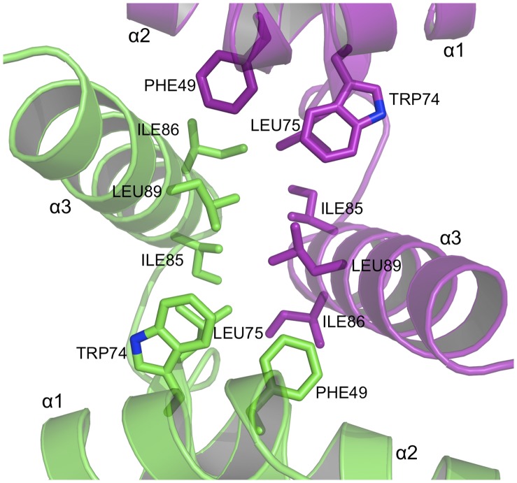
Human paternally expressed gene 3 protein (PEG3) is a large multi-domain entity with diverse biological functions, including acting as a transcription factor. PEG3 contains twelve Cys2-His2 type zinc finger domains, extended regions of predicted disorder and at the N-terminus a SCAN domain. PEG3 has been identified as partner of the E3 ubiquitin-protein ligase Siah1, an association we sought to investigate. An efficient bacterial recombinant expression system of the human PEG3-SCAN domain was prepared and crystals appeared spontaneously when the protein was being concentrated after purification. The structure was determined at 1.95 Å resolution and reveals a polypeptide fold of five helices in an extended configuration. An extensive dimerization interface, using almost a quarter of the solvent accessible surface, and key salt bridge interactions explain the stability of the dimer. Comparison with other SCAN domains reveals a high degree of conservation involving residues that contribute to the dimer interface. The PEG3-SCAN domain appears to constitute an assembly block, enabling PEG3 homo- or heterodimerization to control gene expression in a combinatorial fashion.


Conflict of interest statement

Competing Interests: The authors have declared that no competing interests exist.

Figures

Figure 1
Figure 1. Crystals of PEG3-SCAN.
Crystals grown in 50 mM Tris-HCl pH 7.5 and 150 mM NaCl.
Figure 2
Figure 2. The primary and secondary structure of PEG3-SCAN.
Five α-helices are shown as cylinders (purple) and are numbered accordingly. Multiple sequence alignment of PEG3-SCAN with other SCAN proteins from PDB was performed with ClustalW2 . PEG3-SCAN residues that are strictly conserved in Zfp206 (PDB: 4E6S), Znf24 (PDB: 3LHR), Znf42 (PDB: 2FI2) and Znf174 (PDB: 1Y7Q) are encased in black, while residues sharing similar properties in five proteins are encased in grey. The numbers that are shown above the secondary structure mark residues in the full length PEG3 protein (UniProt: Q9GZU2).
Figure 3
Figure 3. Overall structure of PEG3-SCAN.
The homodimer is shown as ribbons with one subunit green, the partner purple. The N- and C- termini as well as the five α-helices of each monomer are labeled accordingly.
Figure 4
Figure 4. Superposition of the PEG3-SCAN homodimer (purple) with other SCAN structures.
Zfp206 (PDB 4E6S), Znf24 (PDB 3LHR), Znf42 (PDB 2FI2) and Znf174 (PDB 1Y7Q) are shown in cyan, green, yellow and grey, respectively. Superposition was calculated using secondary structure matching .
Figure 5
Figure 5. The dimer interface of PEG3-SCAN (I).
A hydrogen-bonding network is formed between conserved residues lining the subunit-subunit interface. Water molecules are shown as red spheres, N and O positions are colored blue and red respectively, C positions are purple or green depending on the subunit to which they belong, hydrogen bonds are depicted as dashed lines. The same color scheme is used in Figures 6, 7 and 8.
Figure 6
Figure 6. The dimer interface of PEG3-SCAN (II).
A second cluster of hydrogen bonding and salt bridge interactions at the subunit-subunit interface.
Figure 7
Figure 7. The dimer interface of PEG3-SCAN (III).
A pronounced hydrophobic patch occurs at each end of the assembly to stabilize the dimer. The conserved Tyr94 extends across the dimer interface, contributes to hydrophobic interations and donates a hydrogen bond to the carbonyl of Pro60.
Figure 8
Figure 8. The dimer interface of PEG3-SCAN (IV).
A group of conserved, aliphatic and aromatic residues form a hydrophobic core to stabilize the dimer.

References

    1. Relaix F, Weng X, Marazzi G, Yang E, Copeland N, et al. (1996) Pw1, a novel zinc finger gene implicated in the myogenic and neuronal lineages. Dev Biol 177: 383–396. - PubMed
    1. Miller J, McLachlan AD, Klug A (1985) Repetitive zinc-binding domains in the protein transcription factor III A from Xenopus oocytes. EMBO J 4: 1609–1614. - PMC - PubMed
    1. Berg JM (1988) Proposed structure for the zinc-binding domains from tran- scription factor IIIA and related proteins. Proc Natl Acad Sci U S A 85: 99–102. - PMC - PubMed
    1. Lee MS, Gippert GP, Soman KV, Case DA, Wright PE (1989) Three-dimensional solution structure of a single zinc finger DNA-binding domain. Science 245: 635–637. - PubMed
    1. Dang DT, Pevsner J, Yang VW (2000) The biology of the mammalian Kruppel-like family of transcription factors. Int J Biochem Cell Biol 32: 1103–1121. - PMC - PubMed

Publication types

MeSH terms

LinkOut - more resources