. 2013 Jul 30;8(7):e69986.
doi: 10.1371/journal.pone.0069986. Print 2013.

Cep63 and cep152 cooperate to ensure centriole duplication

Affiliations

Cep63 and cep152 cooperate to ensure centriole duplication

Nicola J Brown et al. PLoS One. .

Abstract

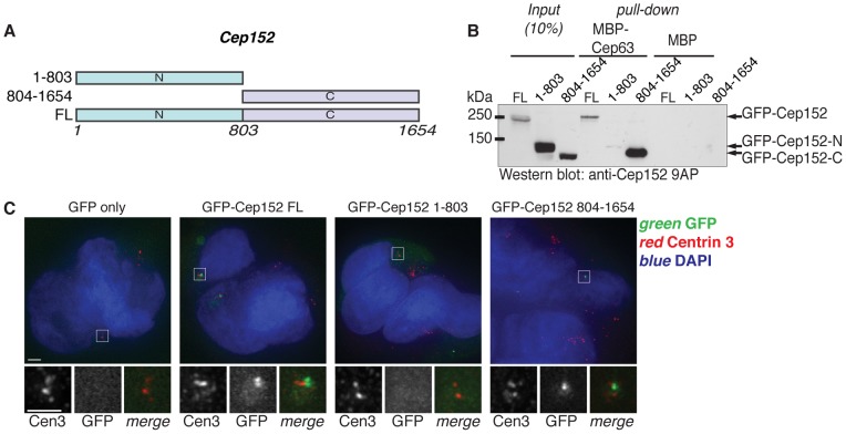
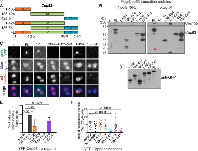
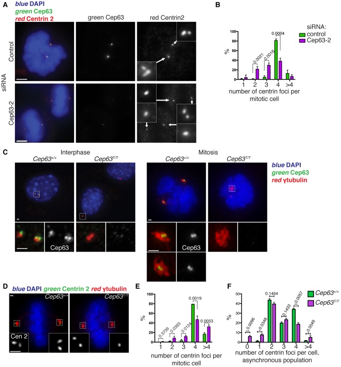
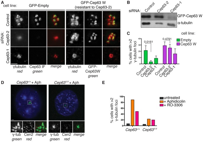
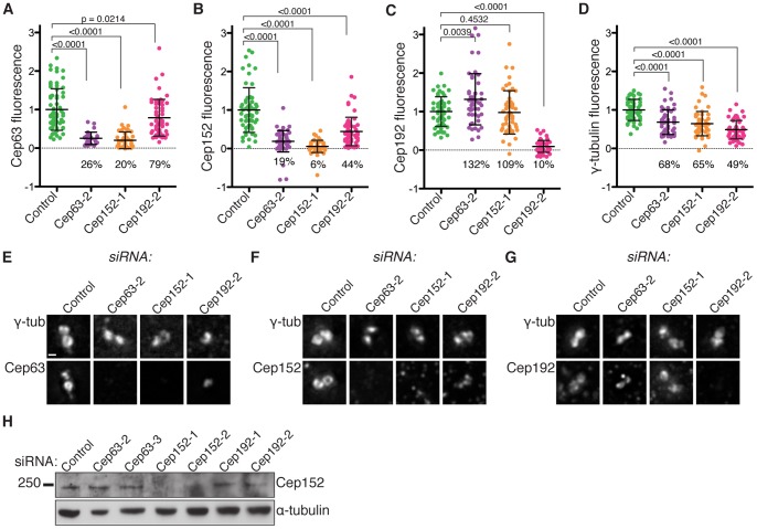
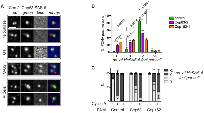
Centrosomes consist of two centrioles embedded in pericentriolar material and function as the main microtubule organising centres in dividing animal cells. They ensure proper formation and orientation of the mitotic spindle and are therefore essential for the maintenance of genome stability. Centrosome function is crucial during embryonic development, highlighted by the discovery of mutations in genes encoding centrosome or spindle pole proteins that cause autosomal recessive primary microcephaly, including Cep63 and Cep152. In this study we show that Cep63 functions to ensure that centriole duplication occurs reliably in dividing mammalian cells. We show that the interaction between Cep63 and Cep152 can occur independently of centrosome localisation and that the two proteins are dependent on one another for centrosomal localisation. Further, both mouse and human Cep63 and Cep152 cooperate to ensure efficient centriole duplication by promoting the accumulation of essential centriole duplication factors upstream of SAS-6 recruitment and procentriole formation. These observations describe the requirement for Cep63 in maintaining centriole number in dividing mammalian cells and further establish the order of events in centriole formation.


Conflict of interest statement

Competing Interests: The authors have declared that no competing interests exist.

Figures

Figure 1
Figure 1. The C-terminal half of Cep152 is required for Cep63 binding and centrosomal localisation.
(A) Diagram of Cep152 full length and truncation proteins used in the following experiments. Numbers indicate amino acid positions. (B) Cep63 interacts with the C-terminal half of Cep152. MBP or MBP-Cep63 pull down experiments were carried out after incubation in lysates of 293 HEK cells expressing GFP-Cep152 full length (FL), N-terminal half (1–803), or C-terminal half (804–1654), and Cep152 proteins were detected by Western blotting with anti-Cep152 antibodies (Cep152 9AP). Input shows 10% of cell lysate used for pull down experiments. (C) The C-terminal half of Cep152 is required for its centrosomal localisation. 293 HEK cells expressing the GFP-Cep152 proteins used in (B) were stained with DAPI (blue) and anti-Centrin 3 antibodies (red), GFP direct fluorescence is shown in green. Lower panels show magnification of one centrosome (boxed region). Scale bars 5 µm.
Figure 2
Figure 2. Cep63 N-terminus is required for centrosomal localisation of Cep63 and Cep152.
(A) Diagram of Cep63 full length and truncation proteins used in the following experiments. Numbers indicate amino acid positions. (B) Cep63 C-terminus interacts with Cep152. Expression of Flag-Cep63 (FL), truncation proteins, or Flag-empty vector control (e) was induced in 293 FlpIn TREX cell lines by incubation with 2 µg/ml doxycycline for 72 hours, then proteins were immunoprecipitated using anti-Flag resin. Western blots show endogenous Cep152, detected by anti-Cep152 (Bethyl), and Cep63 truncations detected by anti-Cep63 (Millipore). Inputs are 5% of the lysate used for Flag IP. Red arrowhead points to Cep63 truncation 1–135 present in the Flag IP. (C-F) U2OS cells were transfected with YFP-Cep63 (FL), truncation proteins, or YFP-empty vector (e) for 48 hours. (C) Cells were stained with anti-Cep152 (red) and γ-tubulin (blue) antibodies; YFP-tagged proteins were detected by direct fluorescence (green). Scale bar 1 µm. (D) Whole cell lysates from (C) were analysed by Western blot with anti-GFP antibodies to visualise YFP-tagged proteins. (E) The localisation of YFP-Cep63 proteins to the centrosome was scored in 3 independent experiments, n >10. (F) Overexpression of Cep63 425–541 and 136–541 deplete Cep152 from the centrosome. The ratio of Cep152 to γ-tubulin fluorescence intensities at the centrosome was measured for multiple cells from the experiment shown in (C), n >10.
Figure 3
Figure 3. Cep63 is needed for efficient centriole duplication in human and mouse cells.
(A-B) Cep63 depletion by RNA interference (RNAi) led to decreased centriole numbers in U2OS cells. Cells treated with control or Cep63 (Cep63-2) short interfering RNAs (siRNAs) for 4 days were stained with anti-centrin 2 (red) and anti-Cep63 (green) antibodies, plus DAPI (blue) to visualise DNA. Small panels show 3 times enlargements of the centrosome region with centrin 2 staining. Scale bar is 5 µm. (B) The number of centrin foci per mitotic cell was scored in three independent experiments, n>60. (C) Cep63 homozygous gene-trap mouse embryonic fibroblasts (MEFs) are devoid of Cep63 protein. Primary MEFs, at passage 3, were stained with anti-Cep63 (green) and anti-γ-tubulin antibodies (red), and DAPI (blue) to detect centrosomes and DNA, respectively. Representative images are shown of interphase and mitotic cells from two cell lines derived from littermates, one homozygous wild type (Cep63+/+) and the other homozygous gene-trap (Cep63T/T). Scale bars 1 µm. (D-F) Cep63T/T MEFs have reduced centriole numbers. (D) Primary MEFs, passage 3, were stained with anti-centrin2 (green) and anti-γ-tubulin (red) antibodies and DAPI. Scale bars 1 µm. (E) Centrin foci were scored in mitotic cells from 3 different cell lines for each genotype at passage 3, 40<n<100. (F) Centrin foci were scored in all cells (all cell cycle stages) in passage 1 primary MEF cell lines from 3 Cep63+/+ and 3 Cep63T/T embryos, n >100.
Figure 4
Figure 4. Cep63 and Cep152 are required for centrosome reduplication in mammalian cells.
(A) U2OS cells stably expressing GFP or GFP-Cep63 W (resistant to siRNA Cep63-2) were treated with Cep63 siRNAs 1 or 2 and incubated with 1.9 µg/ml aphidicolin (Aph) for 72 hours. GFP cells were stained with anti-Cep63 (green) and γ-tubulin (red). GFP-Cep63 W cells were stained with anti-γ-tubulin (red) and GFP-Cep63 W was detected by direct fluorescence. Scale bar 1 µm. (B) U2OS GFP-Cep63 W cell lysates were analysed by Western blotting with anti-GFP to detect GFP-Cep63 W and α-tubulin antibodies, used as a loading control. (C) Cells with greater than 2 γ-tubulin foci were recorded in 3 independent experiments, n = 150. T-test p values are indicated for comparison of control and siRNA Cep63-2 in both cell lines. (D-E) Cep63 homozygous gene-trap MEFs (Cep63T/T) show reduced centrosome reduplication induced by aphidicolin or Cdk1 inhibitor. (D) Cep63+/+ or Cep63T/T 3T3 immortalised MEFs incubated with aphidicolin (2 µg/ml) for 72 hours, stained with anti- γ-tubulin antibodies (green), centrin 3 antibodies (red) and DAPI (blue). Scale bar 5 µm. (H) Quantification of Cep63+/+ and Cep63T/T MEFs with greater than 2 γ-tubulin foci in untreated, Cdk1 inhibitor (RO-3306, 10 µM), or aphidicolin treated populations, n>150.
Figure 5
Figure 5. Cep63-Cep152 centrosomal recruitment is downstream of Cep192.
(A-D) Control RNAi or RNAi of Cep63, Cep152, or Cep192, was carried out for 4 days in U2OS cells, followed by immunofluorescence on replicate samples, with anti- γ-tubulin and anti-Cep63 (A), Cep152 (B), or Cep192 (C) antibodies. Fluorescence intensity of Cep63, Cep152, Cep192, and γ-tubulin at the centrosome were measured (graphs A-D). All intensity measurements were normalised to the mean of the control population and p values are indicated above (students’ t-test). (E-G) Images of γ-tubulin and Cep63, Cep152, or Cep192 immunofluorescence at the centrosome from the experiment shown in A-D. Scale bar 1 µm. (H) Western blots of whole cell lysates from U2OS cells used in experiments (A-G) using anti-Cep152 (Bethyl) and α-tubulin antibodies.
Figure 6
Figure 6. Lack of centrosomal Cep63–Cep152 causes a delay in HsSAS-6 recruitment.
(A) Cep63 is present at the PCM before HsSAS-6 recruitment. Telophase, G1 phase, S or G2 phase and mitotic HeLa cells, as indicated, were stained with anti-centrin 2 (red), Cep63 (green), and HsSAS-6 (blue) antibodies. Centrosomes from each cell are shown. Scale bar 1 µm. (B) HsSAS-6 foci were counted in U2OS cells with nuclear PCNA foci (a marker of DNA replication) after 96 hours RNAi as indicated, n >150, 3 experiments. P values from a students’ t-test are indicated on the graph. (C) U2OS cells were stained with anti-Cyclin A and HsSAS-6 antibodies after control, Cep63, or Cep152 RNAi. Cyclin A status, negative (-, early G1 phase), dull (+, G1-S), or bright (++, S-G2), and the number of HsSAS-6 foci (0,1,2,>2) were scored in asynchronous populations in three independent experiments, n >150.

References

    1. Gönczy P (2012) Towards a molecular architecture of centriole assembly. Nat Rev Mol Cell Biol 13: 425–435. - PubMed
    1. Nigg EA, Raff JW (2009) Centrioles, centrosomes, and cilia in health and disease. Cell 139: 663–678. - PubMed
    1. Nigg EA (2007) Centrosome duplication: of rules and licenses. Trends Cell Biol 17: 215–221. - PubMed
    1. Strnad P, Gönczy P (2008) Mechanisms of procentriole formation. Trends Cell Biol 18: 389–396. - PubMed
    1. Delattre M, Canard C, Gönczy P (2006) Sequential protein recruitment in C. elegans centriole formation. Curr Biol 16: 1844–1849. - PubMed

Publication types