. 2013 Jul 25;8(7):e70479.
doi: 10.1371/journal.pone.0070479. Print 2013.

Pharmacological induction of transforming growth factor-beta1 in rat models enhances radiation injury in the intestine and the heart

Affiliations

Pharmacological induction of transforming growth factor-beta1 in rat models enhances radiation injury in the intestine and the heart

Marjan Boerma et al. PLoS One. .

Abstract

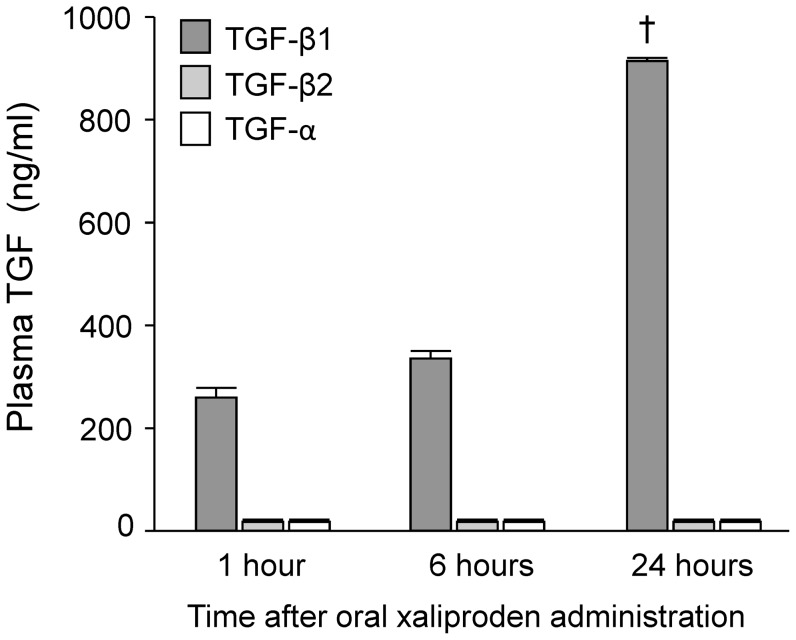
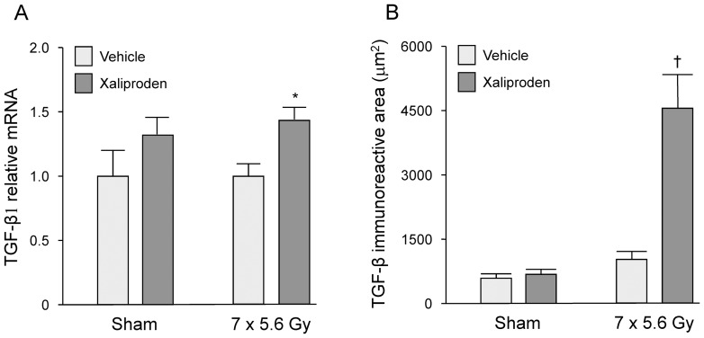
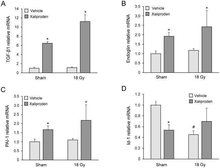
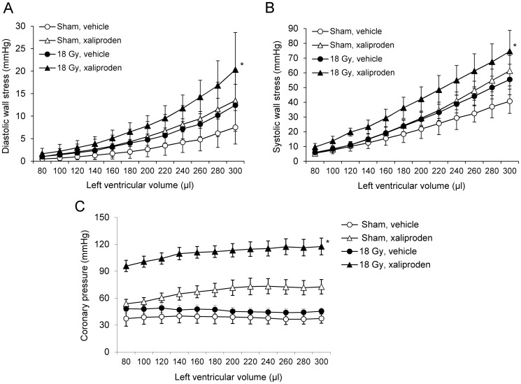
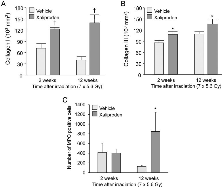
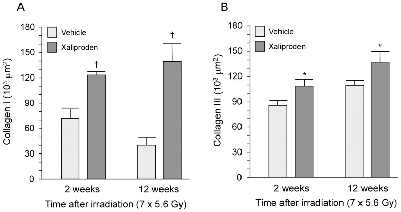
Radiation therapy in the treatment of cancer is dose limited by radiation injury in normal tissues such as the intestine and the heart. To identify the mechanistic involvement of transforming growth factor-beta 1 (TGF-β1) in intestinal and cardiac radiation injury, we studied the influence of pharmacological induction of TGF-β1 with xaliproden (SR 57746A) in rat models of radiation enteropathy and radiation-induced heart disease (RIHD). Because it was uncertain to what extent TGF-β induction may enhance radiation injury in heart and intestine, animals were exposed to irradiation schedules that cause mild to moderate (acute) radiation injury. In the radiation enteropathy model, male Sprague-Dawley rats received local irradiation of a 4-cm loop of rat ileum with 7 once-daily fractions of 5.6 Gy, and intestinal injury was assessed at 2 weeks and 12 weeks after irradiation. In the RIHD model, male Sprague-Dawley rats received local heart irradiation with a single dose of 18 Gy and were followed for 6 months after irradiation. Rats were treated orally with xaliproden starting 3 days before irradiation until the end of the experiments. Treatment with xaliproden increased circulating TGF-β1 levels by 300% and significantly induced expression of TGF-β1 and TGF-β1 target genes in the irradiated intestine and heart. Various radiation-induced structural changes in the intestine at 2 and 12 weeks were significantly enhanced with TGF-β1 induction. Similarly, in the RIHD model induction of TGF-β1 augmented radiation-induced changes in cardiac function and myocardial fibrosis. These results lend further support for the direct involvement of TGF-β1 in biological mechanisms of radiation-induced adverse remodeling in the intestine and the heart.


Conflict of interest statement

Competing Interests: We have read the journal's policy and would like to report the following: Our co-author Jean-Marc Herbert is an employee of the company that produced and provided xaliproden for this study. This does not alter our adherence to all the PLOS ONE policies on sharing data and materials.

Figures

Figure 1
Figure 1. Effects of xaliproden on plasma TGF levels in unirradiated animals.
Xaliproden caused a significant increase in plasma levels of latent TGF-β1, but not of TGF-α or TGF-β2 at 24 hours after oral administration. Average ± SEM, n = 10. p<0.0001.
Figure 2
Figure 2. Effects of radiation and xaliproden on intestinal TGF-β expression.
A 2-weeks administration of xaliproden significantly enhanced relative mRNA levels of TGF-β1 (A) and increased TGF-β immunoreactivity (B) in the irradiated small intestine. Average ± SEM, n = 5 (sham-irradiated) or 13–15 (irradiated). *p<0.05, p<0.0001. Representative micrographs of TGF-β immunohistochemical stainings are shown in Figure S1.
Figure 3
Figure 3. Effects of radiation and xaliproden on left ventricular gene expression of TGF-β1 and TGF-β pathway mediators.
Xaliproden caused a significant increase in left ventricular mRNA of TGF-β1 (A), endoglin (B), PAI-1 (C), and both radiation and xaliproden caused a downregulation of Id-1 mRNA when compared to vehicle-treated rats (D). Average ± SEM, n = 3. #Significant difference with sham-irradiated animals (p<0.05), *Significant difference with vehicle-treated animals (p<0.05).
Figure 4
Figure 4. Ex vivo cardiac function analysis at 6 months after local heart irradiation and xaliproden treatment.
Xaliproden significantly enhanced the effects of radiation on diastolic wall stress (A), systolic wall stress (B), and coronary pressure (C) as measured in Langendorff isolated perfused hearts at 6 months after local heart irradiation. Average ± SEM, n = 3–6. *Significant difference with sham-irradiated vehicle-treated animals (p<0.05).
Figure 5
Figure 5. Immunohistochemical evidence of myofibroblasts after local heart irradiation.
Immunohistochemistry with anti α-SMC antibodies revealed vascular smooth muscle cells with positive staining (arrow) and spindle shaped non-vascular cells with cross-striational α-SMC staining (myofibroblasts, arrowheads) (A). These myofibroblasts were detected in irradiated hearts only, in subendocardial areas near the cardiac valves (B). These areas showed severe fibrosis in the Picrosirius Red+Fast Green staining (C).
Figure 6
Figure 6. Histopathological manifestations of radiation enteropathy.
Xaliproden significantly reduced mucosal surface area (A) at 12 weeks after local irradiation of the small intestine, and enhanced radiation injury score (B), intestinal wall thickness (C) and serosal thickness (D) at 2 weeks and 12 weeks after irradiation. Average ± SEM, n = 5 (sham-irradiated) or 13–15 (irradiated). *p<0.05, p<0.001. Representative micrographs of histopathological stainings are shown in Figure S3.
Figure 7
Figure 7. Immunohistochemical analysis of collagen deposition and MPO positive cells in radiation enteropathy.
Xaliproden caused a significant increase in deposition of collagen types I (A) and III (B) at 2 weeks and at 12 weeks and a significant increase in the number of MPO positive cells (C) at 12 weeks after small intestine irradiation. Average ± SEM, n = 5–15. *p<0.05, p<0.001. Representative micrographs of immunohistochemical stainings are shown in Figure S4 and S5.

Similar articles

Cited by

References

    1. Hauer-Jensen M, Wang J, Boerma M, Fu Q, Denham JW (2007) Radiation damage to the gastrointestinal tract: mechanisms, diagnosis, and management. Curr Opin Support Palliat Care 1: 23–29. - PubMed
    1. Theis VS, Sripadam R, Ramani V, Lal S (2010) Chronic radiation enteritis. Clin Oncol 22: 70–83. - PubMed
    1. Adams MJ, Hardenbergh PH, Constine LS, Lipshultz SE (2003) Radiation-associated cardiovascular disease. Crit Rev Oncol Hematol 45: 55–75. - PubMed
    1. Early Breast Cancer Trialists Collaborative Group (2000) Favourable and unfavourable effects on long-term survival of radiotherapy for early breast cancer: an overview of the randomised trials. Lancet 355: 1757–1770. - PubMed
    1. Darby SC, McGale P, Taylor CW, Peto R (2005) Long-term mortality from heart disease and lung cancer after radiotherapy for early breast cancer: prospective cohort study of about 300,000 women in US SEER cancer registries. Lancet Oncol 6: 557–565. - PubMed

Publication types

MeSH terms