. 2013 Aug 2;8(8):e68004.
doi: 10.1371/journal.pone.0068004. Print 2013.

Upregulated microRNA-92b regulates the differentiation and proliferation of EpCAM-positive fetal liver cells by targeting C/EBPß

Affiliations

Upregulated microRNA-92b regulates the differentiation and proliferation of EpCAM-positive fetal liver cells by targeting C/EBPß

Nian-Song Qian et al. PLoS One. .

Abstract

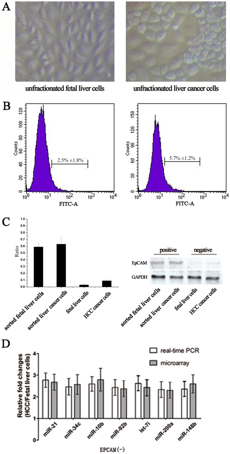
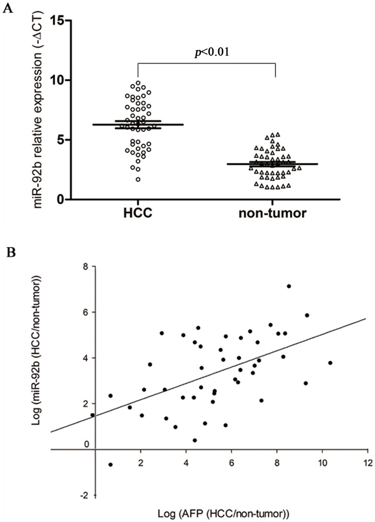
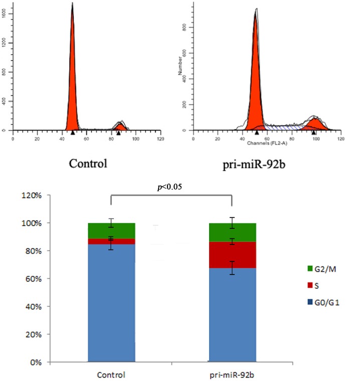
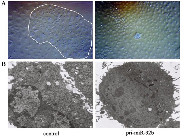
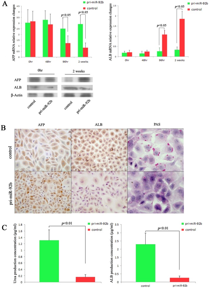
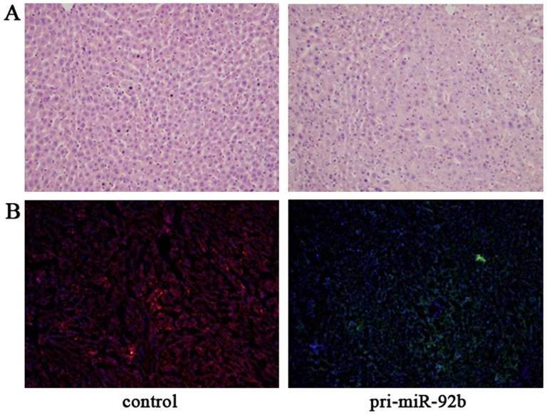
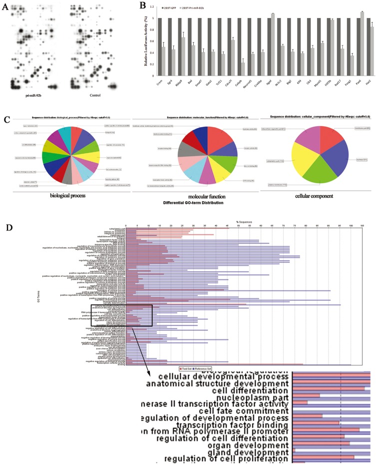
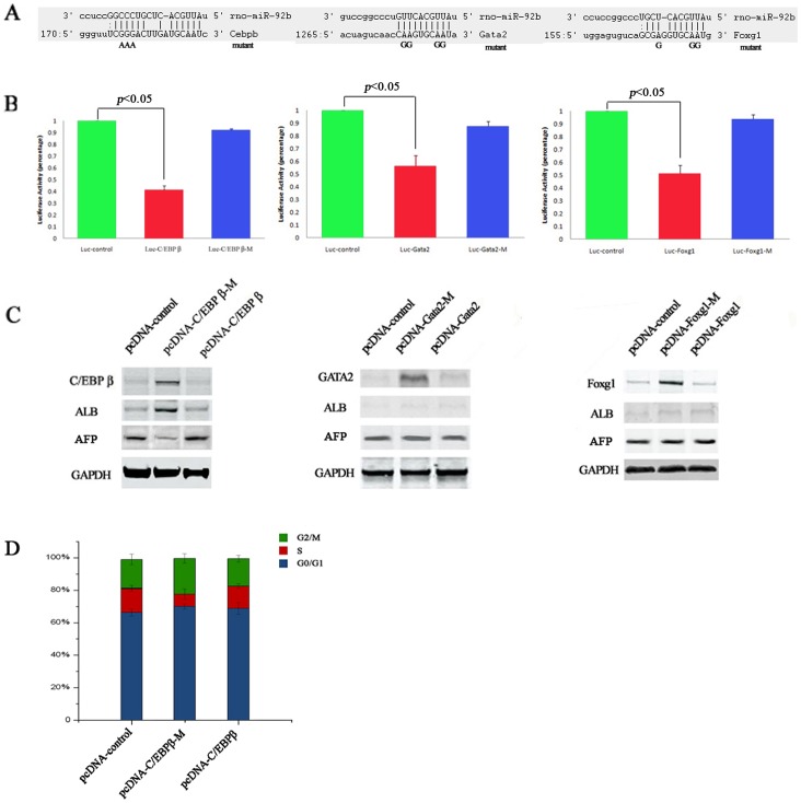
microRNAs (miRNAs) are short noncoding RNAs that negatively regulate gene expression. Although recent evidences have been indicated that their aberrant expression may play an important role in cancer stem cells, the mechanism of their deregulation in neoplastic transformation of liver cancer stem cells (LCSCs) has not been explored. In our study, the HCC model was established in F344 rats by DEN induction. The EpCAM(+) cells were sorted out from unfractionated fetal liver cells and liver cancer cells using the FACS analysis and miRNA expression profiles of two groups were screened through microarray platform. Gain-of-function studies were performed in vitro and in vivo to determine the role of miR-92b on proliferation and differentiation of the hepatic progenitors. In addition, luciferase reporter system and gene function analysis were used to predict miR-92b target. we found that miR-92b was highly downregulated in EpCAM(+) fetal liver cells in expression profiling studies. RT-PCR analysis demonstrated reverse correlation between miR-92b expression and differentiation degree in human HCC samples. Overexpression of miR-92b in EpCAM(+) fetal liver cells significantly increased proliferation and inhibited differentiation as well as in vitro and in vivo studies. Moreover, we verified that C/EBPß is a direct target of miR-92b and contributes to its effects on proliferation and differentiation. We conclude that aberrant expression of miR-92b can result in proliferation increase and differentiation arrest of hepatic progenitors by targeting C/EBPß.


Conflict of interest statement

Competing Interests: The authors have declared that no competing interests exist.

Figures

Figure 1
Figure 1. Primary EpCAM+ cells enrichment and miRNAs microarray analysis.
(A) phase-contrast microscopic observation showed that unfractionated primary fetal liver cells and liver cancer cells had similar appearance; (B) histograms representing the isolated EpCAM+ cells derived from the fetal liver cells and liver cancer cells respectively by using FACS analysis; (C) Real-time RT-PCR and Western Blot analysis showed that none of the EpCAM+ cells were in the EpCAM fraction after enrichment by using FACS sorting; (D) Real-time RT-PCR analysis validated fold changes of partial miRNAs dysregulated in microarray analysis between EpCAM groups.
Figure 2
Figure 2. miR-92b expression in clinical HCC samples.
(A) miR-92b was differentially expressed between HCC and the corresponding nontumoral liver tissues. (B) miR-92b expression demonstrated a significant positive correlation with AFP mRNA (P<0.05).
Figure 3
Figure 3. miR-92b regulated the proliferation of EpCAM+ fetal liver cells.
Cell cycle analysis showed that overexpression of miR-92b in EpCAM+ fetal liver cells would result in the decrease in G1 phase and significantly increase in S and G2/M phase.
Figure 4
Figure 4. Effects of miR-92b on the hepatic differentiation and maturation of EpCAM+ fetal liver cells.
(A) microscopic observation of miR-92b overexpressed and control groups after hepatocyte differentiation induction by using phase-contrast microscope. The control image was showing the mature degree and apoptosis of control cells compared to the mir-92b overexpressed cells, especially the cells in the white lines. However, most of the miR-92b overexpressed cells were similar to immature cells and still at the intermediate stage from hepatic progenitor to mature hepatocyte. (B) electron microscopic appearance of miR-92b overexpressed and control groups after induction. The ultrastructure was more preserved in control cells comparing to the miR-92b overexpressed cells. 100×(A), 8000×(B).
Figure 5
Figure 5. Effects of miR-92b on the differentiation arrest of EpCAM+ fetal liver cells.
(A) AFP and ALB mRNA expressions in miR-92b overexpressed and control groups after induction. The western blotting images showed the most significant difference of APF and ALB expressions between two groups 2 weeks after induction. (B) immunohistochemical staining of AFP, ALB and PAS in miR-92b overexpressed and control groups 2 weeks after induction; (C) detection and analysis of urea and ALB production secreted into the medium in miR-92b overexpressed and control groups. 200×(B).
Figure 6
Figure 6. In vivo differentiation assay of the effect of miR-92b overexpression on liver repopulation capacity of EpCAM+ fetal liver cells.
(A) Histological examination verified the significant differences in the in vivo hepatic differentiation between two groups; (B) Tracing the differentiation of miR-92b overexpressed and control groups in the liver by using fluorescent microscope. Large clusters of red fluorescence-positive cells with hepatocyte morphology were observed in the control group, and only small satellite clusters of GFP positive cells with the small oval cell morphology could be observed in the miR-92b overexpressed group. 200×(A and B).
Figure 7
Figure 7. Predicting functional target genes of miR-92b.
(A) In vivo screening potential targets of miR-92b in EpCAM+ fetal liver cells by using customized microarray analysis. The data showed that 7.96% (34/427) of predicted target genes in miR-92b overexpressed EpCAM+ cells were expressed lower than that in the counterpart. And 22 genes of which that relative fold changes less than 0.5 were considered as potential function targets of miR-92b; (B) Overall, 18 of the 22 3′UTR constructs demonstrated a significant reduction in luciferase activity for 293T cells overexpressing miR-92b (293T-pri-miR-92b), especially C/EBPß; (C) Potential targets could be classified in three aspects: biological process, molecular function and cellular component by using gene function analysis software; (D) Gene ontology analysis of the predicted target genes was performed with Blast2go software. The functions of many target genes were grouped in the categories of cell differentiation, transcription factor, organ development and regulation of cell proliferation, for choosing the genes involved in differentiation that specially mediated by mir-92b.
Figure 8
Figure 8. C/EBPß is a direct functional target of miR-92b.
(A) Diagram depicting the 3′UTR reporter assay. The 3′UTR of the C/EBPß, Gata2 and Foxg1 genes contain putative target sites with the position of seed sequences as indicated; (B) Fetal liver cells transfected with pri-miR-92b and a luciferanse reporter containing a fragment of the 3′UTR harboring either the miR-92b binding site (Luc-C/EBPß, Luc-Gata2, Luc-Foxg1) or a mutant (Luc-C/EBPß-M, Luc-Gata2-M, Luc-Foxg1-M). The assays showed that luciferase activities in the Luc-C/EBPß, Luc-Gata2, Luc-Foxg1 groups were significantly decreased compared to the luciferase activities of the mutant and control groups; (C) When restoring the C/EBPß expression in miR-92b overexpressed fetal liver cells by transfecting pcDNA-C/EBPß-M constructs, increase of ALB production in miR-92b overexpressed fetal liver cells could be observed, while there are no significant changes about ALB production in mir-92b overexpressed fetal liver cells transfected with pcDNA-C/EBPß, pcDNA-Foxg1, pcDNA-Foxg1-M, pcDNA-Gata2 and pcDNA-Gata2-M, respectively. (D) Cell cycle analysis showed the rescue effects of C/EBPß in miR92b overexpressed fetal liver cells. The significantly increase in G1 phase and decrease in S and G2/M phase could be observed when restoring the C/EBPß expression in miR-92b overexpressed fetal liver cells by transfecting pcDNA-C/EBPß-M.

References

    1. Reya T, Morrison SJ, Clarke MF, Weissman IL (2001) Stem cells, cancer, and cancer stem cells. Nature 414: 105–111. - PubMed
    1. Chiba T, Kita K, Zheng YW, Yokosuka O, Saisho H, et al. (2006) Side population purified from hepatocellular carcinoma cells harbors cancer stem cell-like properties. Hepatology 44: 240–251. - PubMed
    1. Yang ZF, Ho DW, Ng MN, Lau CK, Yu WC, et al. (2008) Significance of CD90+ cancer stem cells in human liver cancer. Cancer Cell 13: 153–166. - PubMed
    1. Ma S, Chan KW, Hu L, Lee TK, Wo JY, et al. (2007) Identification and characterization of tumorigenic liver cancer stem/progenitor cells. Gastroenterology 132: 2542–2556. - PubMed
    1. Yamashita T, Ji J, Budhu A, Forgues M, Yang W, et al. (2009) EpCAM-positive hepatocellular carcinoma cells are tumor-initiating cells with stem/progenitor cell features. Gastroenterology 136: 1012–1024. - PMC - PubMed

Publication types

MeSH terms

LinkOut - more resources