

Oro-gustatory perception of dietary lipids and calcium signaling in taste bud cells are altered in nutritionally obesity-prone Psammomys obesus
- PMID: 23936306
- PMCID: PMC3731325
- DOI: 10.1371/journal.pone.0068532
Oro-gustatory perception of dietary lipids and calcium signaling in taste bud cells are altered in nutritionally obesity-prone Psammomys obesus
Abstract
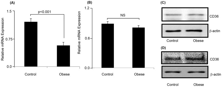
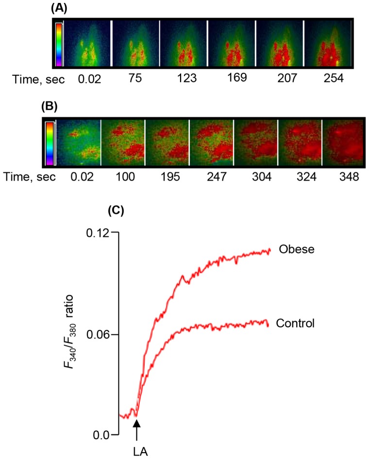
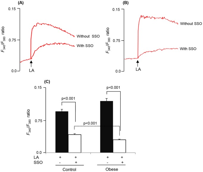
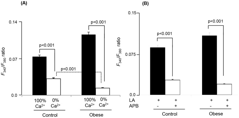
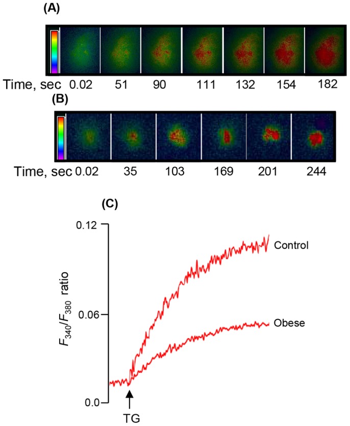
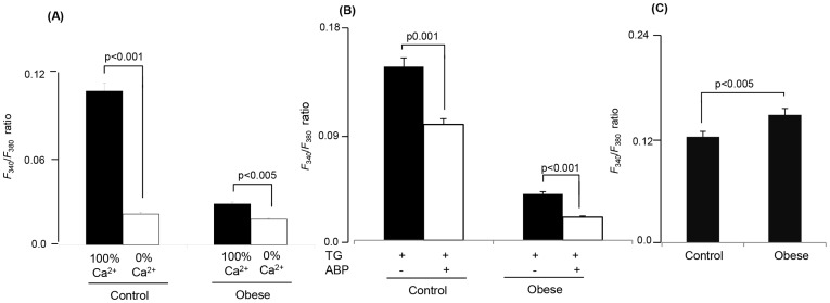
Since the increasing prevalence of obesity is one of the major health problems of the modern era, understanding the mechanisms of oro-gustatory detection of dietary fat is critical for the prevention and treatment of obesity. We have conducted the present study on Psammomys obesus, the rodent desert gerbil which is a unique polygenic natural animal model of obesity. Our results show that obese animals exhibit a strong preference for lipid solutions in a two-bottle test. Interestingly, the expression of CD36, a lipido-receptor, in taste buds cells (TBC), isolated from circumvallate papillae, was decreased at mRNA level, but remained unaltered at protein level, in obese animals. We further studied the effects of linoleic acid (LA), a long-chain fatty acid, on the increases in free intracellular calcium (Ca(2+)) concentrations, [Ca(2+)]i, in the TBC of P. obesus. LA induced increases in [Ca(2+)]i, largely via CD36, from intracellular pool, followed by the opening of store-operated Ca(2+) (SOC) channels in the TBC of these animals. The action of this fatty acid on the increases in [Ca(2+)]i was higher in obese animals than that in controls. However, the release of Ca(2+) from intracellular stores, studied also by employing thapsigargin, was lower in TBC of obese animals than control rodents. In this study, we show, for the first time, that increased lipid intake and altered Ca(2+) signaling in TBC are associated with obesity in Psammomys obesus.
Conflict of interest statement
Figures
Similar articles
-
STIM1 regulates calcium signaling in taste bud cells and preference for fat in mice.J Clin Invest. 2012 Jun;122(6):2267-82. doi: 10.1172/JCI59953. Epub 2012 May 1. J Clin Invest. 2012. PMID: 22546859 Free PMC article.
-
CD36- and GPR120-mediated Ca²⁺ signaling in human taste bud cells mediates differential responses to fatty acids and is altered in obese mice.Gastroenterology. 2014 Apr;146(4):995-1005. doi: 10.1053/j.gastro.2014.01.006. Epub 2014 Jan 9. Gastroenterology. 2014. PMID: 24412488 Free PMC article.
-
Ca2+ signaling in taste bud cells and spontaneous preference for fat: unresolved roles of CD36 and GPR120.Biochimie. 2014 Jan;96:8-13. doi: 10.1016/j.biochi.2013.06.005. Epub 2013 Jun 15. Biochimie. 2014. PMID: 23774298 Review.
-
Cell mechanisms of gustatory lipids perception and modulation of the dietary fat preference.Biochimie. 2014 Dec;107 Pt A:11-4. doi: 10.1016/j.biochi.2014.06.018. Epub 2014 Jul 2. Biochimie. 2014. PMID: 24997404 Review.
-
ERK1/2 activation in human taste bud cells regulates fatty acid signaling and gustatory perception of fat in mice and humans.FASEB J. 2016 Oct;30(10):3489-3500. doi: 10.1096/fj.201600422R. Epub 2016 Jun 29. FASEB J. 2016. PMID: 27358389 Free PMC article.
Cited by
-
Vimentin Deficiency Prevents High-Fat Diet-Induced Obesity and Insulin Resistance in Mice.Diabetes Metab J. 2021 Jan;45(1):97-108. doi: 10.4093/dmj.2019.0198. Epub 2020 Jun 15. Diabetes Metab J. 2021. PMID: 32602277 Free PMC article.
References
-
- World Health Organization (2000) Obesity: preventing and managing the global epidemic. Report of a WHO consultation. World Health Organ Tech Rep Ser 894: 1–253. - PubMed
-
- Blundell JE, MacDiarmid JI (1997) Fat as a risk factor for overconsumption: satiation, satiety, and patterns of eating. J Am Diet Assoc 97: S63–S69. - PubMed
-
- Lanfer A, Knof K, Barba G, Veidebaum T, Papoutsou S, et al. (2012) Taste preferences in association with dietary habits and weight status in European children: results from the IDEFICS study. Int J Obes (Lond) 36: 27–34. - PubMed
-
- Stewart JE, Newman LP, Keast RS (2011) Oral sensitivity to oleic acid is associated with fat intake and body mass index. Clin Nutr 30: 838–844. - PubMed
Publication types
MeSH terms
Substances
LinkOut - more resources
Full Text Sources
Other Literature Sources
Medical
Miscellaneous
