. 2013 Aug 2;8(8):e69025.
doi: 10.1371/journal.pone.0069025. Print 2013.

Adenoviral transduction of human acid sphingomyelinase into neo-angiogenic endothelium radiosensitizes tumor cure

Affiliations

Adenoviral transduction of human acid sphingomyelinase into neo-angiogenic endothelium radiosensitizes tumor cure

Branka Stancevic et al. PLoS One. .

Abstract

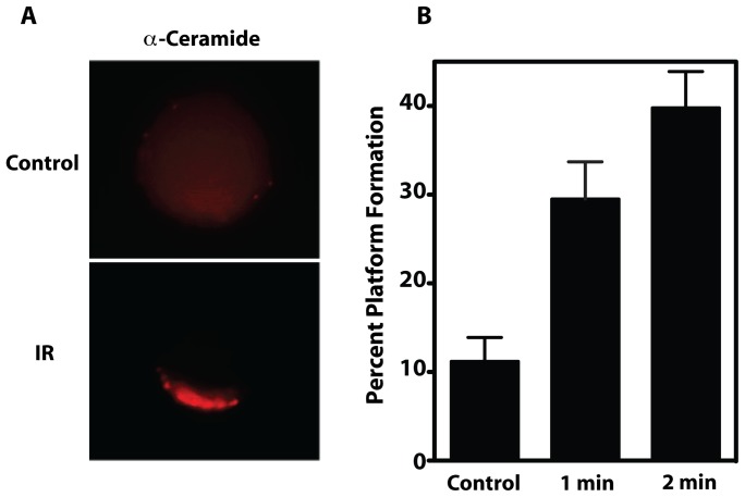
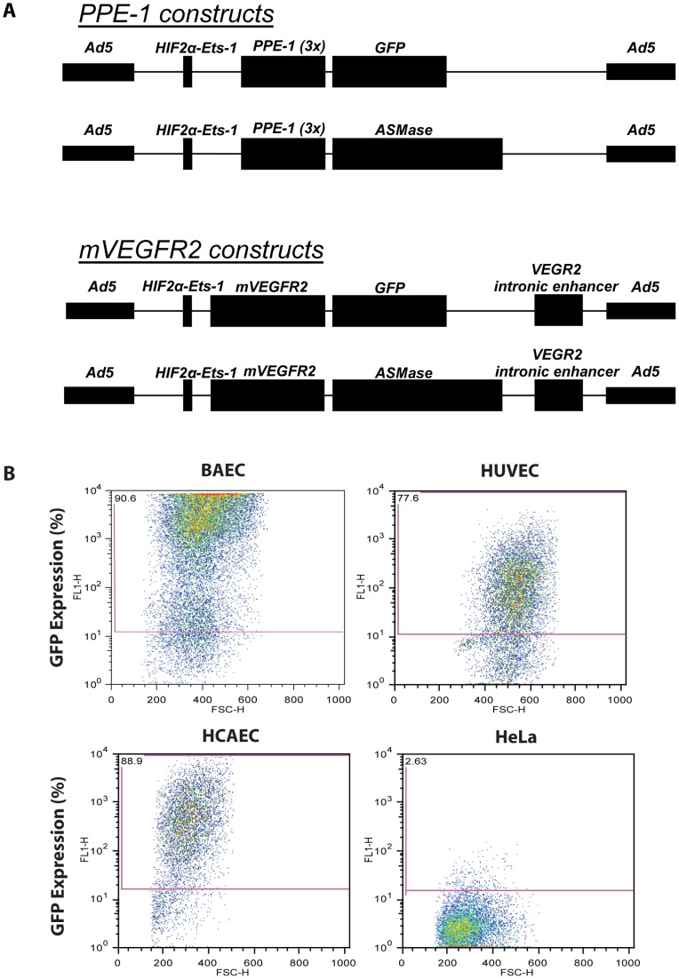
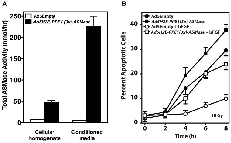
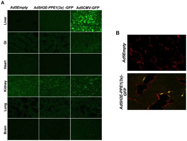
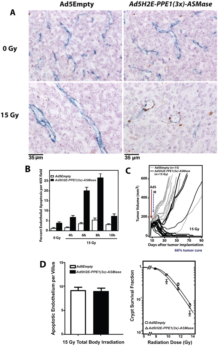
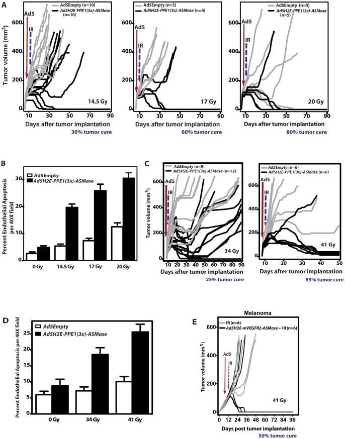
These studies define a new mechanism-based approach to radiosensitize tumor cure by single dose radiotherapy (SDRT). Published evidence indicates that SDRT induces acute microvascular endothelial apoptosis initiated via acid sphingomyelinase (ASMase) translocation to the external plasma membrane. Ensuing microvascular damage regulates radiation lethality of tumor stem cell clonogens to effect tumor cure. Based on this biology, we engineered an ASMase-producing vector consisting of a modified pre-proendothelin-1 promoter, PPE1(3x), and a hypoxia-inducible dual-binding HIF-2α-Ets-1 enhancer element upstream of the asmase gene, inserted into a replication-deficient adenovirus yielding the vector Ad5H2E-PPE1(3x)-ASMase. This vector confers ASMase over-expression in cycling angiogenic endothelium in vitro and within tumors in vivo, with no detectable enhancement in endothelium of normal tissues that exhibit a minute fraction of cycling cells or in non-endothelial tumor or normal tissue cells. Intravenous pretreatment with Ad5H2E-PPE1(3x)-ASMase markedly increases SDRT cure of inherently radiosensitive MCA/129 fibrosarcomas, and converts radiation-incurable B16 melanomas into biopsy-proven tumor cures. In contrast, Ad5H2E-PPE1(3x)-ASMase treatment did not impact radiation damage to small intestinal crypts as non-dividing small intestinal microvessels did not overexpress ASMase and were not radiosensitized. We posit that combination of genetic up-regulation of tumor microvascular ASMase and SDRT provides therapeutic options for currently radiation-incurable human tumors.


Conflict of interest statement

Competing Interests: The authors have the following interests. Dror Harats is employed by Vascular Biogenics Ltd (VBL). Dror Harats and VBL provided us the preproendothelin promoter and advice as to how to insert it into our construct. All work, however, was performed in the corresponding author's laboratory at the Sloan-Kettering Institute in New York. Pending patent application: Method and Composition to Increase Radiation-Induced Tumor Therapeutic Effect PCT/US2010/059204 filed on 12/7/10. Richard N Kolesnick, Branka Stancevic, Michel W J Sadelain, Zvi Fuks, Nira Varda-Bloom and Dror Harats are inventors. Memorial Sloan-Kettering Cancer Center and VBL are assignees. There are National phase filings in the US, Australia, Canada, & Europe (US 13/513,890; AU 2010/328335; CA 2,783,010; and EP 10891023.2). There are no further patents, products in development or marketed products to declare. This does not alter the authors' adherence to all the PLOS ONE policies on sharing data and materials, as detailed online in the guide for authors.

Figures

Figure 1
Figure 1. IR induces CRP formation on the surface of purified tumor endothelium.
(A) Representative image of the clustering of ceramide into platforms on the outer leaflet of the plasma membrane of purified tumor endothelial cells at 1 min post 15 Gy. For these studies, irradiated cells were stained with Texas Red-labeled anti-ceramide antibody, and platforms identified as in Materials and Methods. Images are representative of over 30% of cells from 3 experiments in which over 100 cells were analyzed. (B) Time-dependent generation of platforms in tumor endothelial cells. CRPs in tumor endothelial cells irradiated ex vivo with 15 Gy were quantified as in Materials and Methods. Data (mean ± 95% CI) are collated from 3 experiments in which 200 cells were analyzed per point.
Figure 2
Figure 2. Infection with Ad5H2E-PPE1(3x)-GFP confers GFP expression specific to endothelial cells.
(A) Schematic representation of gene therapy vectors used to overexpress the GFP reporter or human ASMase. (B) Primary cultures of bovine and human endothelial cells (BAEC, HUVEC and HCAEC) and non-endothelial cells (HeLa) were infected with Ad5H2E-PPE1(3x)-GFP. GFP expression was measured in live cells following detachment 72 h post-infection by flow cytometry.
Figure 3
Figure 3. Overexpression of human ASMase via Ad5H2E-PPE1(3x)-ASMase increases lysosomal and secretory ASMase activity, radiosensitizes BAEC, and attenuates bFGF protection against radiation-induced apoptosis.
(A) Cellular homogenates and serum-free conditioned media were harvested from BAEC transduced with Ad5Empty or Ad5H2E-PPE1(3x)-ASMase and assayed for ASMase activity using [14C-methylcholine]sphingomyelin as substrate in the presence of 1 mM EDTA (cellular homogenates) or 0.1 mM Zn2+ (conditioned media). (B) BAEC transduced with Ad5Empty or Ad5H2E-PPE1(3x)-ASMase were pre-treated with 1 ng/ml bFGF 15 minutes prior to 10 Gy irradiation. Apoptosis was assessed at various time points after IR by morphologic analysis following bis-benzimide staining. Data (mean ± SEM) are collated from 3 experiments performed in triplicate in which 400 nuclei were analyzed per sample.
Figure 4
Figure 4. Intravenous administration of Ad5H2E-PPE1(3x)-GFP results in GFP expression selectively in tumor endothelium.
1×10¹⁰ PFU of Ad5Empty, Ad5H2E-PPE1(3x)-GFP or Ad5CMV-GFP were intravenously administered to MCA/129 fibrosarcoma-bearing sv129/BL6 mice. Five days post viral administration, normal tissues (A) and tumor tissue (B) were excised and GFP expression was visualized by standard fluorescence microscopy following staining with anti-GFP (green; A and B) and anti-Meca-32 (red; B) antibodies, as described in Materials and Methods. Shown are representative 200× images of 20 fields analyzed per sample. Note background autofluorescence in the kidney specimens.
Figure 5
Figure 5. Expression of ASMase in endothelium of MCA/129 fibrosarcomas implanted in asmase−/− mice restores radiation-induced endothelial apoptosis and tumor cure.
MCA/129 fibrosarcoma cells (106, resuspended in PBS) were injected intra-dermally into the right hind limb of asmase−/− mice (A, B, C) and tumor volume (based on caliper measurements) was calculated daily according to the formula by Kim et al. . At 90–130 mm3, 1×10¹⁰ PFU of Ad5Empty or Ad5H2E-PPE1(3x)-ASMase was administered intravenously. 5 days post viral administration tumors were locally irradiated with 15 Gy or left untreated. (A) Representative cross sections of MCA/129 fibrosarcoma excised from unirradiated animals (upper panel) and at 6 hours post 15 Gy (lower panel), and co-stained for an endothelial-specific marker (Meca-32, blue) and apoptosis (TUNEL; brown). (B) Quantitation of the effect of Ad5H2E-PPE1(3x)-ASMase treatment on radiation-induced endothelial cell apoptosis. Data (mean ± SEM) represent TUNEL-positive endothelial cells quantified from 20 fields/tumor (400× magnification) and 2 tumors per group. (C) Impact of treatment with Ad5Empty (gray lines) or Ad5H2E-PPE1(3x)-ASMase (black lines) followed by 15 Gy on MCA/129 fibrosarcoma response. N equals number of animals per group. Tumors were measured daily up to 40 days and twice weekly thereafter. (D) Lack of impact of Ad5H2E-PPE1(3x)-ASMase on small intestinal radiation sensitivity. Mice, pre-treated for 5 days with Ad5Empty or Ad5H2E-PPE1(3x)-ASMase, were subjected to 15 Gy (left) or 8–14 Gy (right) total body irradiation known to cause GI damage. Mice were sacrificed at 4 h (left) for endothelial apoptosis measurement or after 3.5 days (right) for Crypt Microcolony Assay as standardized in , . Endothelial apoptosis was identified by microscopic co-detection of TUNEL (brown) and MECA-32 (blue) staining. Data (mean ± SEM) were compiled from 2 mice each, analyzing apoptotic cells in the lamina propria of 20 intact crypt-villus units per mouse. Crypt survival curves were calculated using 2 mice per dose analyzing 5–10 crypt-villus units per mouse by least square regression analysis, with a modification of the FIT software program . The program fits curves by iteratively weighted least squares to each set of dose–survival data, estimates covariates of survival curve parameters and corresponding confidence regions, and plots the survival curve.
Figure 6
Figure 6. ASMase overexpression in tumor endothelium radiosensitizes MCA/129 fibrosarcomas and B16 melanomas.
1×10¹⁰ PFU of Ad5Empty or Ad5H2E-PPE1(3x)-ASMase was administered intravenously to MCA/129 fibrosarcoma- (A,B) and B16 melanoma (C,D)-bearing sv129/BL6JAX mice, as in Figure 5. Five (A,B) or four (C,D) days post virus administration tumors were locally irradiated with 14.5, 17 Gy and 20 Gy (A,B), or 34 and 41 Gy (C,D). (A,C) Response of MCA/129 fibrosarcoma (A) and B16-F1 melanoma (C) to Ad5Empty (gray lines) or Ad5H2E-PPE1(3x)-ASMase (black lines) followed by IR. N = animals/group. Tumors were measured daily up to 40 days and twice weekly thereafter. Tumor cure was confirmed by local biopsy. (B,D) Quantitation of the impact of Ad5H2E-PPE1(3x)-ASMase on radiation-induced endothelial cell apoptosis within MCA/129 fibrosarcomas (B) and B16-F1 melanomas (D) implanted into sv129/BL6JAX asmase+/+ mice. Data (mean ± SEM) represent TUNEL-positive endothelial cells quantified from 20 (400× magnification) fields/tumor using 2 tumors/group. (E) ASMase overexpression in tumor endothelium using the murine VEGFR2 promoter radiosensitizes B16 melanoma. 2×10¹⁰ PFU of Ad5H2E-mVEGFR2-ASMase was administered intravenously to B16 melanoma-bearing sv129/BL6JAX mice. Five days thereafter tumors were locally irradiated with 41 Gy. B16 melanoma response to Ad5H2E-mVEGFR2-ASMase and irradiation (black lines) or radiation alone (gray lines) are presented as tumor volumes.

Similar articles

Cited by

References

    1. Hall EJ, Giaccia AJ (2006) In:McAllister L, Bierig L, Barrett K, editors. Radiobiology for the Radiologist. Sixth Edition ed. Philadelphia: Lippincott, Williams & Wilkins. 378–397.
    1. Hoppe RT, Phillips TL, Roach M (2010) Leibel and Phillips textbook of radiation oncology; Saunders E, editor. Philadelphia, PA. 40–54.
    1. Leibel SA, Ling CC, Kutcher GJ, Mohan R, Cordon-Cordo C, et al. (1991) The biological basis for conformal three-dimensional radiation therapy. Int J Radiat Oncol Biol Phys 21: 805–811. - PubMed
    1. Okunieff P, Morgan D, Niemierko A, Suit HD (1995) Radiation dose-response of human tumors. Int J Rad Oncol Biol Phys 32: 1227–1237. - PubMed
    1. Yamada Y, Bilsky MH, Lovelock DM, Venkatraman ES, Toner S, et al. (2008) High-dose, single-fraction image-guided intensity-modulated radiotherapy for metastatic spinal lesions. Int J Radiat Oncol Biol Phys 71: 484–490. - PubMed

Publication types

MeSH terms

Substances