. 2013 Aug 1;8(8):e70353.
doi: 10.1371/journal.pone.0070353. Print 2013.

TACC3 is essential for EGF-mediated EMT in cervical cancer

Affiliations

TACC3 is essential for EGF-mediated EMT in cervical cancer

Geun-Hyoung Ha et al. PLoS One. .

Erratum in

  • PLoS One. 2013;8(9). doi:10.1371/annotation/23c9bde1-5ced-4eb9-8a73-c53f3f2913d4. Breuer, Eun-Kyoung Yim [corrected to Breuer, Eun-Kyoung]

Abstract

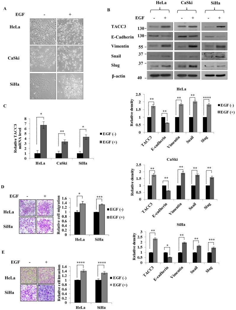
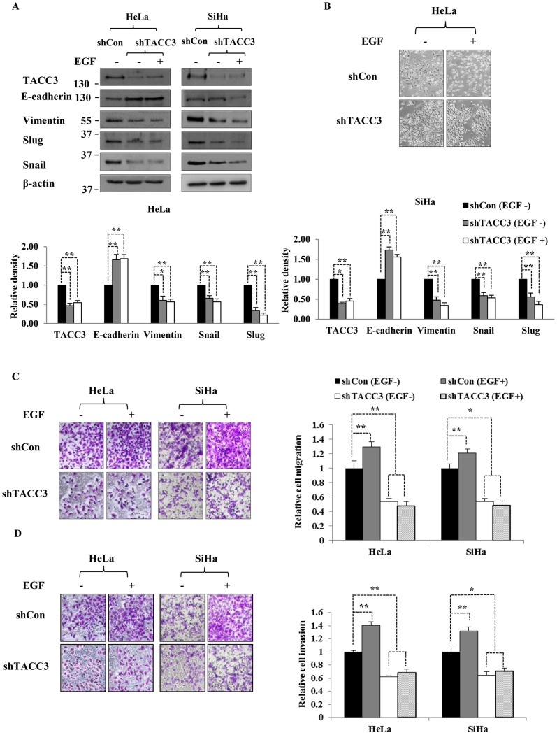
The third member of transforming acidic coiled-coil protein (TACC) family, TACC3, has been shown to be an important player in the regulation of centrosome/microtubule dynamics during mitosis and found to be deregulated in a variety of human malignancies. Our previous studies have suggested that TACC3 may be involved in cervical cancer progression and chemoresistance, and its overexpression can induce epithelial-mesenchymal transition (EMT) by activating the phosphatidylinositol 3-kinase (PI3K)/Akt and extracellular signal-regulated protein kinases (ERKs) signal transduction pathways. However, the upstream mechanisms of TACC3-mediated EMT and its functional/clinical importance in human cervical cancer remain elusive. Epidermal growth factor (EGF) has been shown to be a potent inducer of EMT in cervical cancer and associated with tumor invasion and metastasis. In this study, we found that TACC3 is overexpressed in cervical cancer and can be induced upon EGF stimulation. The induction of TACC3 by EGF is dependent on the tyrosine kinase activity of the EGF receptor (EGFR). Intriguingly, depletion of TACC3 abolishes EGF-mediated EMT, suggesting that TACC3 is required for EGF/EGFR-driven EMT process. Moreover, Snail, a key player in EGF-mediated EMT, is found to be correlated with the expression of TACC3 in cervical cancer. Collectively, our study highlights a novel function for TACC3 in EGF-mediated EMT process and suggests that targeting of TACC3 may be an attractive strategy to treat cervical cancers driven by EGF/EGFR signaling pathways.


Conflict of interest statement

Competing Interests: The authors have declared that no competing interests exist.

Figures

Figure 1
Figure 1. TACC3 is overexpressed in cervical cancer cell lines and tissues.
(A) The expression of TACC3 in Ect1/E6E7 (HPV-immortalized ectocervical epithelial), CaSki (HPV-16), C33A (HPV-negative), SiHa (HPV-16) and HeLa (HPV-18) cell lines was determined by western blot analysis. The expression levels were compared to three normal cervix tissues. β-actin was used as a loading control. The intensity of bands was quantified using imageJ software and normalized to β-actin. Data shown are means ± SD of at least three independent experiments. (B) Representative immunohistochemical staining on cervical cancer tissue microarray. Quantitative analysis of cervical cancer tissue microarrays showed that the expression of TACC3 is higher in cervical cancer than in normal cervix, but its expression does not correlate with tumor stage (C) or grade (D). Data shown are means ± SD of at least three independent experiments. *, p<0.05; **, p<0.001.
Figure 2
Figure 2. EGF stimulation induces the expression of TACC3.
(A) Cervical cancer cells treated with EGF showed a morphological change associated with EMT. (B and C) Both protein (B) and mRNA (C) levels of TACC3 were up-regulated upon EGF stimulation, along with down-regulation of E-cadherin and up-regulation of Vimentin, Snail and Slug. β-actin was used as loading control. The intensity of bands was quantified using imageJ software and normalized to β-actin. The mRNA level of TACC3 was represented relative to β-actin transcripts. Data shown are means ± SD of at least three independent experiments. (D and E) HeLa and SiHa cells treated with or without EGF were subjected to transwell migration (D) and Matrigel invasion assays (E) (see Materials and Methods). Cells were incubated with or without 50 ng/ml of EGF for 24 h. Data shown are means ± SD of at least three independent experiments. *, p<0.05; **, p<0.01; ***, p<0.005; ****, p<0.001.
Figure 3
Figure 3. TACC3 is induced upon EGF stimulation in EGFR-expressing cells.
C33A cells treated with EGF did not show significant changes in cell morphology (A), expression of TACC3 and EMT markers (B), or motility (C). Cells were incubated with or without 50 ng/ml of EGF for 24 h and then subjected to western blot and transwell migration assays. The intensity of bands was quantified using imageJ software and normalized to β-actin. Data shown are means ± SD of at least three independent experiments. (D) The expression of EGFR in Ect1/E6E7, CaSki, C33A, SiHa and HeLa cell lines was determined by western blot analysis. β-actin was used as a loading control. The intensity of bands was quantified using imageJ software and normalized to β-actin. Data shown are means ± SD of at least three independent experiments.
Figure 4
Figure 4. EGF-mediated TACC3 induction is dependent on EGFR activation.
The inhibition of tyrosine kinase activity of EGFR abolished a morphological change associated with EMT (A) and EGF-mediated TACC3 induction (B). Cells were treated with EGF or EGF+AG1478 for 24 h and then subjected to western blot analysis. β-actin was used as a loading control. The intensity of bands was quantified using imageJ software and normalized to β-actin. Data shown are means ± SD of at least three independent experiments. *, p<0.001.
Figure 5
Figure 5. TACC3 is required for EGF-mediated EMT.
In the absence of TACC3, EGF was not able to regulate EMT markers (A), alter cell morphology (B), or enhance cell migration and invasion capabilities (C and D). Cells were transiently transfected with shRNAs against control (scrambled, shCon) or TACC3 (shTACC3) and treated with or without EGF for 24 h. Cells were then subjected to western blot, transwell migration and Matrigel invasion assays. The intensity of bands was quantified using imageJ software and normalized to β-actin. Data shown are means ± SD of at least three independent experiments. *, p<0.01; **, p<0.001.
Figure 6
Figure 6. A correlation between TACC3 and Snail expression in cervical cancer tissue microarray.
(A) Representative immunohistochemical staining of TACC3 and Snail on cervical cancer tissue microarray. Quantitative analysis of cervical cancer tissue microarrays showed that (B) the expression of Snail is higher in cervical cancer than in normal cervix. Data shown are means ± SD of at least three independent experiments. *, p<0.001 (C) Snail expression correlates with TACC3 expression (r = 0.80383, p<0.0001).

Similar articles

Cited by

References

    1. Turley EA, Veiseh M, Radisky DC, Bissell MJ (2008) Mechanisms of disease: epithelial-mesenchymal transition–does cellular plasticity fuel neoplastic progression? Nat Clin Pract Oncol 5: 280–290. - PMC - PubMed
    1. Kalluri R, Weinberg RA (2009) The basics of epithelial-mesenchymal transition. J Clin Invest 119: 1420–1428. - PMC - PubMed
    1. Huber MA, Kraut N, Beug H (2005) Molecular requirements for epithelial-mesenchymal transition during tumor progression. Curr Opin Cell Biol 17: 548–558. - PubMed
    1. Thiery JP, Acloque H, Huang RY, Nieto MA (2009) Epithelial-mesenchymal transitions in development and disease. Cell 139: 871–890. - PubMed
    1. Tanos B, Rodriguez-Boulan E (2008) The epithelial polarity program: machineries involved and their hijacking by cancer. Oncogene 27: 6939–6957. - PubMed

MeSH terms