. 2013 Aug 6;8(8):e70373.
doi: 10.1371/journal.pone.0070373. Print 2013.

Identification and immunocytochemical characterization of Piccolino, a novel Piccolo splice variant selectively expressed at sensory ribbon synapses of the eye and ear

Affiliations

Identification and immunocytochemical characterization of Piccolino, a novel Piccolo splice variant selectively expressed at sensory ribbon synapses of the eye and ear

Hanna Regus-Leidig et al. PLoS One. .

Abstract

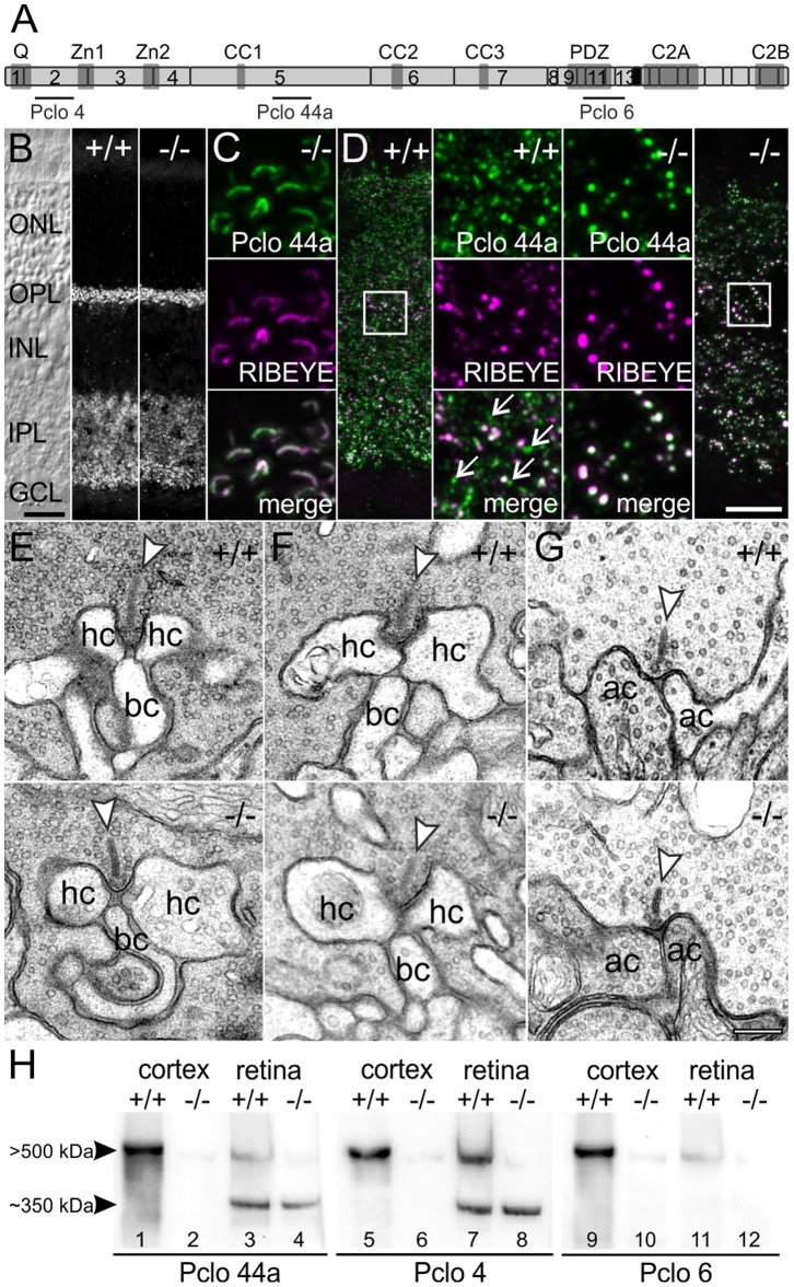
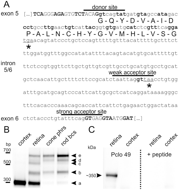
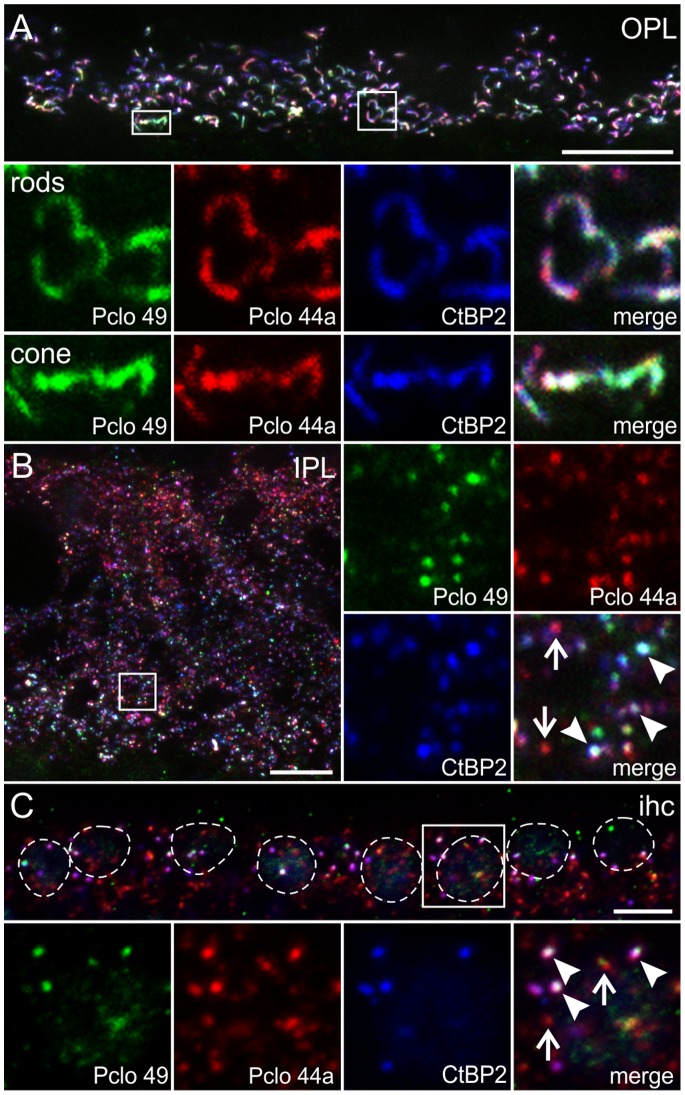
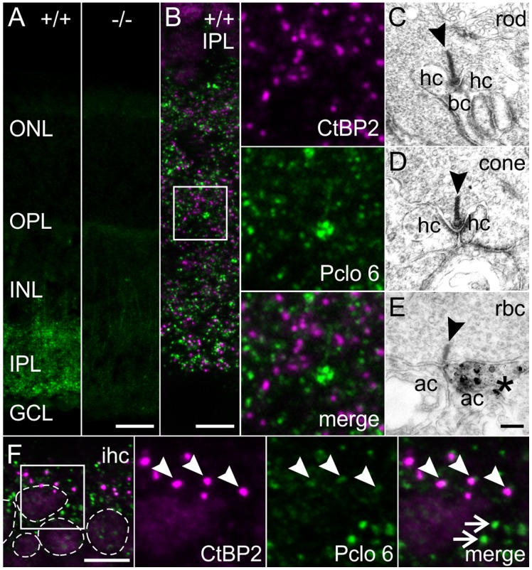
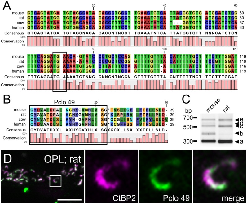
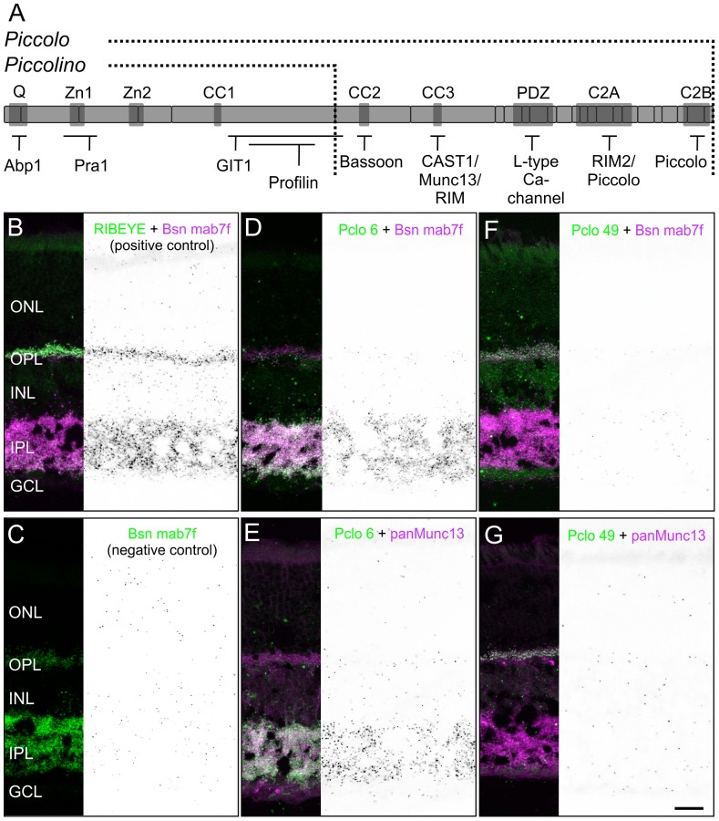
Piccolo is one of the largest cytomatrix proteins present at active zones of chemical synapses, where it is suggested to play a role in recruiting and integrating molecules relevant for both synaptic vesicle exo- and endocytosis. Here we examined the retina of a Piccolo-mutant mouse with a targeted deletion of exon 14 in the Pclo gene. Piccolo deficiency resulted in its profound loss at conventional chemical amacrine cell synapses but retinal ribbon synapses were structurally and functionally unaffected. This led to the identification of a shorter, ribbon-specific Piccolo variant, Piccolino, present in retinal photoreceptor cells, bipolar cells, as well as in inner hair cells of the inner ear. By RT-PCR analysis and the generation of a Piccolino-specific antibody we show that non-splicing of intron 5/6 leads to premature translation termination and generation of the C-terminally truncated protein specifically expressed at active zones of ribbon synapse containing cell types. With in situ proximity ligation assays we provide evidence that this truncation leads to the absence of interaction sites for Bassoon, Munc13, and presumably also ELKS/CAST, RIM2, and the L-type Ca(2) (+) channel which exist in the full-length Piccolo at active zones of conventional chemical synapses. The putative lack of interactions with proteins of the active zone suggests a function of Piccolino at ribbon synapses of sensory neurons different from Piccolo's function at conventional chemical synapses.


Conflict of interest statement

Competing Interests: The authors have declared that no competing interests exist.

Figures

Figure 1
Figure 1. Presence of Pclo at retinal ribbon synapses in the Pclo-mutant mouse.
A: Schematic representation of Pclo. The exons (numbered light gray boxes), interaction domains (dark gray boxes), and epitope locations for the three polyclonal antibodies Pclo 4, Pclo 44a, and Pclo 6 are shown. Exon 14 (black box) is deleted in the Pclo-mutant (−/−) mouse. B: Nomarski micrograph and images of vertical sections through wild-type (+/+) and −/− retina stained with Pclo 44a. C: Synaptic ribbons in the outer plexiform layer (OPL) of the −/− retina double labeled for Pclo (Pclo 44a; green) and RIBEYE (magenta). D: Inner plexiform layer (IPL) in the +/+ and −/− retina double labeled for Pclo (Pclo 44a; green) and RIBEYE (magenta). Arrows depict single Pclo positive puncta. E-G: Electron micrographs of rod (E) and cone (F) photoreceptor, and rod bipolar cell (G) ribbon synapses from +/+ and −/− retina. Arrowheads point to synaptic ribbons. H: Western blots of cortex and retina synaptosomal fractions from +/+ and −/− mice probed with the three different Pclo antibodies. ONL: outer nuclear layer; INL: inner nuclear layer; GCL: ganglion cell layer; hc: horizontal cell; bc: bipolar cell; ac: amacrine cell; kDa: kilo-Dalton. Scale bar in B: 20 µm, D: 10 µm, E–G: 200 nm.
Figure 2
Figure 2. Intron retention generates a C-terminally truncated ribbon synapse specific Pclo variant.
A: Nucleotide sequence of intron 5/6 in the Pclo gene (lower case letters) with flanking exon regions (capital letters). Codons are demarcated through alternating bold and non-bold letters, and the conventionally used donor and strong acceptor site, and a hypothetical alternative weak acceptor site are indicated with black lines. Utilization of the weak acceptor site as well as complete intron retention would result in in-frame stop codons (asterisks). The amino acid sequence used for the generation of Pclo 49 is displayed beneath the nucleotide sequence. B: RT-PCR of cDNA from cortex, retina, isolated cone photoreceptor (cone phrs) and rod bipolar cells (rod bcs) with primers flanking intron 5/6 in the Pclo gene. C: Western blot of wild-type retina and cortex synaptosomal fractions probed with Pclo 49 against the first 23 amino acids of intron 5/6 in the Pclo gene. Pclo 49 labels a ∼350 kDa band in the retina, but not in cortex (left panel). Pre-incubation of Pclo 49 with the antigenic peptide completely abolished the labeling (right panel). bp: base pairs; kDa: kilo-Dalton.
Figure 3
Figure 3. Localization of Piccolino at different types of ribbon synapses in the mouse.
A: Outer plexiform layer (OPL) of +/+ retina triple labeled with Pclo 49 (labels Piccolino; green), Pclo 44a (labels Pclo and Piccolino; red), and an antibody against CtBP2/RIBEYE (blue). B: Inner plexiform layer (IPL) of +/+ retina triple labeled with Pclo 49 (labels Piccolino; green), Pclo 44a (labels Pclo and Piccolino; red), and an antibody against CtBP2/RIBEYE (blue). Arrowheads point to ribbon synapses, arrows demarcate Pclo 44a single stained conventional chemical synapses. C: Inner hair cells (ihc) triple labeled with Pclo 49 (labels Piccolino; green), Pclo 44a (labels Pclo and Piccolino; red), and an antibody against CtBP2/RIBEYE (blue). Nuclei (stained with DAPI, not shown) are circled with dotted lines. Arrowheads point to ribbon synapses, arrows demarcate conventional chemical synapses. Scale bar in A,B: 10 µm, C: 5 µm.
Figure 4
Figure 4. Localization of full-length Pclo at different types of ribbon synapses.
A: Wild-type (+/+) and Pclo-mutant (−/−) retinae stained with the C-terminally binding Pclo 6 against full-length Pclo. B: Inner plexiform layer (IPL) of +/+ retina double labeled for full-length Pclo (Pclo 6; green) and CtBP2/RIBEYE (magenta). C–E: Pre-embedding immunoelectron micrographs of a rod photoreceptor (C), cone photoreceptor (D), and rod bipolar cell (rbc) ribbon synapse (E) in the +/+ retina stained with Pclo 6. Only amacrine cell synapses (E; asterisk) and never ribbon synapses (CE; arrowheads) were stained for full-length Pclo. F: Inner hair cells (ihc) double labeled for full-length Pclo (Pclo 6; green) and CtBP2/RIBEYE (magenta). Nuclei (stained with DAPI, not shown) are circled with dotted lines. Arrowheads point to ribbon synapses, arrows demarcate conventional chemical synapses. ONL: outer nuclear layer; OPL: outer plexiform layer; INL: inner nuclear layer; GCL: ganglion cell layer. hc: horizontal cell; bc: bipolar cell; ac: amacrine cell. Scale bar in A,B: 20 µm, C-E: 200 nm, F: 5 µm.
Figure 5
Figure 5. Piccolino and full-length Pclo expression in different species.
A: Sequence comparison of the first 120 nucleotides of the Pclo intron 5/6 between mouse, rat, cow, and human. Note the 100% conservation of the stop codon in all four species (TGA; boxed region). B: Amino acid sequence comparison of the translation product derived from (A) between mouse, rat, cow, and human. The homology of the translated sequence (boxed region) ranges from 59% between mouse and cow, and 86% between mouse and rat. C: Comparative RT-PCR of mouse and rat retinal cDNA with primers flanking intron 5/6 of the Pclo gene (see also Figure 2). Like in the mouse retina, also in the rat retina four additional amplicons (b–e) were detected in addition to the strongly expressed conventionally spliced Pclo transcript (a), with (e) representing the completely retained intron 5/6 of the Pclo gene. D: Representative image of the outer plexiform layer (OPL) of PFA-fixed vertical sections through rat retina double stained with antibodies against CtBP2/RIBEYE (magenta) and Piccolino (Pclo 49; green). Scale bar in D: 5 µm.
Figure 6
Figure 6. Scotopic and photopic ERG recordings from wild-type (+/+) and Pclo-mutant (−/−) mice.
A: The mean (± sd) amplitude of the scotopic a-wave of +/+ (gray) and −/− mice (filled circles) increased with increasing flash intensity. There was no difference between +/+ and −/− mice. B: The mean (± sd) latency of the scotopic a-wave of +/+ and −/− mice decreased with increasing flash intensity. There was no significant difference between +/+ and −/− mice. C: The mean (± sd) amplitude of the scotopic b-wave of +/+ and −/− mice increased with increasing flash intensity in both +/+ and −/− mice. D: The mean latency of the scotopic b-wave decreased with increasing flash intensity in both +/+ and −/− mice. The asterisk indicates a significant difference between +/+ and −/− mice at a flash strength of 0.0002 cd.s/m2 (p<0.05). E: The mean (± sd) amplitude of the photopic b-wave increased with increasing flash intensity. There was no difference between +/+ and −/− mice. F: The mean latency of the photopic b-wave increased with increasing flash intensity. The b-wave latency of −/− mice was significantly increased (p<0.0001) by approximately 2 ms.
Figure 7
Figure 7. Missing interactions of Piccolino with Bsn and Munc13.
A: Schematic representation of full-length Pclo with its interaction domains (dark gray boxes) and known binding partners. The C-terminally truncated Piccolino lacks the C-terminal interactions. B–G: In situ proximity ligation assays (PLA) on vertical sections through wild-type retina (black and white panels) with corresponding fluorescence stainings. Positive control: interaction of RIBEYE and Bsn with the antibodies RIBEYE (green) and Bsn mab7f (magenta; B). Negative control: antibody Bsn mab7f (green) alone (C). Interaction of full-length Pclo with Bsn (D) and Munc13 (E) probed with the antibodies Pclo 6 (green), Bsn mab7f (magenta), and panMunc13 (magenta). Interaction of Piccolino with Bsn (F) and Munc13 (G) probed with the antibodies Pclo 49 (green), Bsn mab7f (magenta), and panMunc13 (magenta). ONL: outer nuclear layer; OPL: outer plexiform layer; INL: inner nuclear layer; IPL: inner plexiform layer; GCL: ganglion cell layer. Scale bar: 20 µm.

References

    1. Gundelfinger ED, Fejtová A (2012) Molecular organization and plasticity of the cytomatrix at the active zone. Curr Opin Neurobiol 22: 423–30. - PubMed
    1. Südhof TC (2012) The presynaptic active zone. Neuron 75: 11–25. - PMC - PubMed
    1. Zhai RG, Bellen HJ (2004) The architecture of the active zone in the presynaptic nerve terminal. Physiology (Bethesda) 19: 262–70. - PubMed
    1. tom Dieck S, Brandstätter JH (2006) Ribbon synapses of the retina. Cell Tissue Res 326: 339–46. - PubMed
    1. Matthews G, Fuchs P (2010) The diverse roles of ribbon synapses in sensory neurotransmission. Nat Rev Neurosci 11: 812–22. - PMC - PubMed

Publication types

MeSH terms

LinkOut - more resources