. 2013 Aug 2;8(8):e71410.
doi: 10.1371/journal.pone.0071410. Print 2013.

Aging does not enhance experimental cigarette smoke-induced COPD in the mouse

Affiliations

Aging does not enhance experimental cigarette smoke-induced COPD in the mouse

Steven Zhou et al. PLoS One. .

Abstract

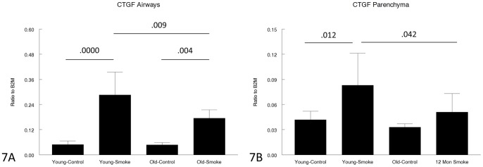
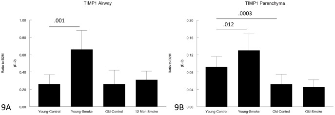
It has been proposed that the development of COPD is driven by premature aging/premature senescence of lung parenchyma cells. There are data suggesting that old mice develop a greater inflammatory and lower anti-oxidant response after cigarette smoke compared to young mice, but whether these differences actually translate into greater levels of disease is unknown. We exposed C57Bl/6 female mice to daily cigarette smoke for 6 months starting at age 3 months (Ayoung@) or age 12 months (Aold@), with air-exposed controls. There were no differences in measures of airspace size between the two control groups and cigarette smoke induced exactly the same amount of emphysema in young and old. The severity of smoke-induced small airway remodeling using various measures was identical in both groups. Smoke increased numbers of tissue macrophages and neutrophils and levels of 8-hydroxyguanosine, a marker of oxidant damage, but there were no differences between young and old. Gene expression studies using laser capture microdissected airways and parenchyma overall showed a trend to lower levels in older animals and a somewhat lesser response to cigarette smoke in both airways and parenchyma but the differences were usually not marked. Telomere length was greatest in young control mice and was decreased by both smoking and age. The senescence marker p21(Waf1) was equally upregulated by smoke in young and old, but p16(INK4a), another senescence marker, was not upregulated at all. We conclude, in this model, animal age does not affect the development of emphysema and small airway remodeling.


Conflict of interest statement

Competing Interests: The authors have declared that no competing interests exist.

Figures

Figure 1
Figure 1. Measures of emphysema.
1A: Mean airspace size is increased by a similar amount in young and old animals. 1B. Surface to volume ratio is decreased by a similar amount in young and old animals. Data are mean ± SD. N = 5 to 7 animals/group. P values as shown.
Figure 2
Figure 2. Measures of small airway remodeling.
There are no differences in airway wall thickness (2A), wall area per unit basement membrane length (2B) or collagen area per unit basement membrane length (2C) comparing young and old animals. Data are mean ± SD. N = 5 to 7 animals/group. P values as shown.
Figure 3
Figure 3. Tissue (parenchymal) inflammatory cells.
Tissue neutrophils (3A) are increased by smoke in both young and old animals, but to a slightly lesser extent in old animals. Tissue macrophages (3B) are increased to the same extend by smoke in young and old animals. Data are mean ± SD. N = 5 animals/group. P values as shown.
Figure 4
Figure 4. Small airway oxidative damage measured by 8-OH Guanosine staining.
Smoke causes a similar increase in both young and old animals. Data are mean ± SD. N = 5 animals/group. P values as shown.
Figure 5
Figure 5. Gene expression of collagen 1α1 in laser capture microdissected airways (bronchioles) (5A) and parenchyma (5B).
Levels of collagen 1α1 are lower in the old animals. Data are mean ± SD. N = 5 animals/group. P values as shown.
Figure 6
Figure 6. Gene expression of collagen 3α1 in laser capture microdissected airways (bronchioles) (6A) and parenchyma (6B).
Levels of collagen 3α1 are lower in the old animals. The decrease in parenchymal collagen 3α1 with smoke exposure suggests that the parenchyma fails to repair smoke-induced damage. Data are mean ± SD. N = 5 animals/group. P values as shown.
Figure 7
Figure 7. Gene expression of CTGF in laser capture microdissected airways (bronchioles) (7A) and parenchyma (7B).
CTGF functions as a proximate mediator of TGFβ-induced matrix production, and is increased by smoke in both young and old animals, albeit to a lesser level in old animals. Data are mean ± SD. N = 5 animals/group. P values as shown.
Figure 8
Figure 8. Gene expression of VEGF in laser capture microdissected airways (bronchioles) (8A) and parenchyma (8B).
VEGF is believed to be a trophic factor for normal lung parenchymal maintenance and is decreased old compared to young animals. Data are mean ± SD. N = 5 animals/group. P values as shown.
Figure 9
Figure 9. Gene expression of TIMP1 in laser capture microdissected airways (bronchioles) (9A) and parenchyma (9B).
TIMP1 is upregulated by smoke in young animals, but not upregulated at all in old animals. Data are mean ± SD. N = 5 animals/group. P values as shown.
Figure 10
Figure 10. Gene expression of MMP-12 in laser capture microdissected airways (bronchioles) (10A) and parenchyma (10B).
MMP-12, which appears play an important role in the pathogenesis of both emphysema and small airway remodeling (see text) is upregulated by smoke in young animals and not upregulated at all in old animals. Data are mean ± SD. N = 5 animals/group. P values as shown.
Figure 11
Figure 11. Gene expression of GM-CSF in laser capture microdissected airways (bronchioles) (11A) and parenchyma (11B).
GM-CSF acts as an inflammatory cell chemoattractant in the lung and is upregulated by smoke in young animals, but not affected by smoke in old animals. Data are mean ± SD. N = 5 animals/group. P values as shown.
Figure 12
Figure 12. Gene expression of KC in laser capture microdissected airways (bronchioles) (12A) and parenchyma (12B).
KC is a neutrophil chemoattractant and is upregulated by smoke in both young and old animals, but to a lesser extent in old animals. Data are mean ± SD. N = 5 animals/group. P values as shown.
Figure 13
Figure 13. Gene expression of MIP-2 in laser capture microdissected airways (bronchioles) (13A) and parenchyma (13B).
MIP-2 is a neutrophil chemoattractant and is upregulated by smoke in both young and old animals. Data are mean ± SD. N = 5 animals/group. P values as shown.
Figure 14
Figure 14. Gene expression of Nrf2 in laser capture microdissected airways (bronchioles) (14A) and parenchyma (14B).
Nrf2 regulates the anti-oxidant response and is upregulated by smoke in both young and old animals, but to a lesser extent in old animals. Data are mean ± SD. N = 5 animals/group. P values as shown.
Figure 15
Figure 15. Gene expression of CYP1B1 in laser capture microdissected airways (bronchioles) (15A) and parenchyma (15B).
CYP1B1 is a phase 1 detoxifying is upregulated by smoke in both young and old animals, but to a lesser extent in the airways in old animals. Data are mean ± SD. N = 5 animals/group. P values as shown.
Figure 16
Figure 16. Gene expression of ALDH3A in laser capture microdissected airways (bronchioles) (16A) and parenchyma (16B).
ALDH3A is a detoxifying enzyme and is upregulated by smoke in the airways in both young and old animals, but to a lesser extent in old animals, and not upregulated in the parenchyma in old animals. Data are mean ± SD. P values as shown.
Figure 17
Figure 17. Gene expression of NQO1 in laser capture microdissected airways (bronchioles) (17A) and parenchyma (17B).
NQO1 is an anti-oxidant/free radical scavenger and is upregulated by smoke in both young and old animals, but to a lesser extent in old animals. Data are mean ± SD. N = 5 animals/group. P values as shown.
Figure 18
Figure 18. Gene expression of p21WAF1 in laser capture microdissected airways (bronchioles) (18A) and parenchyma (18B).
p21WAF1 is believed to be a marker of senescence and is upregulated by smoke to the same extent in young and old animals. Data are mean ± SD. N = 5 animals/group. P values as shown.
Figure 19
Figure 19. Gene expression of p16INK4a in laser capture microdissected airways (bronchioles) (19A) and parenchyma (19B).
p16INK4a prevents cell cycle progression and is believed to be a marker of senescence. Expression is not statistically changes by smoke exposure in young or old animals. Data are mean ± SD. N = 5 animals/group. P values as shown.
Figure 20
Figure 20. Comparison of telomere length between smoked and control mice segregated by age.
Telomere length is greatest in young control animals and is decreased by smoke. Telomere length is also decreased by age but not affected by smoke in old animals. Data are shown as median and interquartile range (box). N = 5 animals/group. * P = 0.0055; † P = 0.0452.

References

    1. MacNee W (2011) Aging, inflammation, and emphysema. Am J Respir Crit Care Med 184: 1327–1329. - PubMed
    1. Tuder RM, Kern JA, Miller YE (2012) Senescence in chronic obstructive pulmonary disease. Proc Am Thorac Soc. 9: 62–63. - PMC - PubMed
    1. Aoshiba K, Nagai A (2009) Senescence hypothesis for the pathogenetic mechanism of chronic obstructive pulmonary disease. Proc Am Thorac Soc. 6: 596–601. - PubMed
    1. Ito K, Barnes PJ (2009) COPD as a disease of accelerated lung aging. Chest. 135: 173–180. - PubMed
    1. Lee J, Sandford A, Man P, Sin DD (2011) Is the aging process accelerated in chronic obstructive pulmonary disease? Curr Opin Pulm Med 17: 90–97. - PubMed

Publication types

MeSH terms