. 2013 Aug 1;8(8):e71542.
doi: 10.1371/journal.pone.0071542. Print 2013.

Crucial role of perilipin-3 (TIP47) in formation of lipid droplets and PGE2 production in HL-60-derived neutrophils

Affiliations

Crucial role of perilipin-3 (TIP47) in formation of lipid droplets and PGE2 production in HL-60-derived neutrophils

Fuyuki Nose et al. PLoS One. .

Abstract

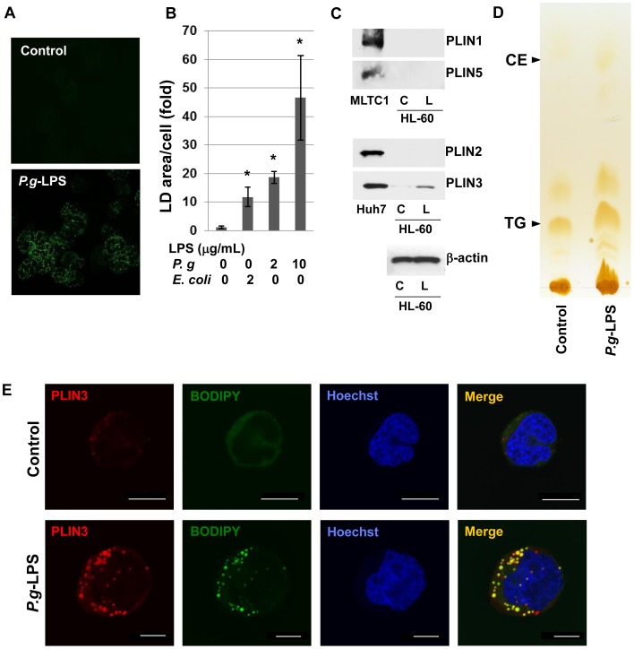
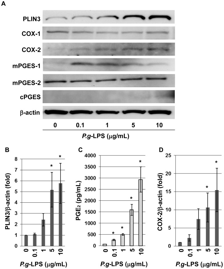
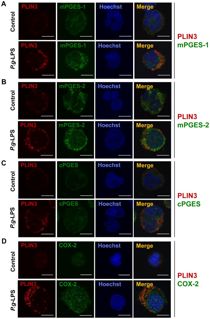
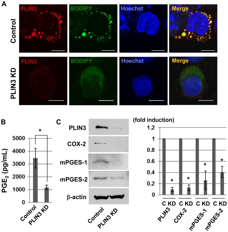
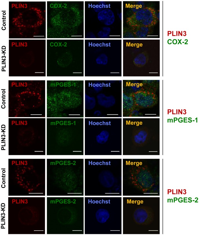
Cytosolic lipid droplets (LDs), which are now recognized as multifunctional organelles, accumulate in leukocytes under various inflammatory conditions. However, little is known about the characteristic features of LDs in neutrophils. In this study, we show that perilipin-3 (PLIN3; formerly called TIP47) is involved in LD formation and the inflammatory response in HL-60-derived neutrophils. HL-60, a promyelocytic cell line, was differentiated into neutrophils via treatment with all-trans retinoic acid. After differentiation, cells were stimulated with Porphyromonas gingivalis lipopolysaccharide (P.g-LPS), a major pathogen in adult periodontitis. When HL-60-derived neutrophils were stimulated with P.g-LPS, LDs increased in both number and size. In the differentiated cells, PLIN3 was induced while PLIN1, PLIN2 and PLIN5 were not detected. PGE2 production and the PLIN3 protein level were increased by the P.g-LPS treatment of the cells in a dose-dependent manner. When PLIN3 was down-regulated with siRNA treatment, LDs essentially disappeared and the level of PGE2 secreted in the cell culture medium decreased by 65%. In addition, the suppression of PLIN3 repressed the PGE2 producing enzymes; i.e., microsomal PGE synthase-1, -2 and cyclooxygenase-2. These findings indicate that PLIN3 has a pivotal role in LD-biogenesis in HL-60-derived neutrophils, and that PLIN3 is associated with the synthesis and secretion of PGE2.


Conflict of interest statement

Competing Interests: The authors have declared that no competing interests exist.

Figures

Figure 1
Figure 1. The size and number of LDs in HL-60-derived neutrophils were increased by P.g-LPS.
A: HL-60 cells were differentiated into neutrophils by treatment with 2 µM AtRA for 4 days and then cultured with or without 10 µg/mL P.g-LPS for 12 h. Cytosolic LDs were labeled with BODIPY493/503 and observed using confocal laser microscopy. B: BODIPY-stained LDs were quantified by the area of fluorescence per cell using NIH ImageJ software. The mean ± S.E. from at least three images were calculated. C: HL-60 neutrophils were incubated in the absence (C) or presence (L) of 10 µg/mL P.g-LPS for 12 h. The expression levels of PLIN1, PLIN2, PLIN3 and PLIN5 were detected by Western blotting. LDs recovered from the MLTC1 cells were used as a positive control for PLIN1 and PLIN5, and Huh7 LDs were for PLIN2 and PLIN3. D: HL-60 neutrophils were incubated in the absence or presence of 10 µg/mL P.g-LPS for 12 h. Total lipids extracted from the cells were separated on thin-layer chromatography. The lipids were visualized with iodine vapor, and the band intensity of TG was calculated using ImageJ. E: Differentiated HL-60 cells were treated with or without 10 µg/mL P.g-LPS for 12 h. Cells were fixed and labeled with BODIPY493/503 (green) and anti-PLIN3 pAb (red). Nuclei were stained with Hoechst33258. The cells were observed under confocal laser microscopy.
Figure 2
Figure 2. PLIN3 and PGE2 production were increased by P.g-LPS treatment in a dose-dependent manner.
HL-60-derived neutrophils were incubated with or without P.g-LPS for 12 h at the concentrations indicated. A: Expression levels of PLIN3, mPGES-1, mPGES-2, COX-1, COX-2, β-actin in HL-60-neutrophils were detected by Western blotting. B: The level of PLIN3 expression was determined using ImageJ software. C: The levels of PGE2 into the media were measured by EIA. D: The level of COX-2 expression was determined using ImageJ. Data are the mean ± SD of three independent experiments. * P<0.05 (Dunnett’s test, vs control).
Figure 3
Figure 3. PLIN3 and PGE2 synthesizing enzymes induced in the P.g-LPS-treated HL-60 neutrophils distribute differently.
Differentiated HL-60 cells were treated with or without 10 µg/mL P.g-LPS for 12 h. Cells were fixed and then double stained with anti-PLIN3 and either anti-mPGES-1, anti-mPGES-2, anti-cPGES or COX-2 pAbs. Nuclei were stained with Hoechst33258. The cells were observed under confocal laser microscopy.
Figure 4
Figure 4. PLIN3 knockdown in HL-60-derived neutrophils suppressed the formation of LDs and production of PGE2.
A: After treatment of HL-60-derived neutrophils with PLIN3 siRNA or control siRNA for 72 h, the cells were stimulated with 10 µg/mL P.g-LPS for 12 h. The cells were fixed and labeled with BODIPY493/503 and anti-PLIN3 pAb. Bar = 10 mm. B: Cells were treated as described for A, and the PGE2 released into the media was measured by EIA. C: Whole cell lysates were subjected to SDS-PAGE and the protein levels of mPGES-1, mPGES-2, COX-2 and β-actin were analyzed by Western blotting. The band intensities were calculated by ImageJ software. Data are the mean ± SD of three independent experiments. *P<0.05.
Figure 5
Figure 5. PLIN3 knockdown reduced the PGE2 synthesizing enzymes.
After PLIN3 or control siRNA treatment of HL-60-derived neutrophils for 2 days, cells were stimulated with P.g-LPS for 12 h, as in Fig. 4. Cells were fixed and then double stained with anti-PLIN3 and either COX-2, anti-mPGES-1, or anti-mPGES-2 pAbs. Nuclei were stained with Hoechst33258. The cells were observed under confocal laser microscopy.

References

    1. Bozza PT, Magalhaes KG, Weller PF (2009) Leukocyte lipid bodies: Biogenesis and functions in inflammation. Biochim Biophys Acta 1791: 540–551. - PMC - PubMed
    1. Tauchi-Sato K, Ozeki S, Houjou T, Taguchi R, Fujimoto T (2002) The surface of lipid droplets is a phospholipid monolayer with a unique fatty acid composition. J Biol Chem 227: 44507–44512. - PubMed
    1. Martin S, Patron RG (2006) Lipid droplets: a unified view of a dynamic organelle. Nat Rev Mol Cell Biol 7: 373–378. - PubMed
    1. Goodman JM (2009) Demonstrated and inferred metabolism associated with cytosolic lipid droplets. J Lipid Res 50: 2148–2156. - PMC - PubMed
    1. Lu X, Gruia-Gray J, Copeland NG, Gilbert DJ, Jenkins NA, et al. (2001) The murine perilipin gene: the lipid droplet-associated perilipins derive from tissue-specific, mRNA splice variants and define a gene family of ancient origin. Mamm Genome 12: 741–749. - PubMed

Publication types

MeSH terms