. 2013:2013:728529.
doi: 10.1155/2013/728529. Epub 2013 Jul 10.

p38 MAPK Signaling in Pemphigus: Implications for Skin Autoimmunity

Affiliations

p38 MAPK Signaling in Pemphigus: Implications for Skin Autoimmunity

Athanasios Mavropoulos et al. Autoimmune Dis. 2013.

Abstract

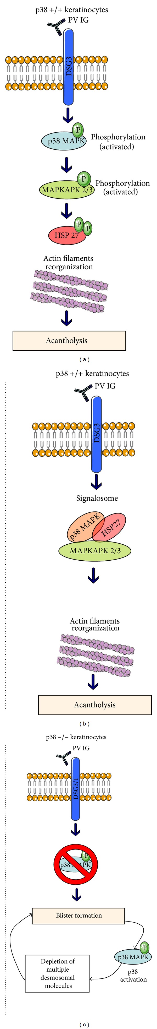
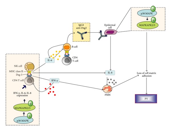
p38 mitogen activated protein kinase (p38 MAPK) signaling plays a major role in the modulation of immune-mediated inflammatory responses and therefore has been linked with several autoimmune diseases. The extent of the involvement of p38 MAPK in the pathogenesis of autoimmune blistering diseases has started to emerge, but whether it pays a critical role is a matter of debate. The activity of p38 MAPK has been studied in great detail during the loss of keratinocyte cell-cell adhesions and the development of pemphigus vulgaris (PV) and pemphigus foliaceus (PF). These diseases are characterised by autoantibodies targeting desmogleins (Dsg). Whether autoantibody-antigen interactions can trigger signaling pathways (such as p38 MAPK) that are tightly linked to the secretion of inflammatory mediators which may perpetuate inflammation and tissue damage in pemphigus remains unclear. Yet, the ability of p38 MAPK inhibitors to block activation of the proapoptotic proteinase caspase-3 suggests that the induction of apoptosis may be a consequence of p38 MAPK activation during acantholysis in PV. This review discusses the current evidence for the role of p38 MAPK in the pathogenesis of pemphigus. We will also present data relating to the targeting of these cascades as a means of therapeutic intervention.


Figures

Figure 1
Figure 1
The role of p38 MAPK in the induction of pemphigus vulgaris (PV). There are at least three potential p38-MAPK-related mechanisms involved in the pathogenesis and/or the progression of PV. (a) The binding of pathogenic autoantibodies targeting Dsg3 in keratinocytes initiates an array of signals leading to the activation of p38 MAPK cascade with subsequent phosphorylation of MAPKAPK (mitogen-activated protein kinase-activated protein kinases 2/3) and heat shock protein 27 (Hsp 27). The final outcome of these events is actin filaments reorganization and induction of acantholysis [19]; (b) p38 MAPK, MAPKAPK, and Hsp27 may form a complex (signalosome) that regulates the reorganization of actin filaments and the induction of acantholysis [20]; (c) studies in p38−/− keratinocytes demonstrate a p38 MAPK-independent blister formation. The subsequent activation of this pathway, however, can lead to de novo depletion of multiple desmosomal molecules, further facilitating spontaneous blister formation [21]. This latter hypothesis indicates that p38 MAPK signaling may not be responsible for the induction of PV but could play a role for the progression of the disease.
Figure 2
Figure 2
Schematic representation of the suggested interplay between natural killer (NK), CD4+ T, B cells, and polymorphonuclear cells (PMN) during pemphigus pathogenesis [22]. NK cells accumulate from the bloodstream to the epidermis and act as antigen-presenting cells by introducing desmoglein (Dsg3) peptides to CD4+ T cells. p38 MAPK phosphorylation within lymphoid subpopulations such as NK and CD4+ T cells induces expression of IFN-γ, IL-6, and IL-8 that amplify the inflammatory response, MHC-II presentation, and autoantibody production by B cells. The activated T cells further enhance B-cell anti-Dsg3 secretion and presentation to epidermal cells. Anti-Dsg3 activates the p38 MAPK pathway within keratinocytes leading to MAPK-activated protein kinase 2 (MK-2) mediated heat shock protein 27 (Hsp27) phosphorylation, actin microfilament reorganization, and acantholysis.

Similar articles

Cited by

References

    1. Dhabhar FS. Psychological stress and immunoprotection versus immunopathology in the skin. Clinics in Dermatology. 2013;31(1):18–30. - PubMed
    1. Moens E, Veldhoen M. Epithelial barrier biology: good fences make good neighbours. Immunology. 2012;135(1):1–8. - PMC - PubMed
    1. Eckert RL, Adhikary G, Balasubramanian S, et al. Biochemistry of epidermal stem cells. Biochim Biophys Acta. 2013;1830(2):2427–2434. - PMC - PubMed
    1. Metz-Boutigue M, Shooshtarizadeh P, Prevost G, Haikel Y, Chich J. Antimicrobial peptides present in mammalian skin and gut are multifunctional defence molecules. Current Pharmaceutical Design. 2010;16(9):1024–1039. - PubMed
    1. Bautista DM, Pellegrino M, Tsunozaki M. TRPA1: a gatekeeper for inflammation. Annual Review of Physiology. 2013;75:181–200. - PMC - PubMed