. 2013:2013:262410.
doi: 10.1155/2013/262410. Epub 2013 Jul 8.

The Correlation between Chronic Periodontitis and Oral Cancer

Affiliations

The Correlation between Chronic Periodontitis and Oral Cancer

Maximilian Krüger et al. Case Rep Dent. 2013.

Abstract

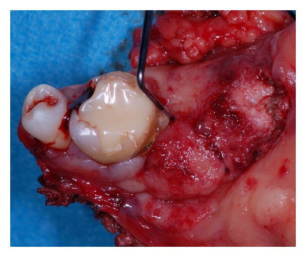
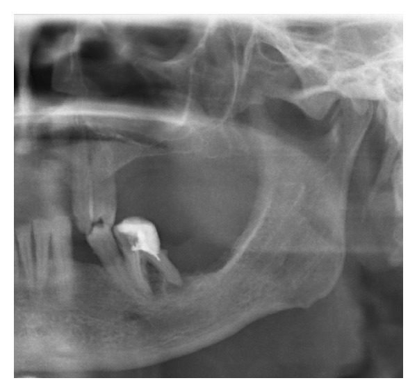
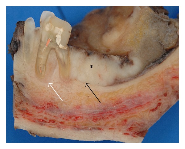
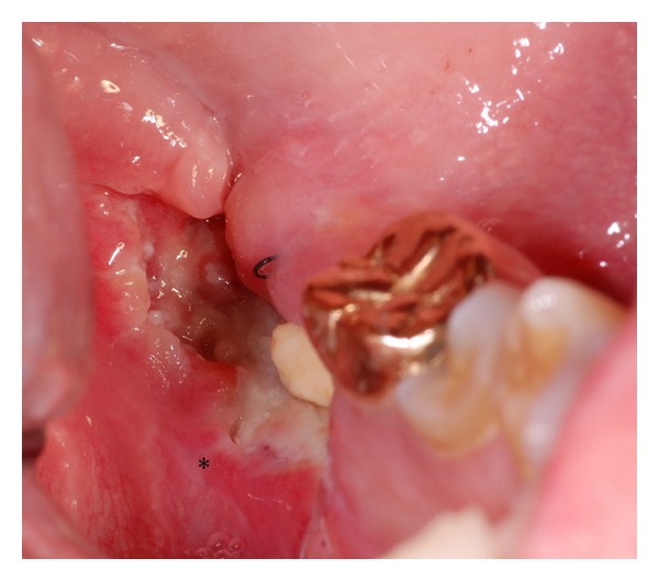
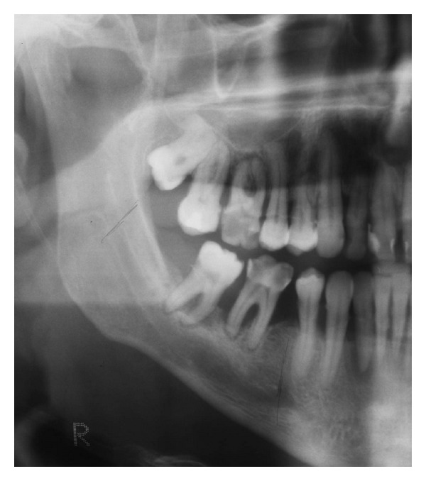
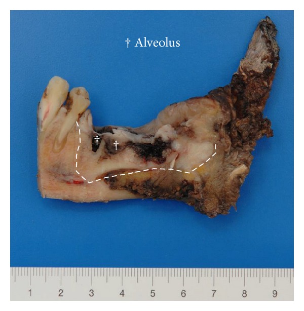
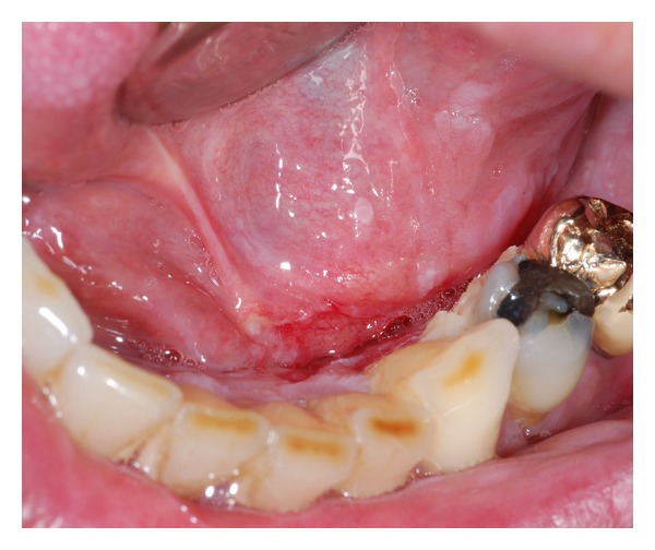
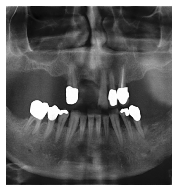
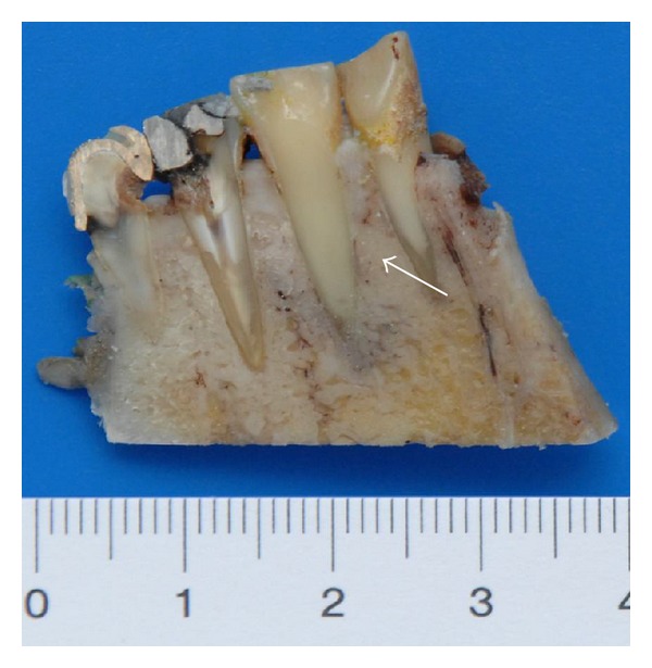
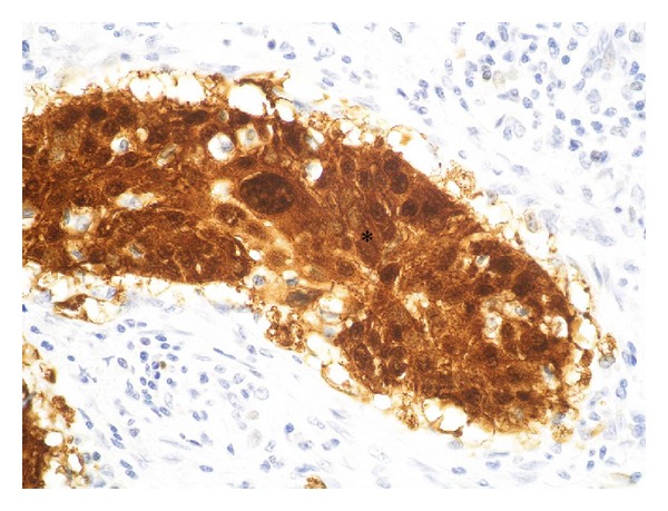
Infections are increasingly considered as potential trigger for carcinogenesis apart from risk factors like alcohol and tobacco. The discussion about human papilloma virus (HPV) in oral squamous cell carcinoma (OSCC) points at a general role of infection for the development of oral carcinomas. Furthermore, first studies describe a correlation between chronic periodontitis and OSCC, thus, characterizing chronic inflammation as being a possible trigger for OSCC. In front of this background, we present four well-documented clinical cases. All patients showed a significant anatomical relation between OSCC and clinical signs of chronic periodontitis. The interindividual differences of the clinical findings lead to different theoretical concepts: two with coincidental appearance of OSCC and chronic periodontitis and two with possible de novo development of OSCC triggered by chronic inflammation. We conclude that the activation of different inflammatory cascades by chronic periodontitis negatively affects mucosa and bone. Furthermore, the inflammatory response has the potential to activate carcinogenesis. Apart from a mere coincidental occurrence, two out of four patients give first clinical hints for a model wherein chronic periodontitis represents a potential risk factor for the development of OSCC.


Figures

Figure 1
Figure 1
Case  1—resection specimen of the mandible. The probe reveals the loss of clinical attachment within the periodontal space, and the tumor seems to emerge from the periodontal compartment.
Figure 2
Figure 2
Case  1—the preoperative OPTG exhibits severe horizontal and vertical bone loss of the jaws. In addition, peri- and interradicular osteolytic lesions around tooth 36 are present. A soft radiopacity as projection of the tumor mass can be identified along the intact alveolar crest distal of 36. Radiolucency, suspect for bone invasion is solely present at the interradicular and distal aspect of the roots.
Figure 3
Figure 3
Case  1—cross section specimen. The black arrow highlights the close relation of the distal root to the tumor mass (∗) with preserved cortical bone distal to the tooth. The white arrow points at a tumor formation along the mesial root filling out the interradicular space.
Figure 4
Figure 4
Case  2—the clinical aspect consists of a fibrin-coated ulcer in region of the wisdom tooth at the left lower mandible. The adjacent mucosa shows perifocal signs of inflammation (∗).
Figure 5
Figure 5
Case  2—Preoperative OPTG—A large formation of enhanced translucency is found in projection of the radices 36 and 37 as also at the pericoronal aspect of the partially retained wisdom tooth.
Figure 6
Figure 6
Case  2—cross section specimen with a sectional view from distal after removal of the bony aspect that contained the wisdom tooth. The tumor is in broad contact to the periodontium (arrow) with infiltration of the adjacent cancellous bone and the floor of the mouth.
Figure 7
Figure 7
Case  2—histological view of the cross section specimen after Haematoxglin-Eosin staining. The arrows mark the arrosion of the bone (∗) by the tumor cells.
Figure 8
Figure 8
Case  3—clinical situation. A partially ulcerous tumour formation covered the lingual aspect of the alveolar crest adjacent to the two distal molars (46 and 47). Nearly all teeth of this quadrant show clearly the sequelae of nearly nonexistent oral hygiene habits. Striae of leukoplakia are found on the wall of the ulcer, the tongue, and also along the vestibular papillae.
Figure 9
Figure 9
Case  3—preoperative OPTG. Beside multiple carious lesions the present X-ray examination reveals signs of chronic periodontal disease with a general loss of horizontal bone level, liberation of both dental roots, and bifurcations. The local maximum of destruction is found in the region of the last two molars. The retromolar triangle, however, appears to be intact.
Figure 10
Figure 10
Case  3—the sagittal split preparation of the resection specimen from a lingual view clearly shows the association of the tumor (borders marked with lines) to the dental alveoli (†) of the distal molars. The teeth 36 and 37 are lost due to the preparation process.
Figure 11
Figure 11
Case  4—a superficial ulcerous lesion atop of a mass at the left sublingual space with extension to the lingual aspect of the alveolar crest comprising the teeth 31 to 34 and surrounding leukoplakia.
Figure 12
Figure 12
Case  4—preoperative OPTG. A general horizontal loss of bone in both jaws with additional vertical translucencies in the front aspect of the mandible and signs of erosion of the alveolar crest at the left lower quadrant.
Figure 13
Figure 13
Case  4—the split resection specimen from a lingual view demonstrates the association of tumor formation along the root of the left lower canine (white arrows) as also further invasion of the cancellous bone.
Figure 14
Figure 14
Case  4—histological view of the tumor after immune-histochemical staining against p16. The brown signal (∗) proves the infection with HPV.

References

    1. Landis SH, Murray T, Bolden S, Wingo PA. Cancer Statistics, 1999. CA: A Cancer Journal for Clinicians. 1999;49(1):8–31. - PubMed
    1. Parkin DM, Bray F, Ferlay J, Pisani P. Global cancer statistics, 2002. CA: A Cancer Journal for Clinicians. 2005;55(2):74–108. - PubMed
    1. Rusin P, Markiewicz L, Majsterek I. Genetic predeterminations of head and neck cancer. Postępy Higieny i Medycyny Doświadczalnej. 2008;62:490–501. - PubMed
    1. Rödström P-O, Jontell M, Mattsson U, Holmberg E. Cancer and oral lichen planus in a Swedish population. Oral Oncology. 2004;40(2):131–138. - PubMed
    1. Pelucchi C, Gallus S, Garavello W, Bosetti C, Vecchia CL. Alcohol and tobacco use, and cancer risk for upper aerodigestive tract and liver. European Journal of Cancer Prevention. 2008;17(4):340–344. - PubMed

LinkOut - more resources