. 2013:2013:474272.
doi: 10.1155/2013/474272. Epub 2013 Jul 7.

Licochalcone A-induced human bladder cancer T24 cells apoptosis triggered by mitochondria dysfunction and endoplasmic reticulum stress

Affiliations

Licochalcone A-induced human bladder cancer T24 cells apoptosis triggered by mitochondria dysfunction and endoplasmic reticulum stress

Xuan Yuan et al. Biomed Res Int. 2013.

Abstract

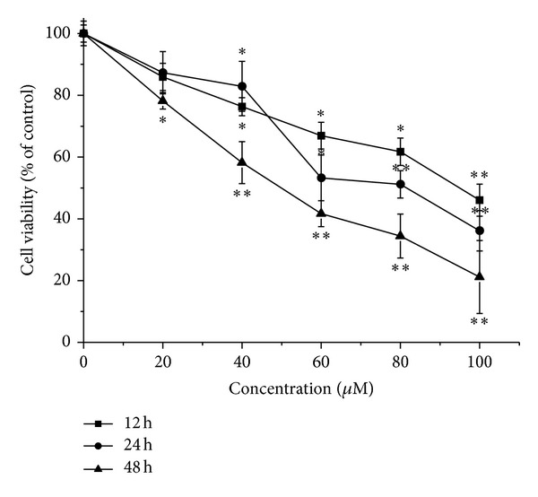
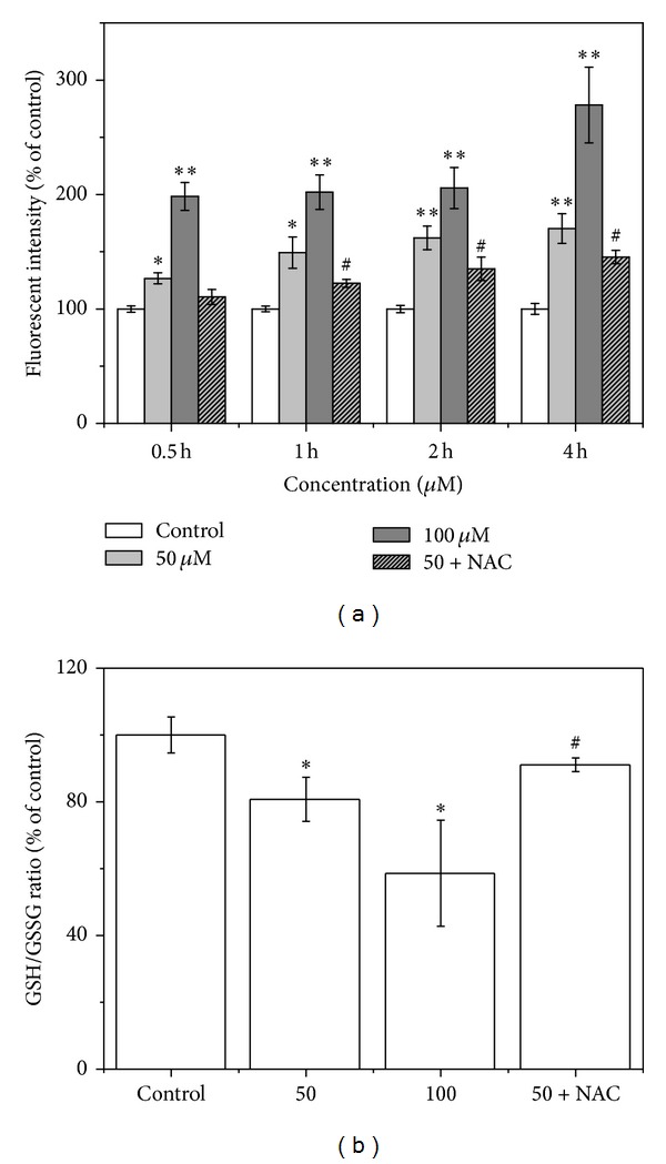
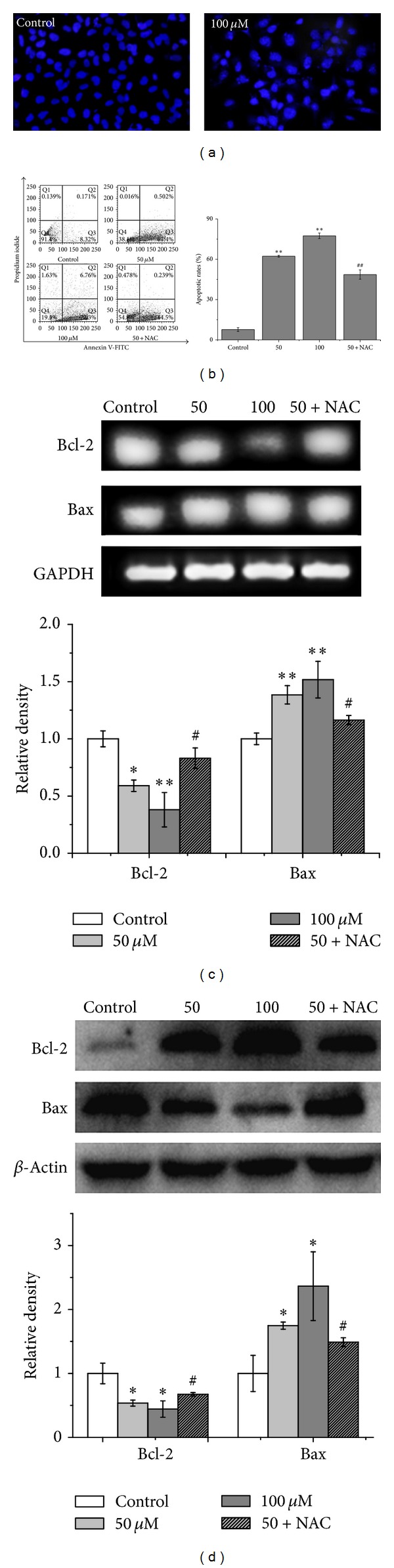
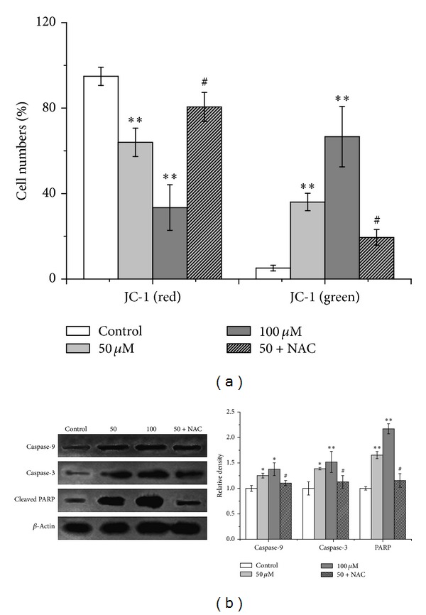
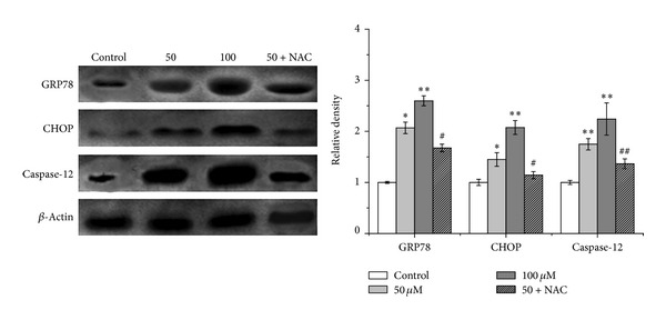
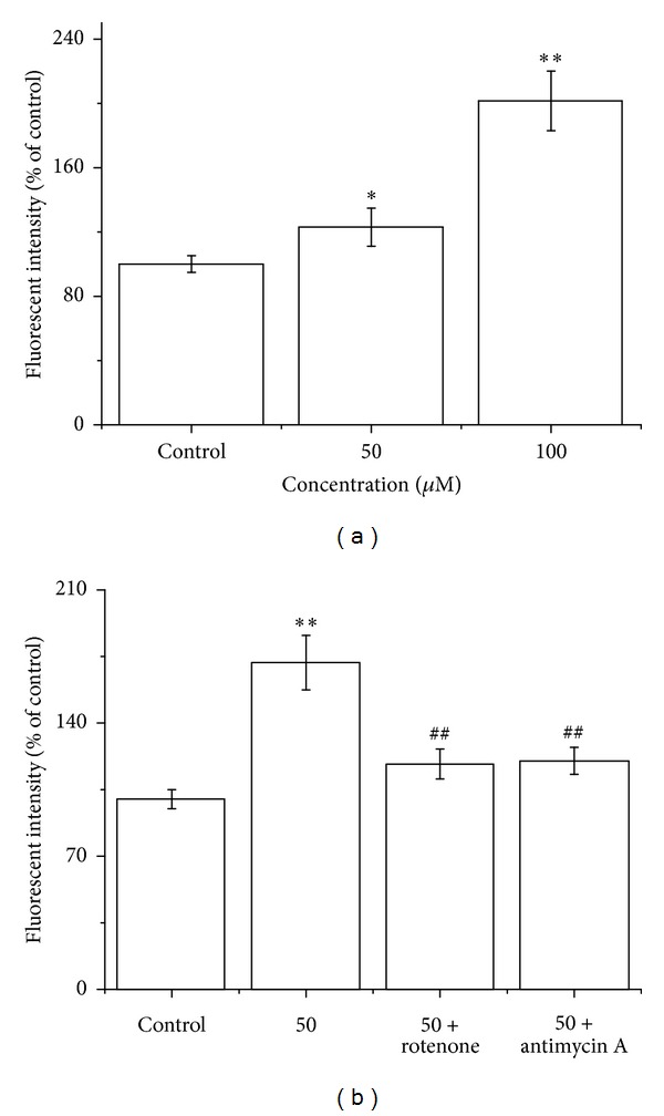
Licochalcone A (LCA), a licorice chalconoid, is considered to be a bioactive agent with chemopreventive potential. This study investigated the mechanisms involved in LCA-induced apoptosis in human bladder cancer T24 cells. LCA significantly inhibited cells proliferation, increased reactive oxygen species (ROS) levels, and caused T24 cells apoptosis. Moreover, LCA induced mitochondrial dysfunction, caspase-3 activation, and poly-ADP-ribose polymerase (PARP) cleavage, which displayed features of mitochondria-dependent apoptotic signals. Besides, exposure of T24 cells to LCA triggered endoplasmic reticulum (ER) stress; as indicated by the enhancement in 78 kDa glucose-regulated protein (GRP 78), growth arrest and DNA damage-inducible gene 153/C/EBP homology protein (GADD153/CHOP) expression, ER stress-dependent apoptosis is caused by the activation of ER-specific caspase-12. All the findings from our study suggest that LCA initiates mitochondrial ROS generation and induces oxidative stress that consequently causes T24 cell apoptosis via the mitochondria-dependent and the ER stress-triggered signaling pathways.


Figures

Figure 1
Figure 1
Effects of LCA on cell viability of T24 cells. Cell viability was determined by using an MTT staining assay. Treatment of T24 cells with varying LCA concentrations (0, 20, 40, 60, 80, or 100 μM) for 12, 24, or 48 h resulted in a significant concentration-dependent and time-dependent reduction in cell viability. The data represent the means ± SD of the three independent experiments. *P < 0.05,**P < 0.01 compared with the LCA-untreated control group cell.
Figure 2
Figure 2
Effects of LCA on ROS level and GSH/GSSG ratio in T24 cells. The cells were treated with LCA (0, 50, or 100 μM) with or without NAC (500 μM). (a) ROS level was determined via flow cytometry. (b) Oxidative stress was measured by GSH/GSSG ratio. Data are presented as the mean ± SD of the three independent experiments. *P < 0.05, **P < 0.01 compared with the control group; # P < 0.05 compared with the LCA group alone (50 μM).
Figure 3
Figure 3
LCA induced apoptosis in T24 cells. The cells were treated with or without the indicated amounts of LCA for 48 h with or without NAC (500 μM). (a) Morphologic measurements in T24 cells were carried out via Hoechst fluorescence staining. (b) Detection of apoptotic rates conducted via flow cytometry. (c) Bcl-2 and Bax expressions were analyzed via RT-PCR (upper), and quantitative analysis of Bcl-2 and Bax expressions mRNA levels (lower). (d) Bcl-2 and Bax expressions were analyzed via western blot (upper), and quantitative analysis of Bcl-2 and Bax protein expressions (lower). Control group (LCA-untreated group) level was accepted to be “1.0”. *P < 0.05, **P < 0.01 compared with the control group; # P < 0.05, ## P < 0.01 compared with the LCA group alone (50 μM).
Figure 4
Figure 4
LCA induced mitochondrial dysfunction, caspase cascade activation, and cleavage of poly(ADP-ribose) polymerase (PARP) in T24 cells. The cells were treated with or without LCA (0, 50, and 100 μM) with or without NAC (500 μM). (a) Mitochondrial membrane potential depolarization was determined by flow cytometry. The number of cells with normal polarized mitochondrial membranes (red) and cells with depolarized mitochondrial membranes (green) is expressed as a percentage of total cell number. (b) Caspase-9, caspase-3 expression, and PARP cleavage were examined via western blot analysis (left), and quantitative analysis of caspase-9, caspase-3 and PARP protein levels (right). Control group (LCA-untreated group) level was accepted to be “1.0”. *P < 0.05,**P < 0.01 compared with the LCB-untreated control group; # P < 0.05 compared with the LCA group alone (50 μM).
Figure 5
Figure 5
Effects of LCA on ER stress marker expression in T24 cells. The expression of GRP78, CHOP, and caspase-12 was analyzed via western blot analysis. Control group (LCB-untreated group) level was accepted to be “1.0”. *P < 0.05, **P < 0.01compared with the LCB-untreated control group; # P < 0.05, ## P < 0.01 compared with the LCA group alone (50 μM).
Figure 6
Figure 6
LCA induced mitochondrial ROS generation in T24 cells. (a) The cells were treated with LCA (0, 50, or 100 μM), and then mitochondrial ROS level was measured using DHR. (b) The cells were treated with LCA (0 or 50 μM) with or without mitochondrial complex I inhibitor rotenone (100 μM) or complex III inhibitor antimycin A (20 μM), and then ROS levels were measured using H2DCFDA. Data are presented as the means ± SD of the three independent experiments. *P < 0.05, **P < 0.01 compared with the control group; ## P < 0.01 compared with the LCA group alone (50 μM).

References

    1. Zieger K. High throughput molecular diagnostics in bladder cancer—on the brink of clinical utility. Molecular Oncology. 2008;1(4):384–394. - PMC - PubMed
    1. Parkin DM, Bray F, Ferlay J, Pisani P. Global cancer statistics, 2002. CA: A Cancer Journal for Clinicians. 2005;55(2):74–108. - PubMed
    1. Clark PE. Bladder cancer. Current Opinion in Oncology. 2007;19(3):241–247. - PubMed
    1. Pichu S, Krishnamoorthy S, Shishkov A, Zhang B, McCue P, Ponnappa BC. Knockdown of Ki-67 by dicer-substrate small interfering RNA sensitizes bladder cancer cells to curcumin-induced tumor inhibition. PLoS ONE. 2012;7(11)e48567 - PMC - PubMed
    1. Tan W, Lu J, Huang M, et al. Anti-cancer natural products isolated from chinese medicinal herbs. Chinese Medicine. 2011;6(article 27) - PMC - PubMed

Publication types

MeSH terms