. 2013:2013:535796.
doi: 10.1155/2013/535796. Epub 2013 Jul 8.

Cytotoxicity of biologically synthesized silver nanoparticles in MDA-MB-231 human breast cancer cells

Affiliations

Cytotoxicity of biologically synthesized silver nanoparticles in MDA-MB-231 human breast cancer cells

Sangiliyandi Gurunathan et al. Biomed Res Int. 2013.

Abstract

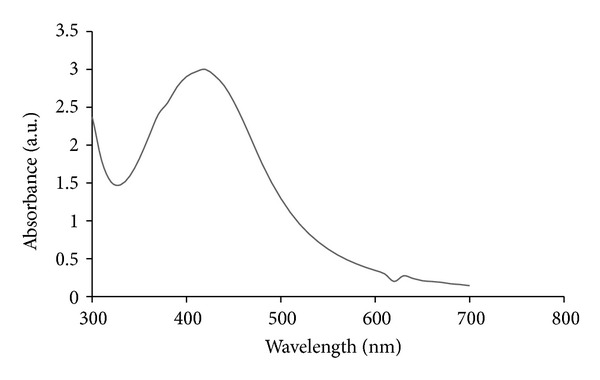
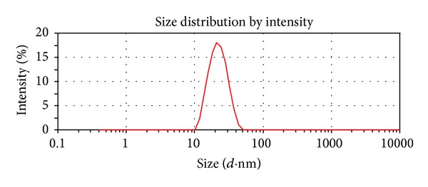
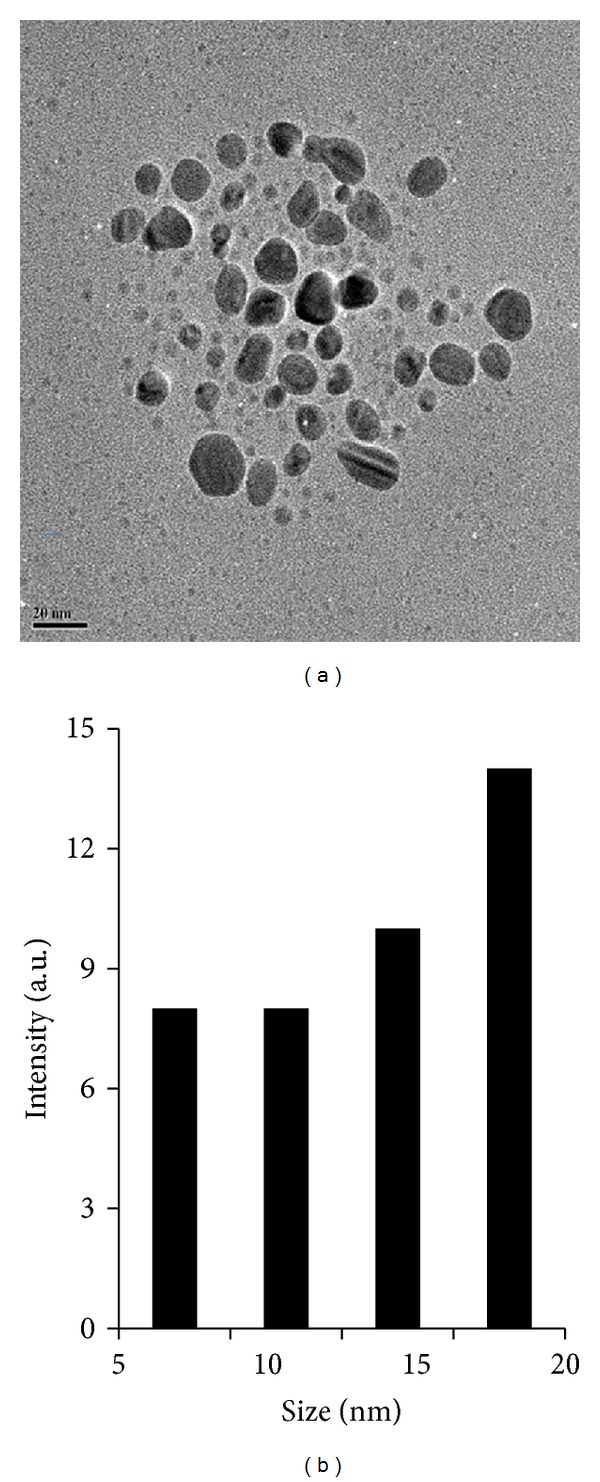
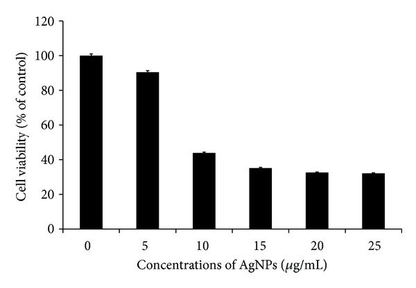
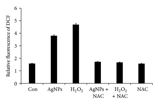
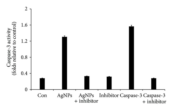
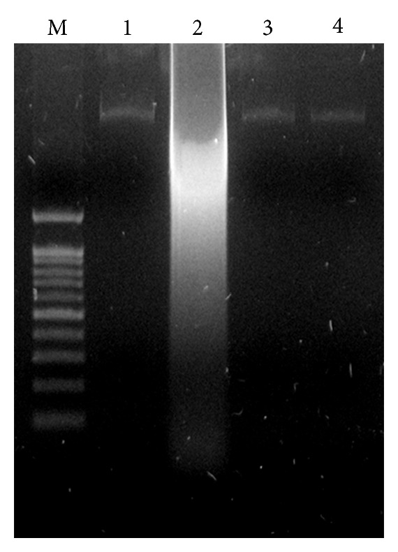
Silver nanoparticles (AgNPs) have been used as an antimicrobial and disinfectant agents. However, there is limited information about antitumor potential. Therefore, this study focused on determining cytotoxic effects of AgNPs on MDA-MB-231 breast cancer cells and its mechanism of cell death. Herein, we developed a green method for synthesis of AgNPs using culture supernatant of Bacillus funiculus, and synthesized AgNPs were characterized by various analytical techniques such as UV-visible spectrophotometer, particle size analyzer, and transmission electron microscopy (TEM). The toxicity was evaluated using cell viability, metabolic activity, and oxidative stress. MDA-MB-231 breast cancer cells were treated with various concentrations of AgNPs (5 to 25 μg/mL) for 24 h. We found that AgNPs inhibited the growth in a dose-dependent manner using MTT assay. AgNPs showed dose-dependent cytotoxicity against MDA-MB-231 cells through activation of the lactate dehydrogenase (LDH), caspase-3, reactive oxygen species (ROS) generation, eventually leading to induction of apoptosis which was further confirmed through resulting nuclear fragmentation. The present results showed that AgNPs might be a potential alternative agent for human breast cancer therapy.


Figures

Figure 1
Figure 1
Synthesis of AgNPs by culture supernatant of B. funiculus. The figure shows flask containing samples of AgNO3 after exposure to 60 min (a), AgNO3 with the extracellular culture supernatant of B. funiculus (b), and AgNO3 plus supernatant of B. funiculus (c). It is observed that the color of the solution turned from colorless to brown after 1 h of the reaction, indicating the formation of AgNPs.
Figure 2
Figure 2
The absorption spectrum of AgNPs synthesized by B. funiculus culture supernatant. The absorption spectrum of AgNPs exhibited a strong broad peak at 420 nm, and observation of such a band is assigned to surface plasmon resonance of the particles. The samples were collected and were incubated with 1 mM silver nitrate solution. After the incubation period, the samples were visualized in UV-vis spectra.
Figure 3
Figure 3
Size distribution analysis by DLS. The particle size distribution revealed that the particles range from 10–20 nm. The average particle size was found to be 20 nm.
Figure 4
Figure 4
Size and morphology of AgNPs analysis by TEM. (a) Several fields were photographed and were used to determine the diameter of nanoparticles. The average range of observed diameter was 20 nm. (b) Particle size distributions from TEM image.
Figure 5
Figure 5
Effect of AgNPs on Cell viability of MDA-MB-231 cells. Cells were treated with AgNPs at various concentrations for 24 h, and cytotoxicity was determined by the MTT method. The results represent the means of three separate experiments, and error bars represent the standard error of the mean. Treated groups showed statistically significant differences from the control group by the Student's t-test (P < 0.05).
Figure 6
Figure 6
Effect of AgNPs on LDH activity in MDA-MB-231. LDH activity was measured by changes in optical densities due to NAD+ reduction which were monitored at 490 nm, as described in Materials and Methods Section, using the cytotoxicity detection lactate dehydrogenase kit. The results represent the means of three separate experiments, and error bars represent the standard error of the mean. Treated groups showed statistically significant differences from the control group by the Student's t-test (P < 0.05).
Figure 7
Figure 7
ROS generation in AgNPs treated MDA-MB-231 cells. Relative fluorescence of DCF was measured using a spectrofluorometer with excitation at 485 and emission at 530 nm. The results represent the means of three separate experiments, and error bars represent the standard error of the mean. Treated groups showed statistically significant differences from the control group by the Student's t-test (P < 0.05).
Figure 8
Figure 8
AgNPs induce apoptosis in MDA-MB-231 cells by caspase-3 activation. MDA-MB-231 cells were treated with AgNPs, purified caspase-3, and caspase-3 inhibitor for 24 h. The assay was performed as described in Materials and Methods Section. The caspase-3 activity is based on the hydrolysis of caspase-3 substrate (acetyl-Asp-Glu-Val-Asp p-nitroanilide (Ac-DEVD-pNA) by caspase-3, resulting in the release of the p-nitroaniline (pNA) moiety. The concentration of the pNA released from the substrate is calculated from the absorbance values at 405 nm. The assay was carried out in the presence of purified caspase-3 and caspase-3 inhibitor (Ac-DEVD-CHO) for a comparative analysis. The results represent the means of three separate experiments, and error bars represent the standard error of the mean. Treated groups showed statistically significant differences from the control group by the Student's t-test (P < 0.05).
Figure 9
Figure 9
Effect of AgNPs on DNA fragmentation. MDA-MB-231 Cells were treated with AgNPs for 24 h and DNA fragmentation was analyzed by agarose gel electrophoresis. Lane M, 1 kB ladder; lane 1, control; lane 2, AgNP (8.7 μg/mL); lane 3, AgNP + NAC; lane 4, NAC (5 mM).

References

    1. Chan K, Morris GJ. Chemoprevention of breast cancer for women at high risk. Seminars in Oncology. 2006;33(6):642–646. - PubMed
    1. Jenal A, Thomas A, Murry T. Cancer stastics. CA: A Cancer Journal for Clinicians . 2002;52:23–37. - PubMed
    1. Johnston SRD. Acquired tamoxifen resistance in human breast cancer—potential mechanisms and clinical implications. Anti-Cancer Drugs. 1997;8(10):911–930. - PubMed
    1. Kato S, Endoh H, Masuhiro Y, et al. Activation of the estrogen receptor through phosphorylation by mitogen-activated protein kinase. Science. 1995;270(5241):1491–1494. - PubMed
    1. Lupu R, Cardillo M, Cho C, et al. The significance of heregulin in breast cancer tumor progression and drug resistance. Breast Cancer Research and Treatment. 1996;38(1):57–66. - PubMed

Publication types

MeSH terms