. 2013 Aug 9:14:543.
doi: 10.1186/1471-2164-14-543.

Genome-wide analyses of Epstein-Barr virus reveal conserved RNA structures and a novel stable intronic sequence RNA

Affiliations

Genome-wide analyses of Epstein-Barr virus reveal conserved RNA structures and a novel stable intronic sequence RNA

Walter N Moss et al. BMC Genomics. .

Abstract

Background: Epstein-Barr virus (EBV) is a human herpesvirus implicated in cancer and autoimmune disorders. Little is known concerning the roles of RNA structure in this important human pathogen. This study provides the first comprehensive genome-wide survey of RNA and RNA structure in EBV.

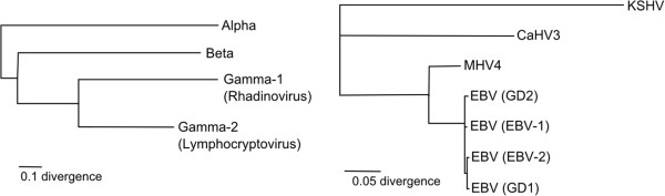
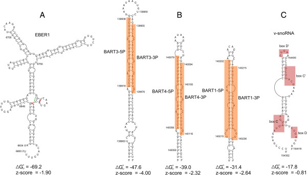
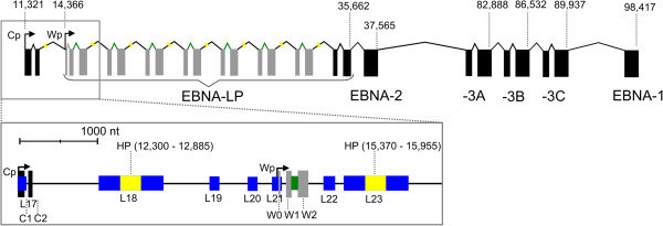
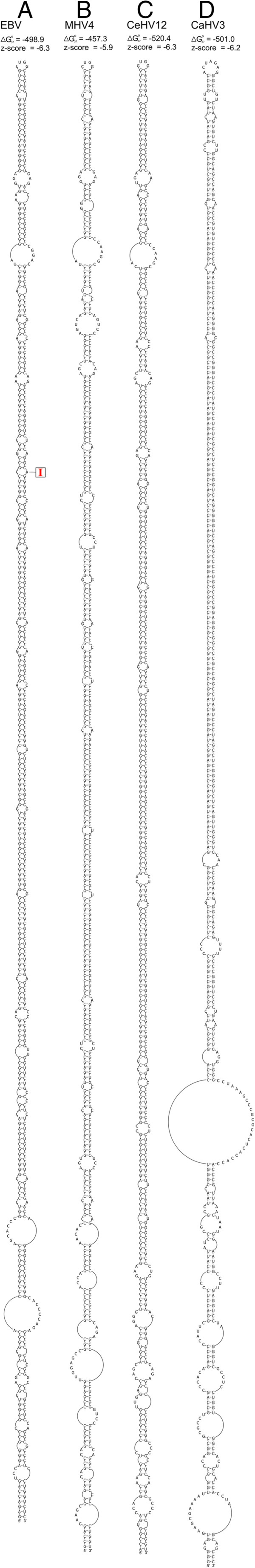
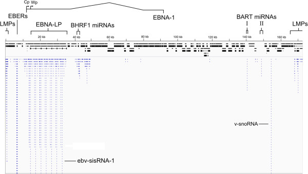
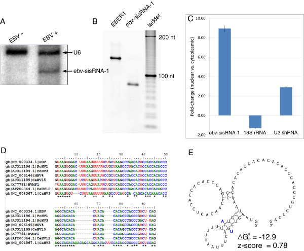
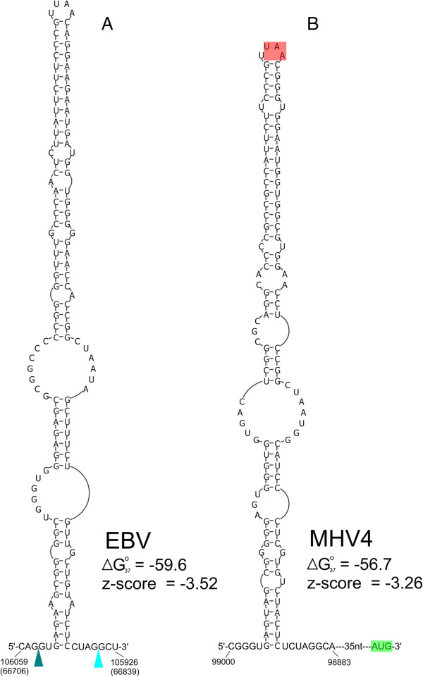
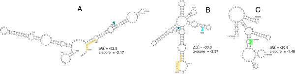
Results: Novel EBV RNAs and RNA structures were identified by computational modeling and RNA-Seq analyses of EBV. Scans of the genomic sequences of four EBV strains (EBV-1, EBV-2, GD1, and GD2) and of the closely related Macacine herpesvirus 4 using the RNAz program discovered 265 regions with high probability of forming conserved RNA structures. Secondary structure models are proposed for these regions based on a combination of free energy minimization and comparative sequence analysis. The analysis of RNA-Seq data uncovered the first observation of a stable intronic sequence RNA (sisRNA) in EBV. The abundance of this sisRNA rivals that of the well-known and highly expressed EBV-encoded non-coding RNAs (EBERs).

Conclusion: This work identifies regions of the EBV genome likely to generate functional RNAs and RNA structures, provides structural models for these regions, and discusses potential functions suggested by the modeled structures. Enhanced understanding of the EBV transcriptome will guide future experimental analyses of the discovered RNAs and RNA structures.


Figures

Figure 1
Figure 1
Phylogenic relationships of analyzed viruses. (Left) Phylogenetic tree showing the three herpesviridae sub-families (alpha, beta, and gamma), where the gamma sub-family is further divided into two genera: gamma-1 (Rhadinovirus) and gamma-2 (Lymphocryptovirus). The tree was generated from data in [51]. (Right) Breakdown of the gamma herpesvirus branch shown on the left. The consensus neighbor-joining tree (100 replicates) was based on the conserved herpesvirus DNA polymerase catalytic subunit (DPOL). Kaposi’s sarcoma-associated herpesvirus (KSHV), a gamma-1 herpesvirus, is used as an outgroup to illustrate the phylogenetic relationships between four EBV strains (EBV-1, EBV-2, GD1, and GD2) and two other lymphocryptoviruses used in this study [Macacine herpesvirus 4 (MHV4) and Callitrichine herpesvirus 3 (CaHV3)]. The tree was generated using the Geneious program.
Figure 2
Figure 2
Structure models for several known EBV ncRNAs. Nucleotide positions for the EBV-2 RefSeq genome sequence are annotated on the structure. Below are the predicted Gibbs folding free energies (in kcal/mol) and z-scores. (A) EBER1 modeled structure. Base pairs that differ from the Rfam model (Rfam ID# RF01789) are covered by red Xs, while the mis-predicted Rfam base pair is indicated with a green line. (B) Modeled structures in RNAz-predicted structured region, locus 209, which contains the BART3, BART4, and BART1 miRNAs (indicated in orange boxes). Sequences are from the EBV-2 RefSeq (NC_009334.1) genome. (C) Modeled structure for the viral snoRNA (v-snoRNA) previously reported [52].
Figure 3
Figure 3
Cartoon of the primary transcript encoding the EBNA proteins. Exons are indicated with black or grey rectangles and the genomic location of the end of each protein-coding region is indicated above. The identical W1 and W2 coding exons that compose most of EBNA-LP are shown in grey. The Cp and upstream Wp promoter sites are indicated by bent arrows with the genome coordinates of the transcription start sites given above. The boxed region zooms in to show the 5′ end of the pre-mRNA, including the locations of the predicted long hairpin (HP) and ebv-sisRNA-1, colored yellow and green, respectively, with their genome coordinates given in parentheses. RNAz-predicted structured regions are shown in blue and labeled L17 to L23 for loci 17 to 23 (genome coordinates for these loci are in Additional file 1).
Figure 4
Figure 4
Structure models for lymphocryptovirus repeat long hairpin RNAs. A reported A-to-I editing site [66] is indicated with a red “I”. Shown are sequences from (A) Epstein-Barr virus (EBV), (B) Macacine herpesvirus 4 (MHV4), (C) Cercopithecine herpesvirus 12 (CeHV12), and (D) Callitrichine herpesvirus 3 (CaHV3).
Figure 5
Figure 5
Small RNA library RNA-Seq reads aligned with the EBV genome. Reads are colored blue; the EBER1, EBER2, and sisRNA peaks are much larger than the represented area (see Table 1). A cartoon of the EBV genome is shown in black (exons as boxes and introns as thin lines). The locations of latently expressed genes [EBNA-LP, LMPs, EBERs (EBER1 and EBER2), BHRF1 miRNAs, and BART miRNAs (clusters I and II)] are indicated at the top of the figure, including the location of the Cp and first Wp promoter sites for producing mRNA for the EBNA-LP and five other EBNA proteins (additional details are in Figure 3). Peaks for the ebv-sisRNA-1 and v-snoRNA are indicated. Images were generated using the integrated genome viewer (IGV, [76,77]) with data from Additional file 5.
Figure 6
Figure 6
Analyses of ebv-sisRNA-1. (A) Northern blot for ebv-sisRNA-1 using RNA from EBV-negative BJAB cells and from isogenic EBV-positive BJAB-B1 cells. The bottom arrow points to the additional band observed in the EBV-positive BJAB-B1 lane that likely corresponds to the 81 nt ebv-sisRNA-1, while the upper arrow indicates the 106 nt human U6 snRNA (present in both cell types). (B) RT-PCR for EBER1 and ebv-sisRNA-1 using cDNA from BJAB-B1 nuclear RNA and primers complementary to the ends of EBER1 and the 5′ end plus nt 52 to 71 of ebv-sisRNA-1. (C) Fold-enrichment in nuclear versus cytoplasmic RNA as measured by RT-qPCR. Shown are the results for the sisRNA and control 18S and U2 snRNA, which are cytoplasmically and nuclearly enriched, respectively. Error bars indicate the standard error of three biological replicates, each with three technical replicates. (D) MAFFT sequence alignment of ebv-sisRNA-1 and those of seven other lymphocryptoviruses: Pongine herpesvirus 1 (PoHV1, AJ311196.1), Pongine herpesvirus 3 (PoHV3, AJ311194.1), Macacine herpesvirus 4 (MHV4, NC_006146), Cercopithicine herpesvirus 15 (CeHV15, AJ311199), Herpesvirus MF-1 from Macaca fascicularis (HVMF1, X77781), Cercopithecine herpesvirus 12 (CeHV12, AF200364.1), and Callitrichine herpesvirus 3 (CaHV3, NC_004367.1). The question mark at the first position in HVMF1 represents missing data, not a true gap. 100% conserved positions are indicated with stars below the alignment. (E) Predicted structure for ebv-sisRNA-1. Compensatory mutations in the upstream hairpin that convert a GC pair to an AU pair (in CaHV3) are indicated with bold blue nts. This sequence is repeated seven times in EBV: genome (NC_009334.1) coordinates 14603 to 14683, 17673 to 17753, 20743 to 20823, 23813 to 23893, 26883 to 26963, 29953 to 30033, and 33023 to 33103.
Figure 7
Figure 7
Hairpin structure predicted in BBLF2/3 mRNA. (A) Hairpin modeled for the EBV BBLF2/3 short intron. Splice donor and acceptor sites are indicated with dark and light blue arrows, respectively. Nucleotide numbers in parentheses correspond to the reverse genome strand models in Additional file 3 (locus 152). The predicted free energy (in kcal/mol) and z-score of the hairpin are shown. (B) Homologous hairpin structure in MHV4. The BBLF2 translation stop codon is shaded in red and the BBLF3 start codon is shown in green.
Figure 8
Figure 8
Predicted RNA structures containing splice sites and a start codon. (A) Structure model for RNAz-predicted structural region locus 1 (in LMP-2B gene). The splice donor site is indicated with a dark blue colored wedge and a putative intronic splicing enhancer is colored yellow. (B) Structure model for locus 2 (in the LMP-2B gene). The splicing acceptor site is indicated with the light blue wedge and the yellow box now indicates a putative exonic splicing enhancer. (C) Structure model for locus 263. The start codon for latent membrane protein 2A (LMP-2A) is indicated by the green box.

Similar articles

Cited by

References

    1. Epstein-Barr virus and infectious mononucleosis. http://www.cdc.gov/ncidod/diseases/ebv.htm. - PubMed
    1. Flavell KJ, Murray PG. Hodgkin’s disease and the Epstein-Barr virus. Mol Pathol: MP. 2000;53(5):262–269. doi: 10.1136/mp.53.5.262. - DOI - PMC - PubMed
    1. Burgos JS. Involvement of the Epstein-Barr virus in the nasopharyngeal carcinoma pathogenesis. Med Oncol. 2005;22(2):113–121. doi: 10.1385/MO:22:2:113. - DOI - PubMed
    1. Akhter S, Liu H, Prabhu R, DeLucca C, Bastian F, Garry RF, Schwartz M, Thung SN, Dash S. Epstein-Barr virus and human hepatocellular carcinoma. Cancer Letters. 2003;192(1):49–57. doi: 10.1016/S0304-3835(02)00695-X. - DOI - PubMed
    1. Begin LR, Eskandari J, Joncas J, Panasci L. Epstein-Barr virus related lymphoepithelioma-like carcinoma of lung. J Surg Oncol. 1987;36(4):280–283. doi: 10.1002/jso.2930360413. - DOI - PubMed

Publication types

LinkOut - more resources