. 2014 Jan;175(1):79-91.
doi: 10.1111/cei.12186.

Influenza infection results in local expansion of memory CD8(+) T cells with antigen non-specific phenotype and function

Affiliations

Influenza infection results in local expansion of memory CD8(+) T cells with antigen non-specific phenotype and function

Gail D Sckisel et al. Clin Exp Immunol. 2014 Jan.

Abstract

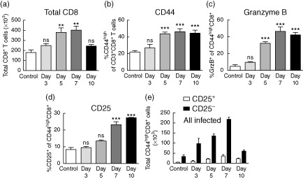
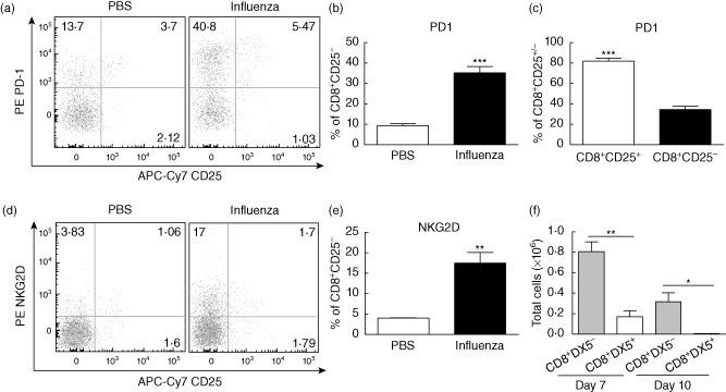
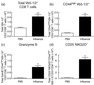
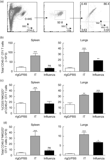
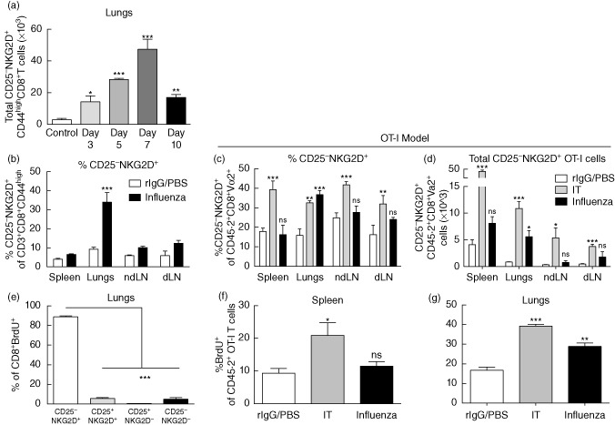
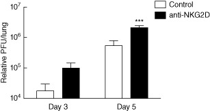
Primary viral infections induce activation of CD8(+) T cells responsible for effective resistance. We sought to characterize the nature of the CD8(+) T cell expansion observed after primary viral infection with influenza. Infection of naive mice with different strains of influenza resulted in the rapid expansion of memory CD8(+) T cells exhibiting a unique bystander phenotype with significant up-regulation of natural killer group 2D (NKG2D), but not CD25, on the CD44(high) CD8(+) T cells, suggesting an antigen non-specific phenotype. We further confirmed the non-specificity of this phenotype on ovalbumin-specific (OT-I) CD8(+) T cells, which are not specific to influenza. These non-specific CD8(+) T cells also displayed increased lytic capabilities and were observed primarily in the lung. Thus, influenza infection was shown to induce a rapid, antigen non-specific memory T cell expansion which is restricted to the specific site of inflammation. In contrast, CD8(+) T cells of a similar phenotype could be observed in other organs following administration of systemic agonistic anti-CD40 and interleukin-2 immunotherapy, demonstrating that bystander expansion in multiple sites is possible depending on whether the nature of activation is either acute or systemic. Finally, intranasal blockade of NKG2D resulted in a significant increase in viral replication early during the course of infection, suggesting that NKG2D is a critical mediator of anti-influenza responses prior to the initiation of adaptive immunity. These results characterize further the local bystander expansion of tissue-resident, memory CD8(+) T cells which, due to their early induction, may play an important NKG2D-mediated, antigen non-specific role during the early stages of viral infection.

Keywords: NKG2D; antigen non-specific; bystander activation; memory CD8; tissue-resident.


Figures

Figure 1
Figure 1
Activation and expansion of CD8+ T cells within the lung after influenza infection. Mice were infected intranasally with 1·2 × 104 plaque-forming units (PFU) A/Mem/71 and harvested at the various time-points indicated. Lungs were processed and stained for flow cytometry to evalulate the following parameters. (a) Total CD8+ T cells. (b) Percentage of CD44high of total CD8+ T cells. (c) Percentage of granzyme B+ and (d) CD25+ of CD44highCD8+ T cells. (e) Absolute numbers of CD25+/−CD44highCD8+ T cells. Data are representative of at least five independent experiments. Statistics performed using one-way analysis of variance (anova) with Bonferroni's post-test where appropriate. *P < 0·05; **P < 0·01; ***P < 0·001.
Figure 2
Figure 2
Characterization of phenotype of CD25CD44highCD8+ T cells. Mice were harvested on day 5 following intranasal inoculation with 1·2 × 104 plaque-forming units (PFU) A/Mem/71 influenza unless noted otherwise. (a) Representative dot-plots depicting programmed death 1 (PD-1) and CD25 expression on CD8+ T cells and (b) percentages depicting PD-1 expression on CD8+CD25 cells. (c) PD-1 expression on CD8+CD25+ and CD8+CD25 populations. (d) Representative dot-plots depicting natural killer group 2D (NKG2D) and CD25 expression on CD3+CD8+ T cells and (e) percentages depicting NKG2D expression on CD3+CD8+CD25 cells. (f) Absolute numbers of DX5+ and DX5CD8+NKG2D+ T cells. Statistics performed using Student's t-test where appropriate. Data are representative of at least three independent experiments. *P < 0·05; **P < 0·01; ***P < 0·001.
Figure 3
Figure 3
Expansion and up-regulation of natural killer group 2D (NKG2D) on CD8+ T cells expressing an irrelevant T cell receptor (TCR). Mice were infected intranasally with 20 plaque-forming units (PFU) A/PR8/8 and harvested at day 7. (a) Total numbers of Vβ 5·1/2+CD8 T cells at day 7 post-influenza infection. Total numbers of (b) CD44high, (c) granzyme B+ and (d) CD25NKG2D CD44highVβ 5·1/2+ CD8 T cells at day 7 post-influenza infection. Data are representative of at least two independent experiments. Statistics performed using one- or two-way analysis of variance (anova) with Bonferroni's post-test where appropriate. *P < 0·05; **P < 0·01; ***P < 0·001.
Figure 4
Figure 4
Expansion and up-regulation of bystander phenotype in ovalbumin (OVA)-specific CD8 T cells (OT-I) following influenza infection. OT-I CD8 T cells (CD45·2) were purified from OT-I T cell receptor (TCR) transgenic mice and transferred adoptively into congenic Ly5·1 (CD45·1) mice. Mice were vaccinated subsequently with OVA in incomplete Freund's adjuvant (IFA) and rested for >30 days. Mice were then challenged with A/PR/8 influenza or subjected to the immunotherapy regimen. For influenza mice were harvested on day 7 and for immunotherapy mice were harvested on day 11. (a) Representative gating schema for OT-I cells from congenic hosts. (b) Total numbers of congenic (CD45·2+) OT-I CD8+ T cells in spleen (left) and lungs (right). (c) Percentages and (d) numbers of bystander phenotype (CD25 natural killer group 2D (NKG2D)+) OT-I CD8+ T cells in spleen (left) and lungs (right). Data are representative of at least two independent experiments. Statistics performed using one- or two-way analysis of variance (anova) with Bonferroni's post-test where appropriate. *P < 0·05; **P < 0·01; ***P < 0·001.
Figure 5
Figure 5
Timing and distribution of expanded CD25 natural killer group 2D (NKG2D)+CD8+ T cells after infection. Mice were infected with A/Mem/71 influenza and harvested on day 5 for wild-type studies. Ovalbumin (OVA)-specific CD8 T cell (OT-I) studies were performed as described in Fig. 4 and Supporting information Fig. S4. (a) Absolute numbers of CD25NKG2D+CD44highCD8+T cells in the lungs at days 3, 5, 7 and 10 within the lung post-influenza infection. (b) Percentages of NKG2D+CD25CD44highCD8+ T cells in the lungs, spleen, mediastinal and non-draining lymph nodes. (c) Percentages and (d) numbers of CD25NKG2D+ OT-I CD8+ T cells in various organs following influenza infection or immunotherapy. (e) Comparison of CD25 and NKG2D expression in the lung CD8+bromodeoxyuridine (BrdU)+ population at day 5 of influenza infection. Percentage of BrdU+ of OT-I CD8+ T cells in (f) spleen and (g) lungs following influenza infection or immunotherapy. Data are representative of at least two independent experiments. Statistics performed using one- or two-way analysis of variance (anova) with Bonferroni's post-test where appropriate. *P < 0·05; **P < 0·01; ***P < 0·001.
Figure 6
Figure 6
Natural killer group 2D (NKG2D)-dependent role in control of early viral replication. Mice were infected with 20 plaque-forming units (PFU) A/PR8 influenza and harvested on days 3 and 5 following infection. Some mice were given 100 μg anti-NKG2D or rat immunoglobulin (Ig)G intranasally on days 0, 2 and 4 following infection. Viral titres as determined by quantitative polymerase chain reaction (qPCR) on days 3 and 5 following influenza infection. Data are representative of at least two independent experiments. Statistics performed using one- or two-way analysis of variance (anova) with Bonferroni's post-test where appropriate. *P < 0·05; **P < 0·01; ***P < 0·001.

References

    1. Bi Z, Barna M, Komatsu T, Reiss CS. Vesicular stomatitis virus infection of the central nervous system activates both innate and acquired immunity. J Virol. 1995;69:6466–6472. - PMC - PubMed
    1. Cousens LP, Orange JS, Biron CA. Endogenous IL-2 contributes to T cell expansion and IFN-gamma production during lymphocytic choriomeningitis virus infection. J Immunol. 1995;155:5690–5699. - PubMed
    1. Murata K, Garcia-Sastre A, Tsuji M, et al. Characterization of in vivo primary and secondary CD8+ T cell responses induced by recombinant influenza and vaccinia viruses. Cell Immunol. 1996;173:96–107. - PubMed
    1. Sunil-Chandra NP, Arno J, Fazakerley J, Nash AA. Lymphoproliferative disease in mice infected with murine gammaherpesvirus 68. Am J Pathol. 1994;145:818–826. - PMC - PubMed
    1. Jacoby RO, Bhatt PN, Brownstein DG. Evidence that NK cells and interferon are required for genetic resistance to lethal infection with ectromelia virus. Arch Virol. 1989;108:49–58. - PubMed

Publication types

MeSH terms