. 2013 Aug 12:11:56.
doi: 10.1186/1478-811X-11-56.

Mena/VASP and αII-Spectrin complexes regulate cytoplasmic actin networks in cardiomyocytes and protect from conduction abnormalities and dilated cardiomyopathy

Affiliations

Mena/VASP and αII-Spectrin complexes regulate cytoplasmic actin networks in cardiomyocytes and protect from conduction abnormalities and dilated cardiomyopathy

Peter M Benz et al. Cell Commun Signal. .

Abstract

Background: In the heart, cytoplasmic actin networks are thought to have important roles in mechanical support, myofibrillogenesis, and ion channel function. However, subcellular localization of cytoplasmic actin isoforms and proteins involved in the modulation of the cytoplasmic actin networks are elusive. Mena and VASP are important regulators of actin dynamics. Due to the lethal phenotype of mice with combined deficiency in Mena and VASP, however, distinct cardiac roles of the proteins remain speculative. In the present study, we analyzed the physiological functions of Mena and VASP in the heart and also investigated the role of the proteins in the organization of cytoplasmic actin networks.

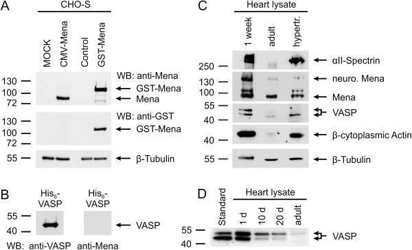
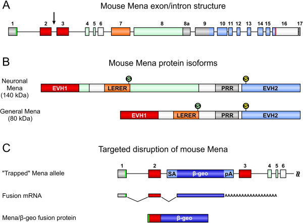
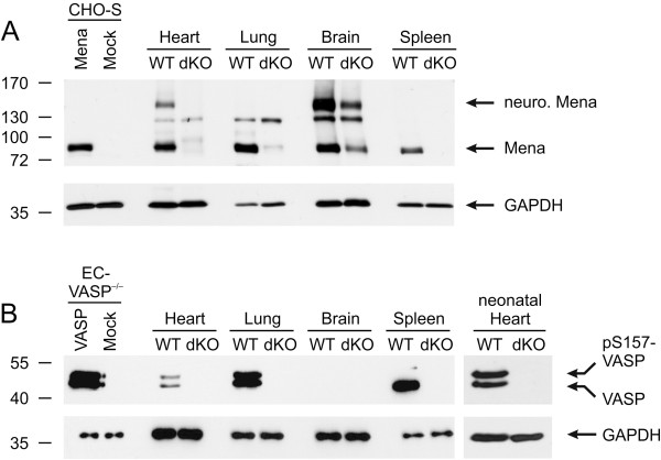
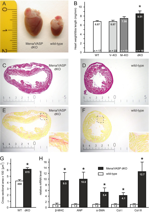
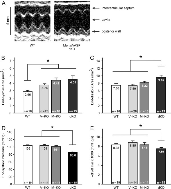
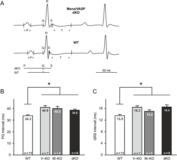
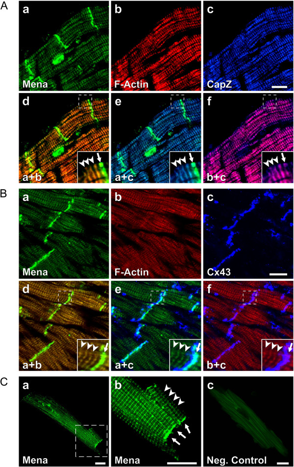
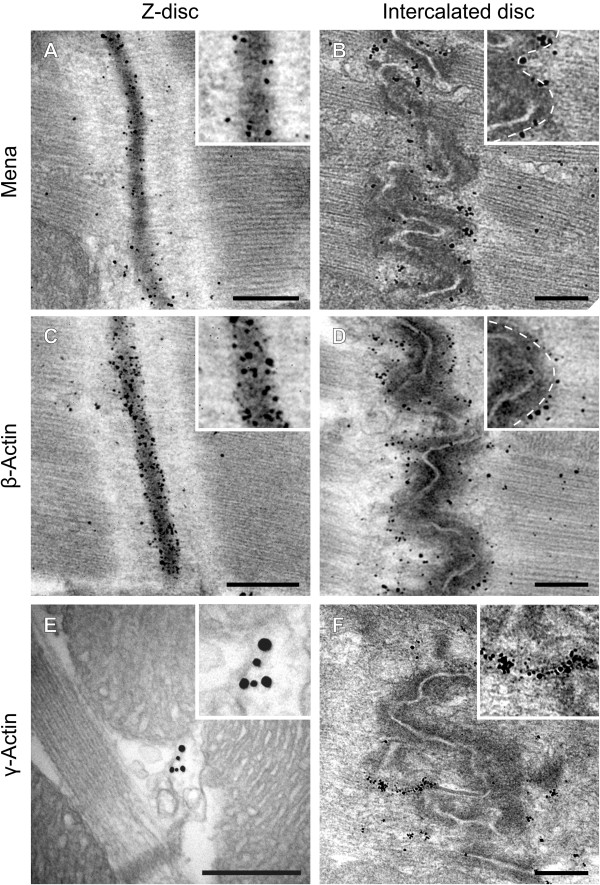
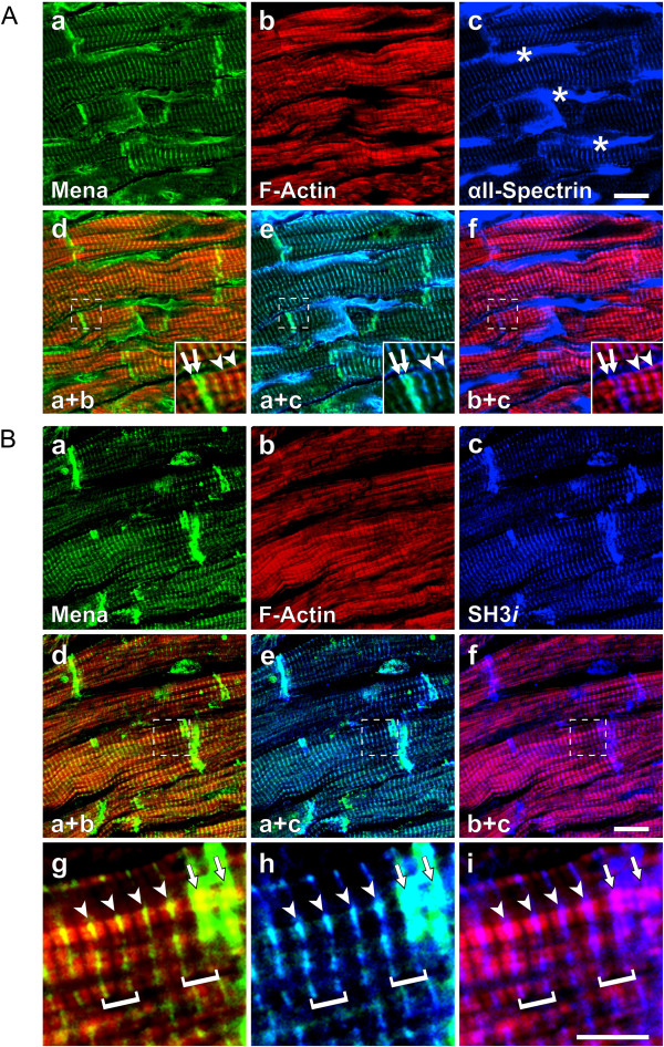
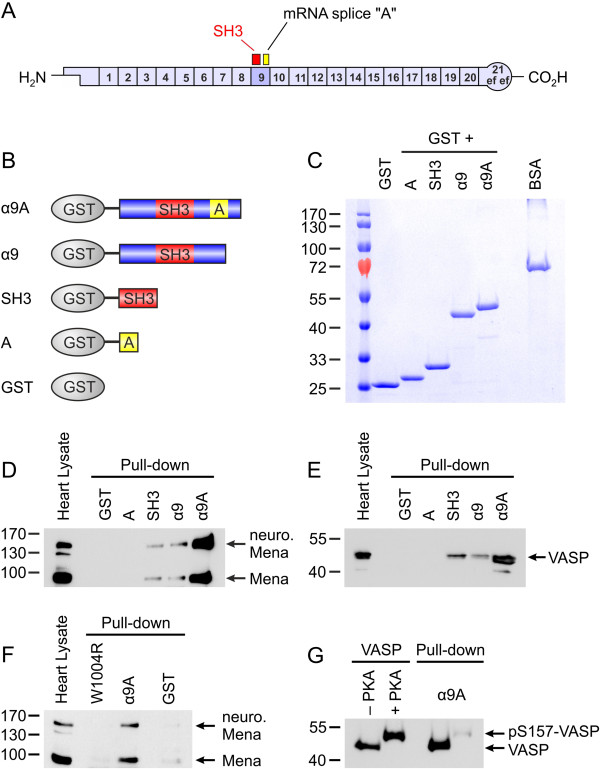
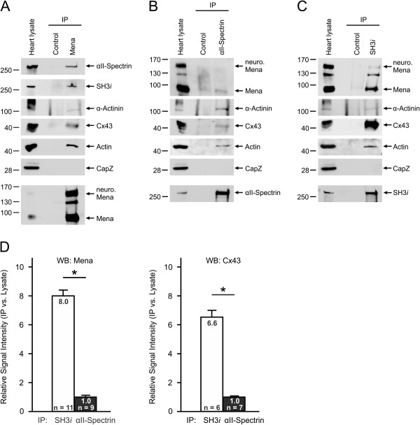
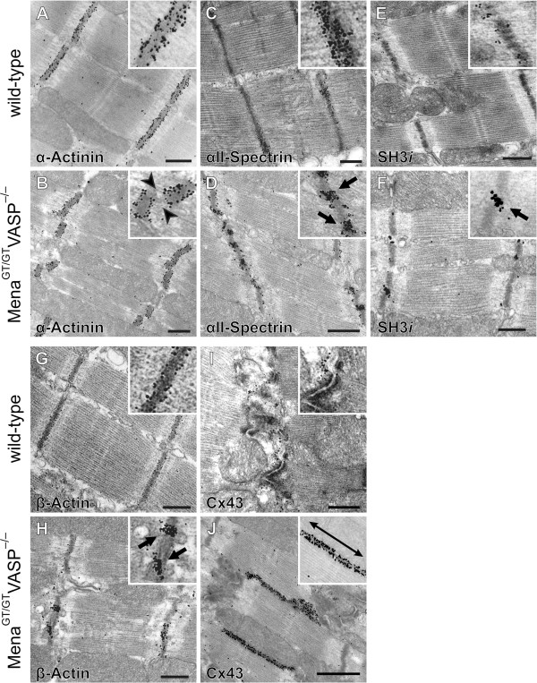
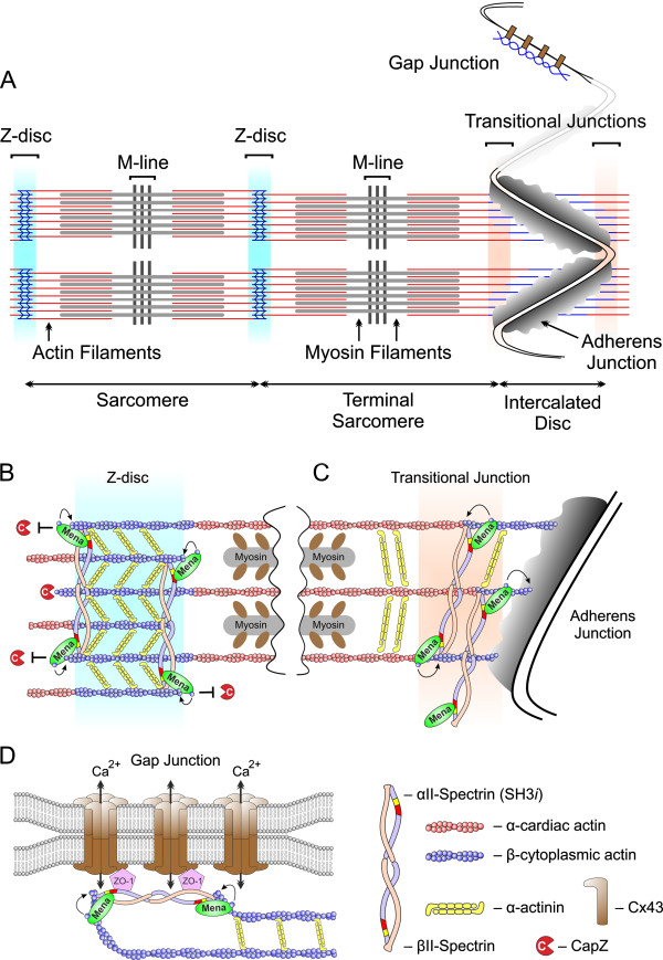
Results: We generated a mouse model, which simultaneously lacks Mena and VASP in the heart. Mena/VASP double-deficiency induced dilated cardiomyopathy and conduction abnormalities. In wild-type mice, Mena and VASP specifically interacted with a distinct αII-Spectrin splice variant (SH3i), which is in cardiomyocytes exclusively localized at Z- and intercalated discs. At Z- and intercalated discs, Mena and β-actin localized to the edges of the sarcomeres, where the thin filaments are anchored. In Mena/VASP double-deficient mice, β-actin networks were disrupted and the integrity of Z- and intercalated discs was markedly impaired.

Conclusions: Together, our data suggest that Mena, VASP, and αII-Spectrin assemble cardiac multi-protein complexes, which regulate cytoplasmic actin networks. Conversely, Mena/VASP deficiency results in disrupted β-actin assembly, Z- and intercalated disc malformation, and induces dilated cardiomyopathy and conduction abnormalities.


Figures

Figure 1
Figure 1
Stage-dependent expression of cytoskeletal proteins in the mouse heart. (A, B) Characterization of Mena-specific antibodies. (A) CHO-S cells, either transiently transfected with murine Mena (CMV-Mena) or stably transfected with GST-tagged Mena (GST-Mena), were lysed and analyzed by Western blotting with Mena-specific (upper panel) or GST-specific (middle panel) antibodies. As control, lysates of MOCK-transfected cells or lysates of CHO-S cells without stable integration of GST-Mena were run on the same gels. β-Tubulin served as loading control (lower panel). (B) 100 ng purified VASP protein was probed by Western blotting with anti-VASP (left panel) and anti-Mena (right panel) antibodies. (C, D) Stage-dependent expression of cytoskeletal proteins in the mouse heart. (C) Lysates of 1-week-old, adult, and hypertrophic adult mouse hearts were probed by Western blotting with antibodies against αII-Spectrin, Mena, VASP, and β-cytoplasmic actin. Blotting for β-Tubulin was used as invariant loading control. Two Mena isoforms are expressed in the mouse heart, the general (~80 kDa) and the neuronal-specific (~140 kDa) form. Depending on its phosphorylation state, VASP migrates at 46 or 50 kDa. (D) VASP expression in the mouse heart at postnatal day 1, 10, and 20 and in the adult heart.
Figure 2
Figure 2
Targeted disruption of the mouse Mena gene. (A) The mouse Mena gene is located on chromosome 1 spanning about 115 kb of genomic sequence. The translational start and termination codons are given in green (exon 1) and red (exon 16), respectively. The neuronal-specific exons are shown in green. Exons 2 and 3, encoding the Ena/VASP homology 1 (EVH1) domain are shown in red. Exons encoding the LERER repeats, the proline-rich region (PRR), and the EVH2 domain are indicated in orange, grey, and light blue, respectively. The insertion of a gene trap vector in intron 2 of the Mena gene is indicated by a black arrow (compare C). (B) Depending on differential splicing, a neuronal-specific and a general Mena protein isoform exist. Mena is phosphorylated at serine 236 by PKA (green “S”) and at serine 376 by PKG (yellow “S”; numbering according to the 541aa general Mena isoform). (C) In the ES cell clone RRG138, the gene trap vector is inserted in Mena intron 2. The gene trap vector consists of a splice acceptor side (SA), a polyadenylation signal (pA), and β-geo, which encodes β-galactosidase and neomycin resistance [26]. Insertion of the gene trap vector disrupts the Mena gene and results in the expression of a Mena/β-geo fusion protein. Because critical parts of the N-terminal EVH1 domain are missing in the fusion protein, Mena/β-geo most likely lacks all known endogenous Mena functions.
Figure 3
Figure 3
Mena/VASP expression in wild-type and MenaGT/GTVASP−/− mouse organs. Western blots to analyze Mena (A) and VASP (B) protein levels in heart, lung, brain, and spleen of wild-type (WT) and MenaGT/GTVASP−/− (dKO) mice. GAPDH and lysates of Mena- or VASP-transfected cells were used as controls. (A) The general Mena protein isoform (~80 kDa) is present in all analyzed organs, the neuronal splice variant (~140 kDa) is only detected in heart and brain. Whereas Mena protein in dKO brain is moderately reduced to ~30% of wild-type levels, Mena is almost undetectable in heart, lung, and spleen. The 130 kDa band seen in heart, lung, and brain likely represents a cross-reaction with an unrelated protein. (B) VASP (46 or 50 kDa, depending on phosphorylation state) expression is high in the neonatal heart and the adult lung and spleen, low in the adult heart, and nearly undetectable in the brain. No VASP protein is detected in dKO organs.
Figure 4
Figure 4
Enlarged and dilated hearts in MenaGT/GTVASP−/− mice. MenaGT/GTVASP−/− (dKO) animals display macroscopically enlarged hearts (A, photography, scale in cm) and significantly increased heart weight to tibia length ratios as compared to wild-type (WT) controls (B; ANOVA, *P<0.05). (C, D) Hematoxilin and eosin stained cross sections of MenaGT/GTVASP−/−(C) and wild-type (D) hearts, demonstrating dilated ventricles in the double-deficient animals. (E, F) Picrosirius red stained heart cross sections of dKO (E) and WT (F) mice to visualize collagen fibers/interstitial fibrosis. Magnified views of the indicated areas are shown as insets. The scales in images C-F are given in mm. (G) Cross sectional areas of cardiomyocytes from WT and MenaGT/GTVASP−/− hearts quantified by morphometric analyses (five animals per group, 25 cardiomyocytes per animal, *P<0.05). (H) Quantitative real-time RT-PCR revealed increased mRNA levels of cardiac hypertrophy markers (β-myosin heavy chain, β-MHC; atrial natriuretic peptide, ANP) and fibrosis markers (α-smooth muscle actin, α-SMA; collagen I, Col I; and collagen III, Col III; *P<0.05; n=6) in dKO mice vs. WT controls.
Figure 5
Figure 5
Mena/VASP double-deficiency induces dilated cardiomyopathy. (A-C) Echocardiography. (A) Representative M-mode recordings at the mid-papillary muscle level of wild-type (WT) and MenaGT/GTVASP−/− (dKO) mice. (B, C) Statistical analyses of the echocardiographic measurements demonstrated a significantly increased left intraventricular end-systolic area in VASP−/− (V-KO), MenaGT/GT (M-KO), and MenaGT/GTVASP−/− (dKO) animals as compared to wild-type controls (B). The left intraventricular end-diastolic area was only significantly increased in the dKO animals (C). (D, E) Left ventricular catheterization revealed a significantly reduced end-systolic pressure (D) and a significantly reduced rate of pressure decline (E) in the left ventricle of dKO mice. Bars represent the mean ± S.E.M (ANOVA, *P<0.05).
Figure 6
Figure 6
Mena/VASP double-deficiency results in conduction abnormalities. Electrocardiography (ECG). (A) Original, high resolution lead I ECG recording of a representative MenaGT/GTVASP−/− (dKO, upper panel) and wild-type (WT, lower panel) mouse. Q, R, and S spikes and the P- and T-waves are indicated. A comparison of the PQ and QRS intervals from dKO and WT mice is shown below the ECG recording of the WT. (B, C) Statistical analysis of the ECG recordings demonstrated significantly prolonged PQ (B) and QRS (C) intervals in VASP−/− (V-KO), MenaGT/GT (M-KO), and MenaGT/GTVASP−/− (dKO) mice. Bars represent the mean ± S.E.M, one-way analysis of variance (ANOVA), *P<0.05).
Figure 7
Figure 7
Mena is localized at Z- and intercalated discs of mouse cardiomyocytes. (A, B) Confocal microscopy images of adult mouse heart cryosections stained for Mena (a, green), non-isoform selective F-actin (b,red), and CapZ (A, image c, blue) or Connexin 43 (Cx43, B, image c, blue). Mena and F-actin colocalized with CapZ at Z-dics (A, merged images d-f, arrowheads) and with Cx43 at intercalated discs (B, merged images d-f, arrows). (C) Adult mouse cardiomyocytes were single-stained with Mena-specific antibodies followed by fluorescence-conjugated secondary antibodies (images a and b). Image b shows a magnified view of the area indicated in image a. Mena localized to Z-discs (arrowheads) and was strongly enriched at the cell termini (arrows). Using identical laser intensities and capture settings, the fluorescent secondary antibodies alone did not reveal any cross-striated signals (image c, negative control). Scale bars 15 μm.
Figure 8
Figure 8
Mena and β-actin localize to the edges of sarcomeres at Z- and intercalated discs. Immuno-electron microscopy of Z- and intercalated disc regions in mouse papillary muscle. Sections were labelled with antibodies to Mena (A, B), β-actin (C, D), and γ-actin (E, F). The dashed line in the insets of B and D indicates the cytoplasmic edge of the dark plaque material that coats the plasma membranes where the thin filaments of the terminal sarcomeres lead into the adherens junctions. (E) Most γ-Actin immunogold labeling was observed at filamentous structures, which project from the upper and lower terminals of Z-discs towards the mitochondria. Scale bars 500 nm.
Figure 9
Figure 9
Mena specifically colocalizes with the αII-Spectrin splice variant SH3i. (A, B) Confocal microscopy images of adult mouse heart cryosections stained for Mena (a, green), F-actin (b, red), and total αII-Spectrin (A, image c, blue) or the αII-Spectrin splice variant SH3i (B, image c, blue). (A) Mena, F-actin, and a portion of total αII-Spectrin colocalized at Z-discs (A, merged images d-f, arrowheads) and intercalated discs (A, merged images d-f, arrows. Detailed analyses revealed a doublet appearance of the proteins across the intercalated disc (A, images d-f, insets). Most αII-Spectrin was detected at the lateral plasma membrane (A, image c, asterisks) and colocalization with Mena and F-actin at these sites was minor. (B) SH3i was exclusively detected at Z- and intercalated discs but was absent from the lateral plasma membrane compare image c of A and B). Merged images d-f and magnified views of the indicated areas therein (images g-i) revealed high degree of colocalization of Mena and SH3i at Z-discs (images g-i, arrowheads) and intercalated discs (arrows), the lateral boundaries of the sarcomeres, which are exemplarily highlighted by brackets. Outside Z- and intercalated discs, colocalization of Mena and SH3i with F-actin was minor. Scale bars in images c and f 15 μm, image i 5 μm.
Figure 10
Figure 10
C-terminal extension of the αII-Spectrin SH3 domain enhances interaction with Mena and VASP. (A) αII-Spectrin is composed of 21 triple-helical repeats. Two αII-Spectrin splice variants exist in the mouse heart, either with or without a 20-amino acids insertion in the 9th repeat (“A”), C-terminal to the SH3 domain (indicated in red). The splice variant with the insertion is termed SH3i. (B, C) Schematic diagram (B) and Coomassie stained gel (C) of the purified GST-αII-Spectrin fusion proteins used for the pull-down experiments shown in D-G. Two microgram bovine serum albumin (BSA) served to calibrate the protein load. (D, E) Lysates of adult (D) and neonatal (E) mouse hearts were incubated with the depicted GST-fusion proteins and the precipitated material was blotted with Mena- or VASP-specific antibodies, respectively. (F) The interaction between Mena and SH3i, requires a functional SH3 domain and mutation of α9A to exchange the conserved Trp1004 residue with an arginine (W1004R) abrogates the binding. (G) The interaction between purified recombinant VASP and SH3i is regulated by phosphorylation. GST-α9A readily precipitated the non-phosphorylated VASP (46 kDa), but not PKA-phosphorylated protein (pS157-VASP, 50 kDa).
Figure 11
Figure 11
Mena and SH3i establish multi-protein complexes at Z- and intercalated discs. Representative Western blots of Immunoprecipiation (IP) studies. Lysates of adult mouse hearts were immunoprecipitated with Mena-specific (A), general αII-Spectrin-specific (B), or SH3i-specific (C) antibodies and the precipitated material was probed with antibodies against total αII-Spectrin, SH3i, Mena, actin, the Z-disc components α-Actinin and CapZ, and the intercalated disc marker Connexin 43 (Cx43). Western blot analysis of the precipitated material with the same antibodies that were used for IP served as control (lowermost panel in A, B, and C). Mena, αII-Spectrin, and SH3i were found in complexes with actin, α-Actinin and Cx43, but not in complexes with CapZ. (D) Statistical analysis of Mena and Cx43 protein amounts in IPs with general αII-Spectrin-specific (black bars) or SH3i-specific antibodies (white bars). SH3i precipitated more Mena and Cx43 than αII-Spectrin. Bars represent the mean ± S.E.M (ANOVA, *P<0.05).
Figure 12
Figure 12
Mena/VASP double deficiency impairs Z- and intercalated disc integrity. Immuno-electron microscopy of Z- and intercalated disc regions in wild-type and MenaGT/GTVASP−/− mouse papillary muscle. Sections were labelled with antibodies to α-Actinin (A, B), αII-Spectrin (C, D), SH3i(E, F), β-Actin (G, H), and Connexin 43 (Cx43, I, J). The Z-discs in the MenaGT/GTVASP−/− appeared fractured (B, arrowheads) and clusters of αII-Spectrin, SH3i, and β-Actin were found within the ruptured Z-discs and often associated with the emerging gaps (D, F, H; arrows). In contrast to the convoluted organization of intercalated discs in wild-type hearts (I), intercalated discs in MenaGT/GTVASP−/− mice displayed a pronounced step-like appearance with elongated and lateralized gap junctions (J, double arrow).
Figure 13
Figure 13
Proposed role of Mena/VASP and αII-Spectrin for cytoplasmic actin assembly in the mammalian heart. (A) Diagram of myofibrils and junctions in two adjacent cardiomyocytes. The longitudinal force generated by sliding of actin and myosin fibers along each other is transduced between adjacent sarcomeres at the Z-disc and between adjacent cardiomyocytes at the adherens junctions of the intercalated disc. In contrast to the transverse adherens junctions, which are located at the end of myofibrils, gap junctions are often found in close association with mitochondria and and propagate electrical signals between cells by exchange of calcium and sodium ions. Whereas actin fibers of the contractile machinery are mainly composed of α-cardiac actin (red), actin filaments at gap junctions and the edges of Z- and intercalated discs, where the thin filaments are anchored, are composed of β-cytoplasmic actin (blue). (B-D) Proposed mechanism how Mena/VASP and αII-Spectrin are thought to regulate cytoplasmic actin networks at Z-discs (B), intercalated discs (C), and gap junctions (D). For simplicity reasons, only Mena is depicted. Complexes of Mena/VASP and SH3i regulate β-actin networks by association at or near the barbed ends of β-actin filaments. Possibly, these complexes protect the barbed ends of thin filaments from capping by CapZ or modulate the geometry of the actin network by the intrinsic actin polymerization/bundling activity of Mena and VASP. Notably, heterotetrameric Spectrins, which are composed of two α- and two β-subunits, also regulate actin networks and are well known F-actin crosslinking proteins. Therefore disrupted Mena/VASP:SH3i complex formation in MenaGT/GTVASP−/− mice may explain the disturbed morphology of Z- and intercalated discs, and the development of dilated cardiomyopathy and conduction abnormalities.

References

    1. Perrin BJ, Ervasti JM. The actin gene family: function follows isoform. Cytoskeleton (Hoboken) 2010;67:630–634. doi: 10.1002/cm.20475. - DOI - PMC - PubMed
    1. Kee AJ, Gunning PW, Hardeman EC. Diverse roles of the actin cytoskeleton in striated muscle. J Muscle Res Cell Motil. 2009;30:187–197. doi: 10.1007/s10974-009-9193-x. - DOI - PubMed
    1. Liew CC, Dzau VJ. Molecular genetics and genomics of heart failure. Nat Rev Genet. 2004;5:811–825. doi: 10.1038/nrg1470. - DOI - PubMed
    1. Bennett PM, Maggs AM, Baines AJ, Pinder JC. The transitional junction: a new functional subcellular domain at the intercalated disc. Mol Biol Cell. 2006;17:2091–2100. doi: 10.1091/mbc.E05-12-1109. - DOI - PMC - PubMed
    1. Balasubramanian S, Mani SK, Kasiganesan H, Baicu CC, Kuppuswamy D. Hypertrophic stimulation increases beta-actin dynamics in adult feline cardiomyocytes. PLoS One. 2010;5:e11470. doi: 10.1371/journal.pone.0011470. - DOI - PMC - PubMed

Publication types

MeSH terms

LinkOut - more resources