

doi: 10.1186/2049-6958-8-54.
The role of chest ultrasonography in the management of respiratory diseases: document I
Affiliations
- PMID: 23937880
- PMCID: PMC3750689
- DOI: 10.1186/2049-6958-8-54
Item in Clipboard
The role of chest ultrasonography in the management of respiratory diseases: document I
Multidiscip Respir Med.
.
Abstract
Chest ultrasonography can be a useful diagnostic tool for respiratory physicians. It can be used to complete and widen the general objective examination also in emergency situations, at the patient's bedside. The aim of this document is to promote better knowledge and more widespread use of thoracic ultrasound among respiratory physicians in Italy. This document I is focused on basic knowledge of chest ultrasonography technique, physical basis, aims and characteristics, fields of application. Document I shows how chest ultrasonography can be useful to detect and monitor pleural diseases, pleural effusions and pneumothorax and how it can assess diaphragmatic kinetics and pathologies.
Figures
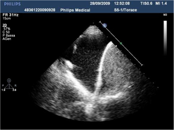
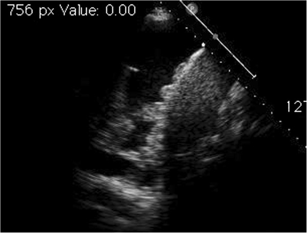
Posterior longitudinal scan in a seated patient, with sector probe showing a free-flowing pleural effusion at the base of the hemithorax.
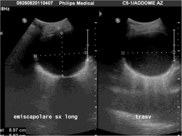
Longitudinal and transverse scan with convex probe of a loculated effusion. It is well demarcated and has a clear contour.
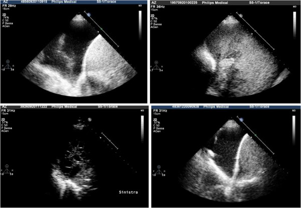
Upper left, anechoic effusion; upper right, echogenic effusion; lower left, complex septated effusion; lower right, complex non-septated effusion.
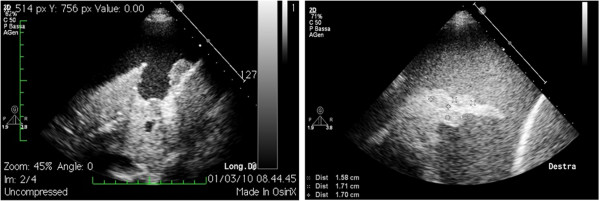
Collateral findings in the diagnosis of pleural effusion: on the left, nodulations of the diaphragmatic profile; on the right, hypoechogenic nodules in the collapsed lung parenchyma.
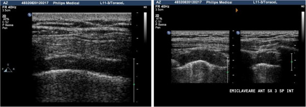
Left, pleural thickening caused by a plaque due to asbestosis: the hypo-anechoic area causes a doubling of the pleural line; right, pleural plaque observed in transverse and longitudinal scan that shows infiltration of the wall muscle layers, suggestive of neoplastic transformation.
Growths of the diaphragmatic pleura observable in the presence of effusion, a sign of secondary involvement of the pleura.
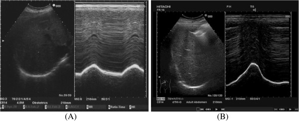
Diaphragm in B- and M-mode in spontaneous breathing (A) and in forced respiration (B).
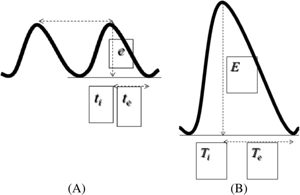
Measure of the diaphragmatic excursion in spontaneous breathing (A), on average 1.8 cm. Measure of the diaphragmatic excursion in forced respiration (B), on average 7.8 cm. ti, inspiratory time; te, expiratory time.
References
-
- Volpicelli G, Lamorte A, Tullio M, Cardinale L, Giraudo M, Stefanone V, Boero E, Nazerian P, Pozzi R, Frascisco MF. Point-of-care multiorgan ultrasonography for the evaluation of undifferentiated hypotension in the emergency department. Intensive Care Med. 2013;39:1290–1298. doi: 10.1007/s00134-013-2919-7. - DOI - PubMed
-
- Soldati G, Testa A. In: Manuale di EcografiaClinica in Urgenza. Testa A, editor. Roma: Verducieditore; 2008. Anatomiaecografia del torace.
-
- Soldati G, Copetti R. In: Ecografiatoracica. Torino CG, editor. Torino: Edizioni Medico Scientifiche; 2012. Semeioticaecograficadeltorace.
LinkOut - more resources
Full Text Sources
Other Literature Sources
Medical
