Review
. 2013 Dec:65:693-703.
doi: 10.1016/j.freeradbiomed.2013.08.003. Epub 2013 Aug 9.

Implications of autophagy for vascular smooth muscle cell function and plasticity

Affiliations
Review

Implications of autophagy for vascular smooth muscle cell function and plasticity

Joshua K Salabei et al. Free Radic Biol Med. 2013 Dec.

Abstract

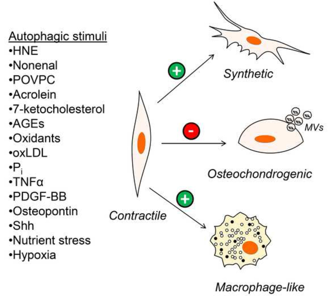
Vascular smooth muscle cells (VSMCs) are fundamental in regulating blood pressure and distributing oxygen and nutrients to peripheral tissues. They also possess remarkable plasticity, with the capacity to switch to synthetic, macrophage-like, or osteochondrogenic phenotypes when cued by external stimuli. In arterial diseases such as atherosclerosis and restenosis, this plasticity seems to be critical and, depending on the disease context, can be deleterious or beneficial. Therefore, understanding the mechanisms regulating VSMC phenotype and survival is essential for developing new therapies for vascular disease as well as understanding how secondary complications due to surgical interventions develop. In this regard, the cellular process of autophagy is increasingly being recognized as a major player in vascular biology and a critical determinant of VSMC phenotype and survival. Although autophagy was identified in lesional VSMCs in the 1960s, our understanding of the implications of autophagy in arterial diseases and the stimuli promoting its activation in VSMCs is only now being elucidated. In this review, we highlight the evidence for autophagy occurring in VSMCs in vivo, elaborate on the stimuli and processes regulating autophagy, and discuss the current understanding of the role of autophagy in vascular disease.

Keywords: Atherosclerosis; Cardiovascular; Free radicals; Hypertension; Oxidative stress; Proliferation; Restenosis.


Figures

Figure 1
Figure 1. Essential steps in the process of autophagy
Once activated, autophagy proceeds through several stages, with each stage requiring specific regulatory proteins and complexes for autophagic procession. In the initiation or nucleation stage, autophagosomal structures are formed from pre-existing plasma or organellar membranes, which leads to the development of an isolation membrane. This stage requires the activation of Atg1/ULK1 and the class III PI3K (PI3KIII)/Beclin 1 (BCL1) complex. In the elongation and maturation stages, the Atg12–Atg5 and LC3-phophatidylethanolamine (PE) conjugation systems collaborate to promote conjugation of LC3 to PE and affix LC3 to the autophagosomal membrane. Specifically, the Atg12–Atg5-Atg16L complex participates in the elongation of the autophagic membrane and Atg4 cleaves Pro-LC3 to generate LC3-I, which exposes a PE conjugation site at the COOH-terminal glycine residue. The conjugation of LC3-I to PE then proceeds from the sequential actions of Atg7 and Atg3. The LC3-II formed is then specifically associated with autophagosomes, and the phagosomal structure closes (maturation). After fusion with lysosomes, the cargo sequestered within autophagosomes is degraded by lysosomal acid hydrolases. The hydrolyzed contents, e.g., amino acids and lipids, can then be released for metabolic recycling.
Figure 2
Figure 2. Working model of the regulation of VSMC phenotype by autophagy
During vascular diseases such as atherosclerosis and hypertension, or upon percutaneous interventions, contractile VSMCs are exposed to autophagic stimuli. These stimuli include reactive species, including 4-hydroxynonenal (HNE), nonenal, 1-palmitoyl-2-(5-oxovaleroyl)-sn-glycero-3-phosphocholine (POVPC), acrolein, 7-ketocholesterol, oxidants such as superoxide and advanced glycation end products (AGEs). In addition, growth factors, cytokines and components of arterial lesions such as platelet-derived growth factor (PDGF)-BB, osteopontin, sonic hedgehog (Shh), tumor necrosis factor-α, oxidized LDL, and inorganic phosphate (Pi) also promote activation of autophagy in VSMCs. Nutrient stress and hypoxia also may stimulate VSMC autophagy. Autophagy induced by some of these factors has been shown to promote the conversion of contractile VSMCs to a synthetic and macrophage-like cell phenotype. In contrast, the osteochondrogenic VSMC phenotype appears to be suppressed by activation of autophagy, which prevents release of matrix vesicles (MVs). Autophagy in VSMCs may be a critical regulator of atherosclerotic plaque stability, the development of hypertension, VSMC proliferation and migration in restenotic lesions, and vascular calcification.

Similar articles

Cited by

References

    1. Lacolley P, Regnault V, Nicoletti A, Li Z, Michel JB. The vascular smooth muscle cell in arterial pathology: a cell that can take on multiple roles. Cardiovasc Res. 2012;95:194–204. - PubMed
    1. Gomez D, Owens GK. Smooth muscle cell phenotypic switching in atherosclerosis. Cardiovasc Res. 2012;95:156–164. - PMC - PubMed
    1. Orford JL, Selwyn AP, Ganz P, Popma P, Rogers C. The comparative pathobiology of atherosclerosis and restenosis. Am J Cardiol. 2000;86:6H–11H. - PubMed
    1. Lee RT, Libby P. The unstable atheroma. Arterioscler Thromb Vasc Biol. 1997;17:1859–1867. - PubMed
    1. Ross R. Atherosclerosis--an inflammatory disease. N Engl J Med. 1999;340:115–126. - PubMed

Publication types

LinkOut - more resources