. 2013 Nov;12(11):3221-36.
doi: 10.1074/mcp.M113.030585. Epub 2013 Aug 12.

Linking spermatid ribonucleic acid (RNA) binding protein and retrogene diversity to reproductive success

Affiliations

Linking spermatid ribonucleic acid (RNA) binding protein and retrogene diversity to reproductive success

Karen M Chapman et al. Mol Cell Proteomics. 2013 Nov.

Abstract

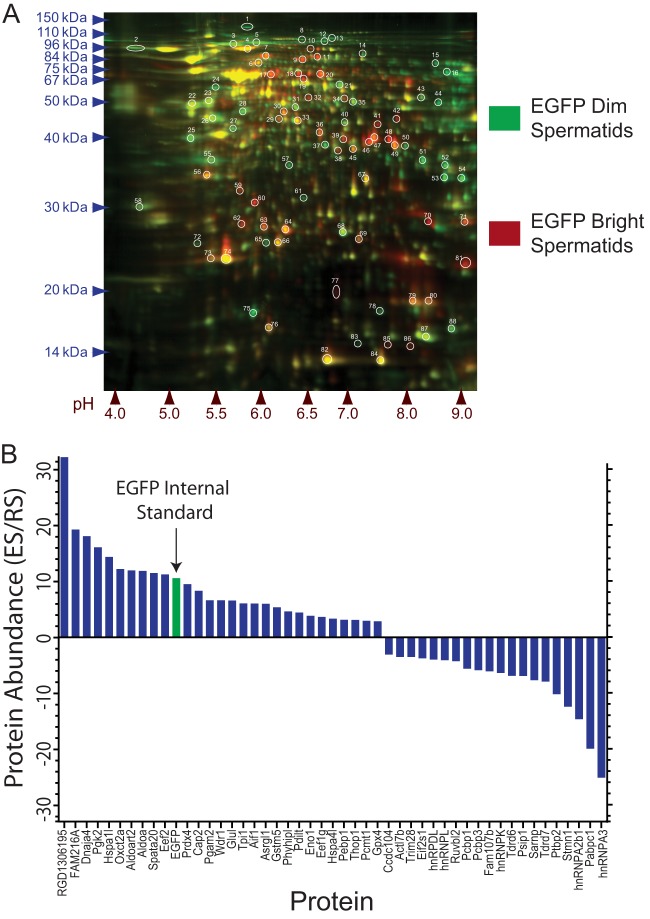
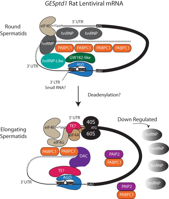
Spermiogenesis is a postmeiotic process that drives development of round spermatids into fully elongated spermatozoa. Spermatid elongation is largely controlled post-transcriptionally after global silencing of mRNA synthesis from the haploid genome. Here, rats that differentially express EGFP from a lentiviral transgene during early and late steps of spermiogenesis were used to flow sort fractions of round and elongating spermatids. Mass-spectral analysis of 2D gel protein spots enriched >3-fold in each fraction revealed a heterogeneous RNA binding proteome (hnRNPA2/b1, hnRNPA3, hnRPDL, hnRNPK, hnRNPL, hnRNPM, PABPC1, PABPC4, PCBP1, PCBP3, PTBP2, PSIP1, RGSL1, RUVBL2, SARNP2, TDRD6, TDRD7) abundantly expressed in round spermatids prior to their elongation. Notably, each protein within this ontology cluster regulates alternative splicing, sub-cellular transport, degradation and/or translational repression of mRNAs. In contrast, elongating spermatid fractions were enriched with glycolytic enzymes, redox enzymes and protein synthesis factors. Retrogene-encoded proteins were over-represented among the most abundant elongating spermatid factors identified. Consistent with these biochemical activities, plus corresponding histological profiles, the identified RNA processing factors are predicted to collectively drive post-transcriptional expression of an alternative exome that fuels finishing steps of sperm maturation and fitness.


Figures

Fig. 1.
Fig. 1.
Transgenic rats produced with lentiviral reporter vector. A, Diagram of self-inactivating lentiviral vector, pHR'CMV-EGFP-SIN18 (107, 108), used to generate transgenic rat lines by in vitro transduction of donor spermatogonia. Depicted are the U3, R, and U5 regions of the 5′ and 3′ long terminal repeat regions (LTRs); surface glycoprotein element (G); rev response element (RRE); splice acceptor sequence (SA); splice donor sequence (S.D.); cytomegalovirus promoter (CMV); enhanced green fluorescent protein (EGFP). Also shown is the region deleted from the 3′ LTR (ΔU3), which helps to disrupt viral replication (107). B, Fluorescence microscopy of EGFP expression (green) in seminiferous tubules of transgenic rat strains GESptd1, GESptd2, and GESptd3 hemizygous for a single copy of the lentiviral construct. Wild-type rat seminiferous tubules illustrate background fluorescence. Right panels show respective bright field images of seminiferous tubules from each rat. Scale bar, 1 mm. C, Lentiviral transgenes integrated between the Trmp6 and Rorb genes in 1q43 of chromosomes 1 in GESptd1 rats, and between the Sema5a and LOC100360282 genes in 2q23 on Chromosome 2 in GESptd2 rats.
Fig. 2.
Fig. 2.
GESptd rats express EGFP specifically in spermatids. A, Phase contrast and green fluorescence microscopy images identify EGFP expression (green) near the lumen of seminiferous tubules in transgenic rat strains GESptd1 and GESptd2. Scale bars, 200 μm. B, Histological cross-section of seminiferous tubules form GESptd1 rats illustrates EGFP expression in elongating spermatids (green). Image overlay shows nuclear with Hoechst 33342 dye in all testis cells (blue). Scale Bar, 200 μm. C, Top: Northern analysis of EGFP expression in tissues of transgenic rat strain, GESptd1. T, testis; M, skeletal muscle; I, small intestine; K, kidney; Lv, liver; Lg, lung; H, heart. Germ Cell Specific EGFP transgenic rat testis (GCS) (109); Wildtype rat testis (WT). Total RNA isolated from day 45 homozygous transgenic and wildtype littermates. Bottom: Same blot after stripping and re-probing for Rn18S (◀ ∼1.9 kb). D, Western analysis of EGFP (◀ ∼27 kDa) and GAPDH (◀ ∼55 kDa) expression in tissues of the transgenic (GESptd1 and GCS) and wildtype rats used in panel “A”.
Fig. 3.
Fig. 3.
GESptd rats robustly express EGFP in elongating spermatids. Testis cross sections from GESptd1 rats show EGFP expressed during elongating steps 11–19 of spermiogenesis (green fluorescence); by comparison, immunolabeling detects the transcription factor, Crem-tau, in round spermatids during Steps 1–11 of spermiogenesis (red fluorescence). Right panel shows respective images of PAS-Feulgen staining in adjacent histological sections to define stages of the seminiferous epithelium cycle (Roman numerals). Scale Bar, 100 μm.
Fig. 4.
Fig. 4.
GESptd1 rats initiate transcription of EGFP in spermatocytes. A, In Situ hybridization of a radio-labeled, EGFP antisense probe to testis cells from GESptd1 rats, but not wildtype rats. Scale Bar, 200 μm. B, Autoradiographic detection of silver grains developing from the EGFP antisense probe most densely formed over late prophase-I spermatocytes (pachytene-secondary) and step 1–17 spermatids. Sertoli Cell (SC); zygotene spermatocyte (Z); pachytene spermatocyte (P); secondary spermatocyte (2nd); round spermatids (RS); elongating spermatids (ES). Scale Bar, 30 μm. C, Illustration of GESptd1 rat transgene expression in spermatozoan progenitor cell types based on in situ hybridization studies in adult rats (top blue and green gradient bars). Note: EGFP transcripts are depicted expressed in GESptd1 rats several days prior to EGFP. As a comparison, arrows at the bottom show relative postnatal days (D) when first generation spermatogenic cell types are initially formed in rat testes. D, Northern analysis of EGFP (◀ ∼1.2 kb), DAZL (◀ ∼3 kb), and Rn18S (◀ ∼1.9 kb) transcripts in GESptd1 rat testes on postnatal days 10, 21, 25, 30, 35, 40, 45; and in D45 testes of wild-type rats (WT). E, Western analysis of EGFP (◀ ∼27 kDa), DAZL (◀ ∼36 kDa) and TUBA1a (◀ ∼55 kDa) in GESptd1 rat testes on postnatal days 21, 25, 30, 35, 40, 45; and in D45 testes of wild-type rats (WT).
Fig. 5.
Fig. 5.
EGFP transcripts localize to mRNP-like particles in GESptd1 rats. A, Northern blot of EGFP transcripts in whole testis lysates from an adult GESptd1 rat after centrifugation into a sucrose density gradient. RNase-H (RH) treated samples after hybridizing with Oligo(dt). Note: a majority of EGFP transcripts are detected in low density mRNP-like particles. Controls (Ct) = testis lysates prepared from GCS-EGFP transgenic (+) and wildtype (−) rats. B, Western blot of MSY2 protein in whole testis lysates (T) from an adult GESptd1 rat after centrifugation into a sucrose density gradient. C, Northern blot of DAZL transcripts in whole testis lysates from an adult GESptd1 rat after centrifugation into a sucrose density gradient. Note: a majority of DAZL transcripts are detected in polysome-like particles. Controls (Ct) = testis lysates prepared from tgGCS-EGFP transgenic (+) and wildtype (−) rats.
Fig. 6.
Fig. 6.
Flow sorting round and elongating spermatids from GESptd1 rats. A, Flow cytometry analysis of EGFP+ testis cells sorted from GESptd1 rats. Left: Total EGFP+ testis cell population gated within region 1 (R1) of forward and side scatter plot (R1 = 31.9 ± 5.7% total) Center: Background green fluorescence gates G1 and G2 set using a wild-type rat littermate; Right: Dominant peaks of EGFP+ GESptd1 rat testis cells sorted from gates G1 and G2 (G1 = 27.8 ± 3.4% R1; G2 = 31.6 ± 0.9% R1); ±S.D. n = 5 sorts. B, Relative purities of “EGFP-Dim” and “EGFP-Bright” GESptd1 rat cells post-sorted from gates G1 and G2, respectively. C, Top: Images of testis cells isolated from wildtype and GESptd1 rats before and after sorting into EGFP-Bright and EGFP-Dim fractions. Bottom: Hoechst 33342 nuclear labeling (blue) of cells in top panels. Scale bars, 8 μm.
Fig. 7.
Fig. 7.
Analysis of differentially expressed rat spermatid proteins. A, Differential display of molecules in round and elongating spermatids from GESptd1 rats after separation by 2D gel electrophoresis. Round spermatids in EGFP Dim fractions were prelabeled with a green fluorescent dye; Elongating spermatids in the EGFP Bright fractions were prelabeled with a red dye. The circled molecules were submitted for sequence analysis by mass spectrometry. B, Relative abundance of fluorescently labeled molecules identified by mass spectrometry in fractions of round spermatids (EGFP Dim Spermatids; green), and elongating spermatids (EGFP Bright Spermatids; red) from GESptd1 rats. Ratios of elongating spermatid/round spermatid molecular fluorescence signal intensities (ES/RS) were plotted for positively identified rat proteins (symbols) changing ≥threefold.
Fig. 8.
Fig. 8.
Mass spectrometry predicts protein localization in rat spermatids. A, Localization of anti-hnRNPK IgG binding (red) in wild-type rat testis. Note: labeling in pachytene spermatocytes (PS) and round spermatids (RS), but not in elongating spermatids (ES). Hoechst 33342 dye labels nuclei (blue) of all testis cells. Scale bar, 30 μm. B, Localization of anti-STMN1 IgG binding (red) in GESptd1 rat testis. Note: labeling in pachytene spermatocytes (PS) and round spermatids (RS), but not in elongating spermatids (ES). Hoechst 33342 dye labels nuclei (blue) of all testis cells. Asterisks marks stage IX seminiferous tubule containing step 9 spermatids starting to elongate. Scale bar, 100 μm. C, Localization of anti-GLUL IgG binding (red) in rat GESptd1 testis. Note: labeling in elongating spermatids (ES), but not in pachytene spermatocytes (PS) or round spermatids (RS). Hoechst 33342 dye labels nuclei (blue) of all testis cells. Scale bar, 100 μm.
Fig. 9.
Fig. 9.
Testicular expression of PCBP1 and EEF2 correlate with mass spectrometry signals. A, Localization of anti-PCBP1 IgG binding (red) in wild-type rat testis. Note: widespread labeling in pachytene spermatocytes (PS) and round spermatids (RS), but predominantly focal localization to granules in elongating spermatids (ES). Hoechst 33342 dye labels nuclei (blue) of all testis cells. Roman numbers estimate spermatogenic stage. Scale bars, 30 μm. B, Localization of Anit-EEF2 IgG Binding in wild-type rat Testis. Round Spermatid (RS); Elongating Spermatid (ES); Pachytene Spermatocyte (Py); Preleptotene Spermatocyte (PL); Intermediate Spermatogonia (Int Spg); Sertoli Cell (SC). Roman numbers estimate spermatogenic stage. Scale bar, 30 μm.
Fig. 10.
Fig. 10.
Post-transcriptional regulation of rat tgGESptd1 expression. Open questions are illustrated on how expression of the GESptd1 rat transgene (tgGESptd1) is regulated post-transcriptionally during spermatid maturation. Here, translation of a tgGESptd1 encoded mRNA upon spermatid elongation is modeled hypothetically in association with hnRNP-family proteins (Fig. 5, Table I), reported small RNA processing pathways (110), poly-A tail de-adenylation (18, 20) and incorporation of processed transcripts into translational complexes (98, 99). Mechanisms controlling relative abundance of hnRNPs upon spermatid elongation remain to be determined. Also illustrated, based on information from other studies, are hypothetical GW182/AGO-like protein components in RNA silencing complex (RISC) (110), associated poly-A tail, de-adenylation complex (DAC) (18, 20), 40S and 60S ribosomal subunits, 5′ and 3′ untranslated regions (UTR), eIF4A, eIF4E, eIF4G and eIF3 elongation initiation factors, poly-A-binding protein C1 (PABPC1), poly(A)-tail binding protein-interacting protein 2 (PAIP2) (19, 102), and additional putative translational enhancers (TE) (19, 102).

Similar articles

Cited by

References

    1. David C. J., Chen M., Assanah M., Canoll P., Manley J. L. (2010) HnRNP proteins controlled by c-Myc deregulate pyruvate kinase mRNA splicing in cancer. Nature 463, 364–368 - PMC - PubMed
    1. Kir S., Beddow S. A., Samuel V. T., Miller P., Previs S. F., Suino-Powell K., Xu H. E., Shulman G. I., Kliewer S. A., Mangelsdorf D. J. (2011) FGF19 as a postprandial, insulin-independent activator of hepatic protein and glycogen synthesis. Science 331, 1621–1624 - PMC - PubMed
    1. Licatalosi D. D., Yano M., Fak J. J., Mele A., Grabinski S. E., Zhang C., Darnell R. B. (2012) Ptbp2 represses adult-specific splicing to regulate the generation of neuronal precursors in the embryonic brain. Genes Dev. 26, 1626–1642 - PMC - PubMed
    1. Zhu H., Shyh-Chang N., Segre A. V., Shinoda G., Shah S. P., Einhorn W. S., Takeuchi A., Engreitz J. M., Hagan J. P., Kharas M. G., Urbach A., Thornton J. E., Triboulet R., Gregory R. I., Altshuler D., Daley G. Q. (2011) The Lin28/let-7 axis regulates glucose metabolism. Cell 147, 81–94 - PMC - PubMed
    1. Rouget C., Papin C., Boureux A., Meunier A. C., Franco B., Robine N., Lai E. C., Pelisson A., Simonelig M. (2010) Maternal mRNA deadenylation and decay by the piRNA pathway in the early Drosophila embryo. Nature 467, 1128–1132 - PMC - PubMed

Publication types

MeSH terms

LinkOut - more resources