. 2013 Aug 12;368(1626):20130047.
doi: 10.1098/rstb.2013.0047. Print 2013 Sep 19.

Functional endogenous viral elements in the genome of the parasitoid wasp Cotesia congregata: insights into the evolutionary dynamics of bracoviruses

Affiliations

Functional endogenous viral elements in the genome of the parasitoid wasp Cotesia congregata: insights into the evolutionary dynamics of bracoviruses

Annie Bézier et al. Philos Trans R Soc Lond B Biol Sci. .

Abstract

Bracoviruses represent the most complex endogenous viral elements (EVEs) described to date. Nudiviral genes have been hosted within parasitoid wasp genomes since approximately 100 Ma. They play a crucial role in the wasp life cycle as they produce bracovirus particles, which are injected into parasitized lepidopteran hosts during wasp oviposition. Bracovirus particles encapsidate multiple dsDNA circles encoding virulence genes. Their expression in parasitized caterpillars is essential for wasp parasitism success. Here, we report on the genomic organization of the proviral segments (i.e. master sequences used to produce the encapsidated dsDNA circles) present in the Cotesia congregata parasitoid wasp genome. The provirus is composed of a macrolocus, comprising two-thirds of the proviral segments and of seven dispersed loci, each containing one to three segments. Comparative genomic analyses with closely related species gave insights into the evolutionary dynamics of bracovirus genomes. Conserved synteny in the different wasp genomes showed the orthology of the proviral macrolocus across different species. The nudiviral gene odv-e66-like1 is conserved within the macrolocus, suggesting an ancient co-localization of the nudiviral genome and bracovirus proviral segments. By contrast, the evolution of proviral segments within the macrolocus has involved a series of lineage-specific duplications.

Keywords: bracovirus; comparative genomics; obligatory mutualism; parasitoid wasp; polydnavirus.


Figures

Figure 1.
Figure 1.
Structural organization of proviral loci within (a) Cotesia congregata and (b) Cotesia sesamiae. Proviral segments are represented as black or red arrows depending on their orientation. CcBV proviral segments have been given the same number as their corresponding circles packaged in virus particles, whereas CsBV segments were numbered based on their CcBV homologues, except for CsBV S20/33 and S37 specific for CsBV. Only partial sequences of CsBV S25, S5 and S18 could be identified from available data. Loci were named based on those previously characterized in G. indiensis and G. flavicoxis [16] except for PL8 and PL9 (specific for C. congregata). The small tree on the right is a schematic of phylogenetic relationships between wasp species indicating the estimated time since the separation of Cotesia and Glyptapanteles lineages. Note the wasp genes in flanking sequences that are conserved in orthologous positions in Glyptapanteles spp. (purple stars) and the gene of the nudiviral machinery involved in particle production (green star) within the macrolocus. Scale is expressed in basepairs. For detailed analysis of proviral loci flanking region synteny, see figure 2, table 2 and electronic supplementary material, S5.
Figure 2.
Figure 2.
Synteny in wasp genes-containing region joining PL1 to PL2 (macrolocus). This region includes the conserved nudiviral odv-e66-like1 gene. Genes are indicated by squares and numbers are those given in GenBank. Their positions on the DNA sequences (following the numbering in GenBank) are indicated above the squares. Gene synteny is highlighted in purple and the nudiviral gene is coloured green. Interruptions in the black lines indicate gaps in the sequence (non-overlapping BACs). White areas correspond to non-homologous sequence or to a lack of data for one species. Proviral segments flanking this region corresponding to the extremities of PL1 and PL2 are shown in red, with arrows indicating their orientation. CcBV sequences were obtained either from overlapping BAC sequencing or PCR fragments as indicated below. Cc, C. congregata; Cs, C. sesamiae; Gi, G. indiensis and Gf, G. flavicoxis; nc, non-coding sequences. CsPL1 and region containing wasp genes (accession number EF710629); CsPL2 (EF710635); GiPL1 (AC191960); GiPL2 (EF710657); GfPL1 (EF710644) and GfPL2 (EF710648).
Figure 3.
Figure 3.
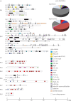
Gene content of CcBV proviral segments within (a) macrolocus and (b) dispersed loci displayed by coloured boxes (macrolocus, 260 genes; dispersed loci, 62 genes). CcBV contains 37 gene families: seven encode proteins with described conserved domains (cystatin, RNaseT2, C-type lectin, Cys-rich, BEN, VANK and ptp) representing approximately 23.5% of the genes and one encoded a baculovirus homologue protein (p94-like). Twenty-nine gene families representing 57% of the genes encode proteins of unknown function conserved in BVs associated with wasps of the Cotesia and Glyptapanteles genera (Ser-rich, EP2-like, EP1-like and BV1BV26 represented by grey boxes, with the number identifying the family indicated above the boxes). Some other previously identified BV gene families are only represented by one member in CcBV (CrV1, histone H4, Duffy and p494). Other genes of unknown function are unique (approx. 14.5%) and some coding DNA sequences are identified as remnants of genes from mobile elements (approx. 4.25%). Note that the ptp gene family constitutes the major part of the genes from isolated loci (see gene distribution pie charts on the right). Unlike in GiBV, no ptp genes are found in the CcBV macrolocus. Other genes such as cystatin, RNaseT2, C-type lectin or cys-rich are found only in the macrolocus, which contains a majority of BV specific genes (table 2). ps34 shown by a dashed line corresponds to a former proviral segment mutated in the 3′DRJ core homologous to CvBV S30 (accession number HQ009553) but no longer producing a circle.
Figure 4.
Figure 4.
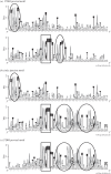
DRJ sequence motifs within C. congregata proviral segments and CcBV circles visualized using WebLogo. Each logo consists of stacks of bases, with one stack for each position in the sequence. The height of the stack at a position indicates the sequence conservation, whereas the height of a base indicates the relative frequency of this base at this position. Note that the circle junction sequence (b) corresponds to a recombined form of the two DRJs within the perfectly conserved DRJ core shown in the black box. Sequences characterizing (a) 3′DRJ and (c) 5′DRJs are circled in black. A 30 bp sequence containing a 13 bp repeat (TTtnAatantGAAyaaAAatnntGAwcAaa) following the 5′DRJ core was found to be conserved, whereas the sequence following the core in 3′DRJ was smaller (TTcnAATTgt). A highly conserved motif (TGAa/tT) was also identified 80 bp upstream of the 3′DRJ core. These graphical representations were generated from independent alignments of 34 5′DRJ, 35 3′DRJ and 35 circle junction sequences (see electronic supplementary material, figure S1).
Figure 5.
Figure 5.
Similarity matrix of the C. congregata proviral macrolocus compared with itself. The main diagonal represents sequence alignment with itself; dotted lines (grey) identify duplicated regions within the sequence analysed. Those parallel to the diagonal correspond to duplicated regions in the same orientation; those antiparallel correspond to inverted sequences (striped arrows). Scale is expressed in kilobase pair. Relative positions of proviral loci 1 and 2 and of each CcBV proviral segment forming the macrolocus are indicated below the dot plot matrix. PL1, proviral locus 1; PL2, proviral locus 2; TE, transposable element. Grey boxes: duplication PL1–Dp1/PL1–Dp2 within PL1; striped boxes: inverted duplication PL1–Inv1/PL2–Inv2; light grey boxes: duplication PL2–Dp1/PL2–Dp2; dark grey boxes: triplication PL2–Tr1/PL2–Tr2/PL2–Tr3. Nucleotide positions of duplication extremities are indicated in the electronic supplementary material, table S6.
Figure 6.
Figure 6.
Proviral segment clustering based on (a) 5′ and (b) 3′DRJ sequences. The trees were obtained from maximum-likelihood phylogenetic inferences based on the alignments of approximately 200 bp sequences of 5′ and 3′DRJs including the DRJ core (‘extended DRJs’). Only SH-like branch values above 50 are indicated. Thick branches highlight 5′DRJs in co-phylogeny with the 3′DRJ of the same segment, produced by complete duplication of proviral segments including their DRJs (duplicated regions containing the DRJs are indicated on the right). The stars indicated the three S28 DRJs. In this segment, the ancestral 3′DRJ S28* (still functional in C. sesamiae) was replaced by a new 3′DRJ recruited during the rearrangement that produced PL2-Tr1. This resulted in a mosaic S28 segment (figure 7).
Figure 7.
Figure 7.
A proposed parsimonious scenario for the complex rearrangement that may have produced PL2-Tr1 based on the analysis of duplications among Cotesia spp. (a) Cotesia congregata and (c) C. sesamiae macrolocus sequences were used to infer the putative organization of this region in their common ancestor (b). In the lineage leading to C. sesamiae, the bv8 gene was lost (or this gene was acquired specifically in the C. congregata lineage). In the lineage leading to C. congregata, a complex rearrangement occurred resulting in inversion and duplication of proviral segment sequences. Inversion: the segment S37 was inverted and its ep1-like6 gene and regular 3′DRJ (black triangle) were incorporated into an enlarged S28. The regular 3′DRJ became that of S28, replacing the former S28 DRJ readily identified by its particular sequence (3′DRJ*, grey triangle). Duplication: the region encompassing S28, S27 and a part of S15 was duplicated and inserted within S37 that was dismantled (dis37). It should be noted that inversion, duplication and dismantlement might have been produced by a single complex rearrangement caused by errors during replication (fork stalling and template switching model). DRJs are indicated by white triangles to delimit the segments.
Figure 8.
Figure 8.
A proposed parsimonious scenario for macrolocus proviral genome evolution in Cotesia and Glyptapanteles lineages based on the analysis of duplications among wasp species. Sequences from C. congregata (a), C. sesamiae (b), G. indiensis and G. flavicoxis (d) were used to infer the putative organization of an ancestral macrolocus (c) containing two proviral regions that would have existed before the separation of the Cotesia and Glyptapanteles lineages over 17 Ma. In the lineage leading to Glyptapanteles spp., new segments 1p–2p–3p, 17p, 18p and 20p (re-integrated) were formed. In the lineage leading to Cotesia spp., inverted duplications of PL1 sequences (hashed boxes) resulted in modifications of the anterior PL2 region. Subsequent rearrangements in the lineage leading to C. sesamiae lead to the fusion of segments S20 and S33 (S20/33) and the loss of segment S15 gene content. In the lineage leading to C. congregata, duplication in the posterior region of PL2 produced PL2–Tr1 and a larger S28. In addition, duplications upstream of PL2 lead to the formation of PL2–Dp1 and PL2–Dp2. Names and box colours for duplications and inversion are the same as in figure 5.

Similar articles

Cited by

References

    1. Herniou EA, Huguet E, Thézé J, Bézier A, Periquet G, Drezen J-M. 2013. When parasitic wasps hijacked viruses: genomic and functional evolution of polydnaviruses. Phil. Trans. R. Soc. B 368, 20130051 (doi:10.1098/rstb.2013.0051) - DOI - PMC - PubMed
    1. Dupuy C, Huguet E, Drezen J-M. 2006. Unfolding the evolutionary story of polydnaviruses. Virus Res. 117, 81–89 (doi:10.1016/j.virusres.2006.01.001) - DOI - PubMed
    1. Murphy N, Banks JC, Whitfield JB, Austin AD. 2008. Phylogeny of the parasitic microgastroid subfamilies (Hymenoptera: Braconidae) based on sequence data from seven genes, with an improved time estimate of the origin of the lineage. Mol. Phylogenet. Evol. 47, 378–395 (doi:10.1016/j.ympev.2008.01.022) - DOI - PubMed
    1. Bézier A, et al. 2009. Polydnaviruses of braconid wasps derive from an ancestral nudivirus. Science 323, 926–930 (doi:10.1126/science.1166788) - DOI - PubMed
    1. Théze J, Bézier A, Periquet G, Drezen J-M, Herniou EA. 2011. Paleozoic origin of insect large dsDNA viruses. Proc. Natl Acad. Sci. USA 108, 15 931–15 935 (doi:10.1073/pnas.1105580108) - DOI - PMC - PubMed

Publication types

LinkOut - more resources