Review
. 2013 Aug 12;368(1626):20130051.
doi: 10.1098/rstb.2013.0051. Print 2013 Sep 19.

When parasitic wasps hijacked viruses: genomic and functional evolution of polydnaviruses

Affiliations
Review

When parasitic wasps hijacked viruses: genomic and functional evolution of polydnaviruses

Elisabeth A Herniou et al. Philos Trans R Soc Lond B Biol Sci. .

Abstract

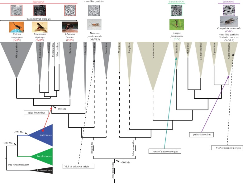
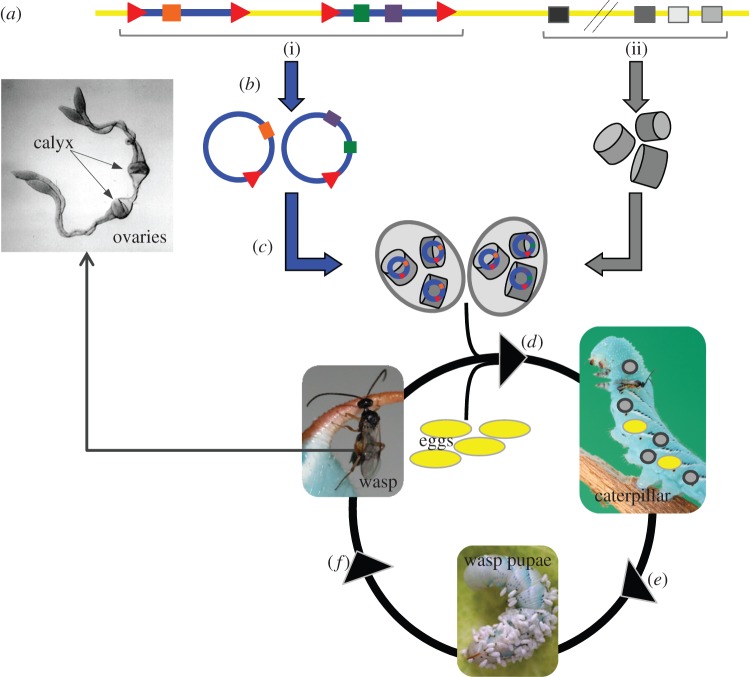
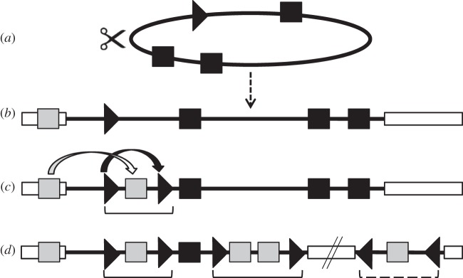
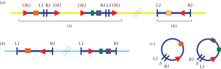
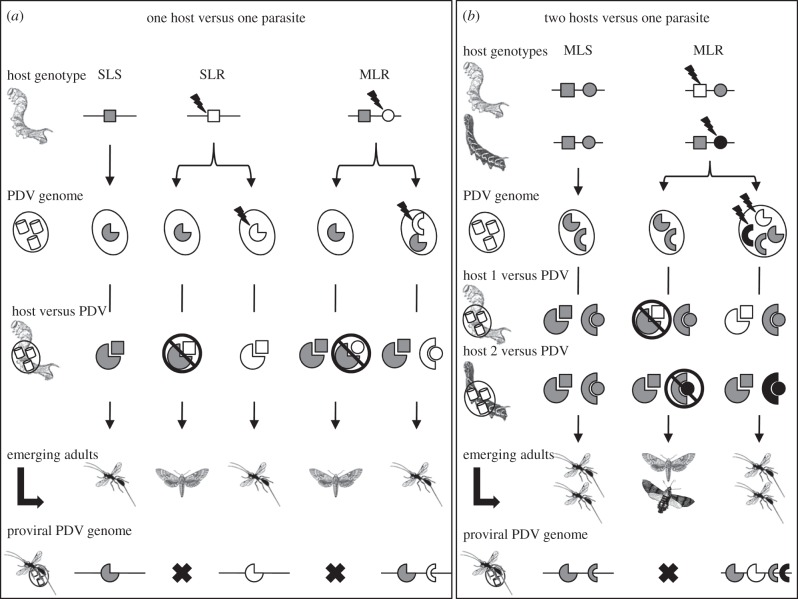
The Polydnaviridae (PDV), including the Bracovirus (BV) and Ichnovirus genera, originated from the integration of unrelated viruses in the genomes of two parasitoid wasp lineages, in a remarkable example of convergent evolution. Functionally active PDVs represent the most compelling evolutionary success among endogenous viral elements (EVEs). BV evolved from the domestication by braconid wasps of a nudivirus 100 Ma. The nudivirus genome has become an EVE involved in BV particle production but is not encapsidated. Instead, BV genomes have co-opted virulence genes, used by the wasps to control the immunity and development of their hosts. Gene transfers and duplications have shaped BV genomes, now encoding hundreds of genes. Phylogenomic studies suggest that BVs contribute largely to wasp diversification and adaptation to their hosts. A genome evolution model explains how multidirectional wasp adaptation to different host species could have fostered PDV genome extension. Integrative studies linking ecological data on the wasp to genomic analyses should provide new insights into the adaptive role of particular BV genes. Forthcoming genomic advances should also indicate if the associations between endoparasitoid wasps and symbiotic viruses evolved because of their particularly intimate interactions with their hosts, or if similar domesticated EVEs could be uncovered in other parasites.

Keywords: Cotesia; genome evolution; obligatory mutualism; parasitoid wasp; polydnavirus; virus adaptation.


Figures

Figure 1.
Figure 1.
Multiple origins of virus symbioses in the Ichneumonoidea. Phylogenies and molecular dating are modified from [–9] for ichneumonoid wasp and [2] for the free insect DNA virus.
Figure 2.
Figure 2.
Bracovirus life cycle and genome organization. (a) The BV genome is integrated in the wasp genome (yellow). It is composed of (i) proviral segments (blue) used to produce the multiple dsDNA circles that encode virulence genes (coloured rectangles) and that are packaged in the particles, and of (ii) BV structural genes (nudiviral genes; black or grey rectangles) that are involved in particle production. (b) Nudiviral gene expression as well as amplification and excision of BV circles occurs in the calyx cells of the wasp ovaries. Direct repeat junctions (DRJs; red triangles) are involved in circularization. (c) DNA circles are packaged into BV particles. (d) BV particles are injected in the lepidopteran host during oviposition of parasitoid eggs and infect many lepidopteran cell types but do not replicate. (e) BV virulence gene expression leads to modifications in lepidopteran host physiology, such as inhibition of wasp egg encapsulation, allowing wasp development. (f) Emergence of adults carrying bracovirus genomes from wasp pupae. This figure is based on the life cycle of CcBV associated with C. congregata parasitoid wasp of M. sexta. (Photographs A. Bézier and A. Wild.)
Figure 3.
Figure 3.
Bracovirus evolutionary model from an ancestral nudivirus. (a) Genome of the ancestral nudivirus; (b) initial nudivirus genome integration into a wasp genome; (c) formation of the first proviral segment and (d) simplified scheme of a present-day BV. Black squares are for nudiviral genes, black arrowhead for nudivirus derived circularization site, white rectangles for wasp genomes, grey squares are for wasp genes. Brackets are for BV proviral segments located in the macrolocus, hashed bracket is for an isolated proviral segment.
Figure 4.
Figure 4.
Bracovirus sequences integrated into insect genomic DNA and encapsidated circles. (a) BV segments integrated in wasp genomic DNA. Within the C. sesamiae wasp genome (in yellow), (i) classical proviral BV segments containing virulence genes (coloured rectangles) delimited by DRJ sequences (red triangles) and (ii) a reintegrated segment can be identified. The reintegrated segment is not delimited by DRJ, but is bordered by left junction (LJ) and right junction (RJ) sequences. (b) BV segments reintegrated in lepidopteran host. Within lepidopteran genomes (in light blue), reintegrated BV segments bordered by LJ and RJ sequences can also be identified. (c) Encapsidated BV circles injected in lepidopteran host. Sequence comparison between circular and reintegrated viral sequences (in wasp or lepidopteran genomes) show that circle reintegration in both cases involves loss of a stretch of viral sequence (indicated by Δ), and is mediated by similar reintegration boundaries (LJ, RJ), suggesting that BV use a specific but unknown mechanism to reintegrate into genomic DNA. See table 2 for sequences involved in BV circle circularization and reintegration.
Figure 5.
Figure 5.
Polydnavirus mediated host parasite coevolution. (a) PDV adaptation to a single host species harbouring either a single locus susceptibility (SLS), a single locus resistance (SLR) or multilocus resistance (MLR); (b) PDV adaptation to two hosts species harbouring either a multilocus susceptibility (MLS), or multilocus resistance (MLR). Pie and semicircle shapes indicate PDV genes; squares and circles host resistance genes. Corresponding shapes and colours indicate an efficient PDV effector targeting of host factor allowing the wasp larvae to develop and the transmission of the PDV gene in their chromosomes. Lightning shapes depict mutations and virulence or resistance gene acquisitions.

References

    1. Emerman M, Malik HS. 2010. Paleovirology: modern consequences of ancient viruses. PLoS Biol. 8, e1000301 (doi:10.1371/journal.pbio.1000301) - DOI - PMC - PubMed
    1. Thézé J, Bézier A, Periquet G, Drezen J-M, Herniou EA. 2011. Paleozoic origin of insect large dsDNA viruses. Proc. Natl Acad. Sci. USA 108, 15 931–15 935 (doi:10.1073/pnas.1105580108) - DOI - PMC - PubMed
    1. Katzourakis A, Gifford RJ, Tristem M, Gilbert MT, Pybus OG. 2009. Macroevolution of complex retroviruses. Science 325, 1512 (doi:10.1126/science.1174149) - DOI - PubMed
    1. Katzourakis A, Gifford RJ. 2010. Endogenous viral elements in animal genomes. PLoS Genet. 6, e1001191 (doi:10.1371/journal.pgen.1001191) - DOI - PMC - PubMed
    1. Strand MR, Burke GR. 2012. Polydnaviruses as symbionts and gene delivery systems. PLoS Pathog. 8, e1002757 (doi:10.1371/journal.ppat.1002757) - DOI - PMC - PubMed

Publication types

LinkOut - more resources