. 2013 Oct;9(10):1467-74.
doi: 10.4161/auto.25687. Epub 2013 Aug 5.

Structural characterization of the Saccharomyces cerevisiae autophagy regulatory complex Atg17-Atg31-Atg29

Affiliations

Structural characterization of the Saccharomyces cerevisiae autophagy regulatory complex Atg17-Atg31-Atg29

Leon H Chew et al. Autophagy. 2013 Oct.

Abstract

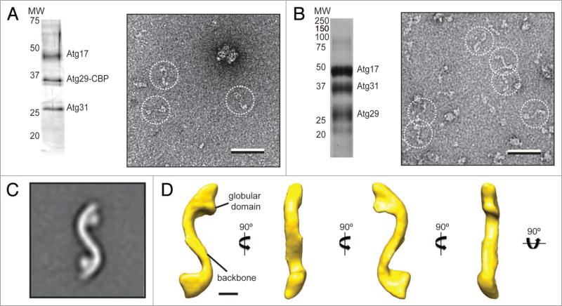
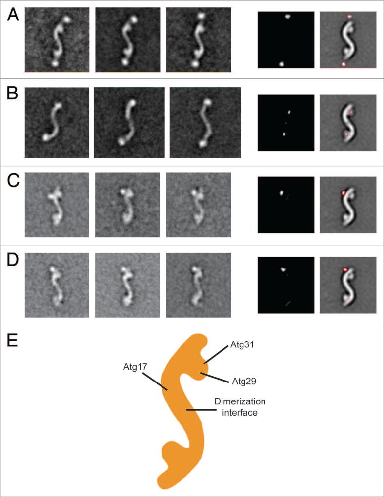
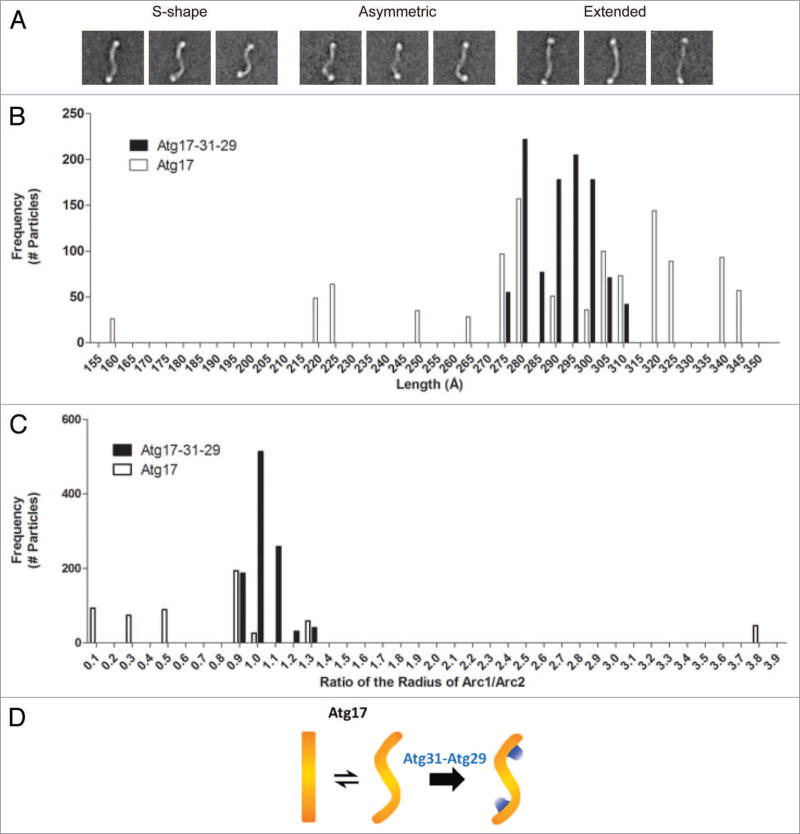
Atg17, in complex with Atg29 and Atg31, constitutes a key module of the Atg1 kinase signaling complex and functions as an important organizer of the phagophore assembly site in the yeast Saccharomyces cerevisiae. We have determined the three-dimensional reconstruction of the full S. cerevisiae Atg17-Atg31-Atg29 complex by single-particle electron microscopy. Our structure shows that Atg17-Atg31-Atg29 is dimeric and adopts a relatively rigid and extended "S-shape" architecture with an end-to-end distance of approximately 345 Å. Subunit mapping analysis indicated that Atg17 mediates dimerization and generates a central rod-like scaffold, while Atg31 and Atg29 form two globular domains that are tethered to the concave sides of the scaffold at the terminal regions. Finally, our observation that Atg17 adopts multiple conformations in the absence of Atg31 and Atg29 suggests that the two smaller components play key roles in defining and maintaining the distinct curvature of the ternary complex.

Keywords: Atg17; Atg29; Atg31; autophagy; single-particle electron microscopy.


Conflict of interest statement

Disclosure of Potential Conflicts of Interest

No potential conflicts of interest were disclosed.

Figures

Figure 1
Figure 1
Purification and negative-stain EM analysis of the S. cerevisiae Atg17-Atg31-Atg29 complex. (A) Silver-stained SDS-PAGE analysis of purified native Atg17-Atg31-Atg29 complex (left). A representative raw image of negatively-stained native Atg17-Atg31-Atg31 (right) with particles in dotted circles (scale bar: 50 nm). (B) Silver-stained SDS-PAGE analysis of purified recombinant Atg17-Atg31-Atg29 complex (left). A representative image of negatively-stained recombinant Atg17-Atg31-Atg29 (right) with particles in dotted circles (scale bar: 50 nm). (C) class average for the top class from classification of 10,251 negatively-stained particles showing an overall S-shape with two circular-shaped domains attached to the terminal regions of the complex (inset). Side length of this panel is 52 nm. (D) The 3D reconstruction of Atg17-Atg31-Atg29 viewed in different orientations (scale bar: 5 nm). Two characteristic features of the reconstruction are labeled.
Figure 2
Figure 2
Subunit organization of the Atg17-Atg31-Atg29 complex. (A) Localization of Atg17. The left panels are representative class averages of negatively- stained His-MBP-tagged Atg17-Atg31-Atg29 (side length: 59 nm). The middle panel is a difference image generated from subtracting the untagged Atg17-Atg31-Atg29 average from the first His-MBP-tagged Atg17-Atg31-Atg29 average. The right panel depicts difference densities with respect to untagged Atg17-Atg31-Atg29. (B) Overall architecture of Atg17. The left panels show averages of negatively-stained His-MBP-tagged Atg17 that have similar overall shapes as Atg17-Atg31-Atg29. Side length of each panel is 64 nm. Difference image (middle panel), and positions of the difference densities (right panel) show that the terminal circular domains are composed of Atg29 and Atg31. (C) Representative class averages of negatively-stained Atg17-Atg31-Atg29 containing C-terminal GFP -tagged Atg29 (left 3 panels). Localizing the GFP density by difference mapping (middle and right panels). (D) Representative class averages of negatively-stained Atg17-Atg31-Atg29 containing C-terminal GFP -tagged Atg31 (left 3 panels). Localizing the GFP density by difference mapping (middle and right panels). (E) Subunit organization map of Atg17-Atg31-Atg29 with labels depicting the positions of the three subunits.
Figure 3
Figure 3
Analysis of the conformational flexibility of Atg17. (A) Structural segregation of class averages from classification of 5,922 negatively-stained His-MBP-tagged Atg17 particles exhibiting a range of conformations Atg17 can adopt: S-shaped, asymmetric, and extended. Side length of each panel is 64 nm. (B) Distribution of junction-to-junction lengths measured for 17 class averages (1,029 total particles) of His-MBP-labeled Atg17-Atg31-Atg29 (black) and 18 class averages (1,099 total particles) of His-MBP-Atg17 (white). Median length was 293 Å and 285 Å for His-MBP-Atg17-Atg31-Atg29 and His-MBP-Atg17, respectively. (C) Distribution of the ratio of curvature measured for the two arcs within each ternary complex and Atg17 dimer. Seventeen class averages (1,029 total particles) of His-MBP-labeled Atg17-Atg31-Atg29 (black) and 9 class averages (539 total particles) of His-MBP-Atg17 particles (white) were analyzed. Median radius was 75 Å and 213 Å for His-MBP-Atg17-Atg31-Atg29 and His-MBP-Atg17, respectively. (D) Schematic depicting the role of Atg29 and Atg31 in defining and stabilizing the distinct curvature of Atg17.

References

    1. Nakatogawa H, Suzuki K, Kamada Y, Ohsumi Y. Dynamics and diversity in autophagy mechanisms: lessons from yeast. Nat Rev Mol Cell Biol. 2009;10:458–67. http://dx.doi.org/10.1038/nrm2708. - DOI - PubMed
    1. Kraft C, Martens S. Mechanisms and regulation of autophagosome formation. Curr Opin Cell Biol. 2012;24:496–501. http://dx.doi.org/10.1016/j.ceb.2012.05.001. - DOI - PubMed
    1. Noda NN, Ohsumi Y, Inagaki F. ATG systems from the protein structural point of view. Chem Rev. 2009;109:1587–98. http://dx.doi.org/10.1021/cr800459r. - DOI - PubMed
    1. Suzuki K, Ohsumi Y. Current knowledge of the pre-autophagosomal structure (PAS) FEBS Lett. 2010;584:1280–6. http://dx.doi.org/10.1016/j.febslet.2010.02.001. - DOI - PubMed
    1. Mizushima N, Yoshimori T, Ohsumi Y. The role of Atg proteins in autophagosome formation. Annu Rev Cell Dev Biol. 2011;27:107–32. http://dx.doi.org/10.1146/annurev-cellbio-092910-154005. - DOI - PubMed

Publication types

MeSH terms

Substances

LinkOut - more resources