. 2013 Nov;11(3):1013-21.
doi: 10.1016/j.scr.2013.07.001. Epub 2013 Jul 16.

Endothelial cells mitigate DNA damage and promote the regeneration of hematopoietic stem cells after radiation injury

Affiliations

Endothelial cells mitigate DNA damage and promote the regeneration of hematopoietic stem cells after radiation injury

Derek K Zachman et al. Stem Cell Res. 2013 Nov.

Abstract

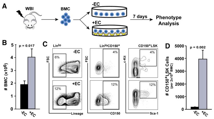
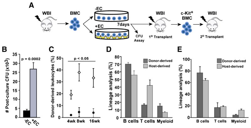
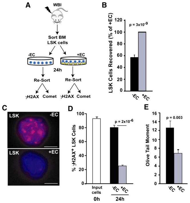
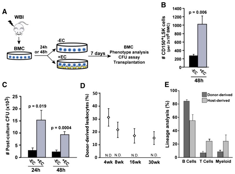
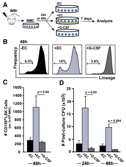
Endothelial cells (ECs) are an essential component of the hematopoietic microenvironment, which maintains and regulates hematopoietic stem cells (HSCs). Although ECs can support the regeneration of otherwise lethally-irradiated HSCs, the mechanisms are not well understood. To further understand this phenomenon, we studied HSC regeneration from irradiated bone marrow using co-culture with human aortic ECs (HAECs). Co-culture with HAECs induced a 24-fold expansion of long-term HSCs (CD150(+), lineage(lo), Sca-1(+), c-Kit(+); CD150(+)LSK cells) in vitro. These cells gave rise to functional hematopoietic stem and progenitor cells (HSPCs) with colony-forming activity, multilineage reconstitution and serial transplantation potential. Furthermore, HAECs significantly reduced DNA damage in irradiated LSK cells within 24h. Remarkably, we were able to delay the exposure of irradiated bone marrow to the regenerative, HAEC-derived signals for up to 48h and still rescue functional HSCs. G-CSF is the gold standard for promoting hematopoietic regeneration in vivo. However, when compared to HAECs, in vitro G-CSF treatment promoted lineage differentiation and regenerated 5-fold fewer CD150(+)LSK cells. Together, our results show that HAECs are powerful, direct mitigators of HSC injury and DNA damage. Identification of the HAEC-derived factors that rescue HSCs may lead to improved therapies for hematopoietic regeneration after radiation injury.


Figures

Figure 1
Figure 1
HAECs promote the regeneration of cells with hematopoietic stem and progenitor phenotypes. (A) Bone marrow cells (BMC) were harvested from the femurs of mice treated with 580 cGy 137Cs whole body irradiation (WBI) and cultured in the absence (−EC, black bars) or presence (+EC, grey bars) of HAEC monolayers (input BMC: 2 × 10⁶ cells). (B) After 7 days in culture, total BMC were counted and (C) HSCs (Linlo, CD150+, Sca-1+, c-Kit+ (CD150+LSK) cells) were identified by FACS. (D) The absolute number of CD150+LSK cells recovered on day 7 from 2 × 10⁶ input BMC is shown. Error bars show SEM of 5 independent experiments.
Figure 2
Figure 2
HAECs rescue long-term repopulating HSCs. (A) BMC harvested from 580 cGy-irradiated mice were cultured for 7 days in the presence or absence of HAECs and then assayed for CFU activity and in vivo hematopoietic potential. (B) BMC were plated in methylcellulose and absolute CFUs per 2 × 10⁶ input BMC was determined (n = 3 independent experiments). (C) Peripheral blood (PB) engraftment of recipients transplanted with BMC cultured in the presence (open diamonds) or absence (closed circles) of HAECs (n = 5 recipients/group). (D) Primary transplant recipient donor- and host-derived, multilineage hematopoietic reconstitution of the PB by BMC cultured in the presence of HAECs. Donor-derived, c-Kit+ cells isolated from the bone marrow of primary recipients were transplanted into irradiated secondary recipients. (E) Multilineage hematopoietic reconstitution in secondary transplant recipients at 16 weeks. Error bars show SEM.
Figure 3
Figure 3
HAECs attenuate DNA damage in LSK cells. (A) LSK cells were FACS-sorted from 580 cGy-irradiated mouse bone marrow and cultured in the presence or absence of HAECs. After 24 h, LSK cells and their progeny were re-sorted based on Sca-1 and CD45 expression, and DNA damage was assessed using γH2AX immunofluorescence and a Comet assay. (B) LSK cells recovered after 24 h, with the + EC group normalized to 100% (n = 9 independent experiments). (C) Re-sorted LSK cells were assessed for DNA double strand breaks using γH2AX immunofluorescence. Representative images of irradiated LSK cells cultured in the absence (top) or presence (bottom) of HAECs (scale bar = 5 μm). (D) Quantification of γH2AX-positive cells (≥5 foci/cell) in LSK cells immediately after irradiation (white bar) and after 24 h culture in the presence (grey bar) or absence (black bar) of HAECs. At least 100 cells were scored per group (combined results from 4 independent experiments). (E) DNA damage in re-sorted LSK cells was also determined using a Comet assay. A representative experiment is shown where >100 LSK cells/group were scored for olive tail moment (n = 3 independent experiments).
Figure 4
Figure 4
HAECs rescue functional hematopoiesis up to 48 h following radiation injury. (A) BMC harvested from 580 cGy-irradiated mice were initially cultured for 24 h or 48 h in control conditions and then cultured in either the presence or absence of HAECs for 7 additional days. (B) Absolute CD150+LSK cells (per 2 × 10⁶ input BMC) recovered after a 48 h post-irradiation delay and 7 days of culture (n = 5 independent experiments). (C) CFU activity (per 2 × 10⁶ input BMC) after a 24 h or 48 h post-irradiation delay (n = 5–7 independent experiments). (D) PB engraftment by 48 h HAEC-rescued BMC for up to 30 weeks following transplantation (n = 5 recipients/group). BMC cultured in the absence of ECs provided no measurable level of engraftment (not detectable, N.D., sensitivity = 0.5%). (E) 30 week multilineage hematopoietic reconstitution of the PB by HAEC-rescued BMC after a 48 h delay. Error bars show SEM.
Figure 5
Figure 5
HAECs are superior to G-CSF for promoting HSPC regeneration for up to 48 h after radiation injury. (A) After a 24 h or 48 h delay, irradiated BMC were cultured in the presence or absence of HAECs, or with G-CSF. (B) Analysis of BMC by FACS after 7 days of culture showed a relative depletion of Linlo BMC in 48 h delayed, G-CSF-treated cultures (representative flow histograms and a mean of 3 experiments are shown). (C) Quantification of CD150+LSK cells following culture in the presence or absence of HAECs, or with G-CSF (n = 3 independent experiments). (D) CFU activity of BMC cultured in the presence or absence of EC, or the presence of G-CSF, after the indicated delay period (n = 4 independent experiments). Error bars show SEM.

References

    1. Bai J, Guo XG, Bai XP. Epidermal growth factor receptor-related DNA repair and radiation-resistance regulatory mechanisms: a mini-review. Asian Pac J Cancer Prev. 2012;13:4879–4881. - PubMed
    1. Chao NJ. Accidental or intentional exposure to ionizing radiation: biodosimetry and treatment options. Exp Hematol. 2007;35:24–27. - PubMed
    1. Chen J, Ellison FM, Keyvanfar K, Omokaro SO, Desierto MJ, Eckhaus MA, Young NS. Enrichment of hematopoietic stem cells with SLAM and LSK markers for the detection of hematopoietic stem cell function in normal and Trp53 null mice. Exp Hematol. 2008;36:1236–1243. - PMC - PubMed
    1. Chen S, Lewallen M, Xie T. Adhesion in the stem cell niche: biological roles and regulation. Development. 2013;140:255–265. - PMC - PubMed
    1. Chitteti BR, Cheng YH, Poteat B, Rodriguez-Rodriguez S, Goebel WS, Carlesso N, Kacena MA, Srour EF. Impact of interactions of cellular components of the bone marrow microenvironment on hematopoietic stem and progenitor cell function. Blood. 2010;115:3239–3248. - PMC - PubMed

Publication types

MeSH terms

Substances

LinkOut - more resources