Review
. 2013 Aug 9;14(8):16532-53.
doi: 10.3390/ijms140816532.

Mechanisms of Lin28-mediated miRNA and mRNA regulation--a structural and functional perspective

Affiliations
Review

Mechanisms of Lin28-mediated miRNA and mRNA regulation--a structural and functional perspective

Florian Mayr et al. Int J Mol Sci. .

Abstract

Lin28 is an essential RNA-binding protein that is ubiquitously expressed in embryonic stem cells. Its physiological function has been linked to the regulation of differentiation, development, and oncogenesis as well as glucose metabolism. Lin28 mediates these pleiotropic functions by inhibiting let-7 miRNA biogenesis and by modulating the translation of target mRNAs. Both activities strongly depend on Lin28's RNA-binding domains (RBDs), an N-terminal cold-shock domain (CSD) and a C-terminal Zn-knuckle domain (ZKD). Recent biochemical and structural studies revealed the mechanisms of how Lin28 controls let-7 biogenesis. Lin28 binds to the terminal loop of pri- and pre-let-7 miRNA and represses their processing by Drosha and Dicer. Several biochemical and structural studies showed that the specificity of this interaction is mainly mediated by the ZKD with a conserved GGAGA or GGAGA-like motif. Further RNA crosslinking and immunoprecipitation coupled to high-throughput sequencing (CLIP-seq) studies confirmed this binding motif and uncovered a large number of new mRNA binding sites. Here we review exciting recent progress in our understanding of how Lin28 binds structurally diverse RNAs and fulfills its pleiotropic functions.


Figures
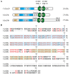
Figure 1
Figure 1
Domain organization of Lin28. (A) Caenorhabditis elegans (Cel) and human (h) Lin28a/Lin28b contain two RNA-binding domains (RBDs): an N-terminal cold-shock domain (CSD) and a C-terminal Zn-knuckle domain (ZKD) comprised of two retroviral type CCHC Zn knuckles (ZnK). Additionally, Lin28 harbors low-complexity sequences, Lys/Arg (K/R)-rich stretches, bipartite nuclear localization signals (NLS) or putative nucleolar localization sequences (NoLS); (B) Sequence alignment of hLin28a and hLin28B. Amino acids belonging to CSD or ZKD are shaded in blue or green, respectively.
Figure 2
Figure 2
Lin28/let-7 regulatory axis. In undifferentiated cells, Lin28 is highly expressed and blocks the biogenesis of let-7 miRNA. By binding to the pre-element of pri- or pre-let-7, neither Drosha nor Dicer can process the corresponding let-7 precursor. In addition, Lin28 recruits TUT4/TUT7 to pre-let-7 and promotes its 3′-end oligo-uridylation. Oligo-uridylated pre-let-7 cannot be cleaved by Dicer and thus serves as a signal for the cellular 3′–5′ exoribonuclease Dis3l2. Upon differentiation, Lin28 expression is reduced, which leads to increased levels of mature let-7. The latter silences gene expression of proto-oncogenes (Ras, c-Myc, Hmga2), cell cycle progression factors (Cyclin D1 and D3, Cdk4), components of the insulin-PI3K-mTOR pathway and Lin28 itself, thereby establishing a positive feedback loop. Besides its role in differentiation, a Lin28/let-7 regulatory network is apparently involved in several cellular processes such as proliferation, oncogenesis, development and physiology, as well as metabolism (recently reviewed in [42]).
Figure 3
Figure 3
Lin28 binds various mRNAs and modulates their translation. Both Lin28 paralogs were shown to influence mRNA processing on several levels. In the nucleus, Lin28 could regulate splicing of bound pre-mRNAs in concert with heterogeneous nuclear ribonucleoproteins (hnRNPs). In the cytosol, Lin28 was shown to interact with an RNA helicase A (RHA) thereby modulating the translation of target mRNAs via interactions with eukaryotic translation initiation factors (eIFS), elongation factors (eEFS) and poly(A)-binding proteins (PABP). Furthermore, Lin28 was found to shuttle mRNAs to poly-ribosomes and, under stress condition, to P-bodies and stress granules, thereby providing a direct link to the miRNA decay machinery. Lin28 binding to mRNAs was typically associated with a globally enhanced protein synthesis. However, in hESCs Lin28 binding repressed translation of bound mRNAs that were destined for the ER.
Figure 4
Figure 4
Co-crystal structure of a minimal mouse Lin28a construct with preE-let-7d derived RNA (PDB ID 3TRZ). The ZKD specifically binds to the conserved GGAG motif, whereas Lin28 CSD establishes extensive interactions with the less conserved terminal hairpin loop.
Figure 5
Figure 5
Lin28 ZKD specifically recognizes single-stranded GGAG or GGAG-like sequences. (A) Sequence alignment of HIV-1 NC, HIV-2 NC, hLin28a and hLin28b ZKDs. The chelating Cys and His residues of the CCHC Zn knuckles (ZnK) are shaded in red. Conserved residues are labeled from light red (100% type-conserved) to dark red (70% type-conserved); (B) Comparison between unbound hLin28a ZKD (green, PDB-ID 2CQF) and AGGAGAU-bound hLin28a ZKD (purple, PDB-ID 2LI8). Upon RNA binding, hLin28a ZKD undergoes a dramatic conformational shift mainly caused by a rotation of the Pro158 ψ angle; (C) In comparison to HIV-1 NC, the inter-knuckle linker of hLin28a ZKD harbors an additional Pro. As a consequence, the knuckles are further apart, thereby explaining why HIV-1 NC ZKD specifically binds G-2 and G-4 while hLin28a ZKD binds G-1 and G-4 of the GGAG motif in a hydrophobic pocket; (D) Structure of mLin28a ZKD bound to GGAG (derived from PDB-ID 3TSO). mLin28a is represented in green cartoon and the bound GGAG motif in purple (G) and pink (A). Tyr140 of the first and His162 of the second ZnK are key residues for the interaction, since they contact each other and stack with the bases, thereby establishing a kinked conformation in the RNA. All three guanosines are specifically recognized via various hydrogen bonds with backbone amide and carbonyl groups. In addition, G-1 and G-4 are bound in a hydrophobic pocket formed by His140, His162, Tyr140 and Met170.
Figure 5
Figure 5
Lin28 ZKD specifically recognizes single-stranded GGAG or GGAG-like sequences. (A) Sequence alignment of HIV-1 NC, HIV-2 NC, hLin28a and hLin28b ZKDs. The chelating Cys and His residues of the CCHC Zn knuckles (ZnK) are shaded in red. Conserved residues are labeled from light red (100% type-conserved) to dark red (70% type-conserved); (B) Comparison between unbound hLin28a ZKD (green, PDB-ID 2CQF) and AGGAGAU-bound hLin28a ZKD (purple, PDB-ID 2LI8). Upon RNA binding, hLin28a ZKD undergoes a dramatic conformational shift mainly caused by a rotation of the Pro158 ψ angle; (C) In comparison to HIV-1 NC, the inter-knuckle linker of hLin28a ZKD harbors an additional Pro. As a consequence, the knuckles are further apart, thereby explaining why HIV-1 NC ZKD specifically binds G-2 and G-4 while hLin28a ZKD binds G-1 and G-4 of the GGAG motif in a hydrophobic pocket; (D) Structure of mLin28a ZKD bound to GGAG (derived from PDB-ID 3TSO). mLin28a is represented in green cartoon and the bound GGAG motif in purple (G) and pink (A). Tyr140 of the first and His162 of the second ZnK are key residues for the interaction, since they contact each other and stack with the bases, thereby establishing a kinked conformation in the RNA. All three guanosines are specifically recognized via various hydrogen bonds with backbone amide and carbonyl groups. In addition, G-1 and G-4 are bound in a hydrophobic pocket formed by His140, His162, Tyr140 and Met170.
Figure 6
Figure 6
The Lin28 CSD can bind to a wide range of different RNA sequences. (A) Superimposition of unbound (skin color, PDB-ID 3ULJ) and heptathymidine-bound XtrLin28b CSD (green, PDB-ID 4A76). Both structures are highly conserved and reveal a pre-formed nucleic-acid binding platform with exposed aromatic residues; (B) Superimposition of XtrLin28b:dT7 (green) and mLin28s:preE-let-7f (RNA: blue, protein: gray, PDB-ID 3TS0). Both Lin28 CSDs bind single-stranded nucleic acids predominantly via base stacking interactions in a defined orientation. The protein nucleic-acid interaction surface is similar for binding subsites 1 to 7. Binding of an additional eighth (U-8) and ninth (U-9) base in mLin28:preE-let-7f is triggered by the formation of a closed RNA loop; (C) Superimposition of bound nucleotides at binding subsite 6 derived from various bacterial and Lin28 CSDs in complex with ssDNA/ssRNA (PDB-IDs 4A76, 4A75, 3TS0, 3TS2, 3PF4, 2HAX). All structures contained T or U nucleotides at this binding pocket. A highly conserved Lys-Asp salt bridge limits the size of the pocket and establishes specific hydrogen bonds with the T/U base; (D) Since few interactions are formed with the sugar-phosphate backbone, the bound oligonucleotides can adopt different backbone conformations to optimize binding with Lin28 CSD. For example, at binding subsite 2, the sugar-phosphate backbone of mLin28a:preE-let-7f is farther displaced from the protein, thereby enabling binding of G (G-2) instead of T (T-2) without disrupting hydrogen bonds.
Figure 7
Figure 7
The pre-elements of let-7 family members are structurally diverse. In six out of eleven human let-7 family members, the conserved GGAG motif (blue) is inaccessible for ZKD binding in the lowest-energy folding state. Secondary structure predictions of human let-7 family members (except miR-98 and miR-202) were calculated and visualized by CLC genomics workbench 3.65. All lowest-energy structures within a ΔΔG range of 1.5 kcal/mol are depicted. For simplicity, only 5 bp of the miRNA stem are shown (labeled in red).

References

    1. Ambros V., Horvitz H. Heterochronic mutants of the nematode Caenorhabditis elegans. Science. 1984;226:409–416. - PubMed
    1. Moss E.G., Lee R.C., Ambros V. The cold shock domain protein LIN-28 controls developmental timing in C. elegans and is regulated by the lin-4 RNA. Cell. 1997;88:637–646. - PubMed
    1. Seggerson K., Tang L., Moss E.G. Two genetic circuits repress the Caenorhabditis elegans heterochronic gene lin-28 after translation initiation. Dev. Biol. 2002;243:215–225. - PubMed
    1. Moss E.G., Tang L. Conservation of the heterochronic regulator Lin-28, its developmental expression and microRNA complementary sites. Dev. Biol. 2003;258:432–442. - PubMed
    1. Darr H., Benvenisty N. Genetic analysis of the role of the reprogramming gene LIN-28 in human embryonic stem cells. Stem Cells. 2009;27:352–362. - PubMed

LinkOut - more resources