. 2013 Oct:63:175-9.
doi: 10.1016/j.yjmcc.2013.07.017. Epub 2013 Aug 9.

BET acetyl-lysine binding proteins control pathological cardiac hypertrophy

Affiliations

BET acetyl-lysine binding proteins control pathological cardiac hypertrophy

Jessica I Spiltoir et al. J Mol Cell Cardiol. 2013 Oct.

Abstract

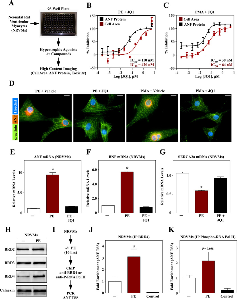
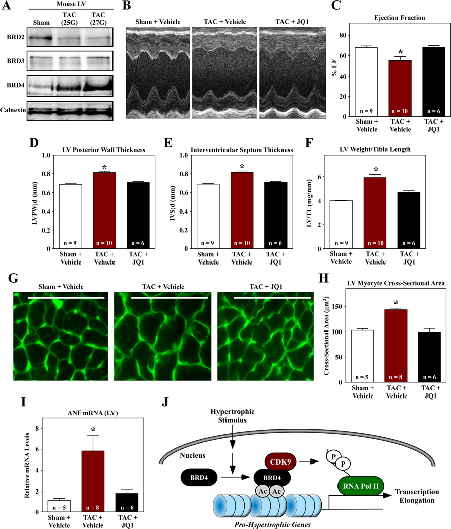
Cardiac hypertrophy is an independent predictor of adverse outcomes in patients with heart failure, and thus represents an attractive target for novel therapeutic intervention. JQ1, a small molecule inhibitor of bromodomain and extraterminal (BET) acetyl-lysine reader proteins, was identified in a high throughput screen designed to discover novel small molecule regulators of cardiomyocyte hypertrophy. JQ1 dose-dependently blocked agonist-dependent hypertrophy of cultured neonatal rat ventricular myocytes (NRVMs) and reversed the prototypical gene program associated with pathological cardiac hypertrophy. JQ1 also blocked left ventricular hypertrophy (LVH) and improved cardiac function in adult mice subjected to transverse aortic constriction (TAC). The BET family consists of BRD2, BRD3, BRD4 and BRDT. BRD4 protein expression was increased during cardiac hypertrophy, and hypertrophic stimuli promoted recruitment of BRD4 to the transcriptional start site (TSS) of the gene encoding atrial natriuretic factor (ANF). Binding of BRD4 to the ANF TSS was associated with increased phosphorylation of local RNA polymerase II. These findings define a novel function for BET proteins as signal-responsive regulators of cardiac hypertrophy, and suggest that small molecule inhibitors of these epigenetic reader proteins have potential as therapeutics for heart failure.

Keywords: Acetyl-lysine; BET protein; Cardiac hypertrophy.


Conflict of interest statement

Disclosures

No conflicts of interest exist for the authors.

Figures

Fig. 1
Fig. 1
A small molecule BET protein inhibitor blocks cardiac hypertrophy. (A) A high throughput assay of cardiomyocyte hypertrophy based on culture of primary neonatal rat ventricular myocytes (NRVMs) on 96-well plates and high content imaging to quantify cell size and expression of atrial natriuretic factor (ANF) protein as a biomarker of pathological cardiac hypertrophy. (B, C) A hit compound, JQ1, dose-dependently blocked hypertrophy in response to treatment with phenylephrine (PE; 10 µM) or phorbol myristate acetate (PMA; 50 nM). (D) Images of ANF and α-actinin staining in NRVMs treated with agonists in the absence or presence of JQ1 (1 µM). Scale bar = 10 µm. (E – G) NRVMs were stimulated for 48 hrs with PE in the absence or presence of JQ1 and quantitative PCR was performed to assess expression of ANF, brain natriuretic peptide (BNP) and sarcoendoplasmic reticulum Ca2+ ATPase 2a (SERCA2a). N = 3 plates of cells per condition; *P<0.05 vs. unstimulated controls. (H) Immunoblot analysis of BET proteins in NRVMs treated with PE for 48 hrs. Calnexin is a loading control. (I) Chromatin immunoprecipitation (ChIP) strategy. (J, K) BRD4 and phospho-RNA Pol II association with the ANF transcription start site. IP with normal IgG was used as a negative control. N = 3 plates of cells per condition (4 pooled plates per N); *P<0.05 vs. unstimulated controls.
Fig. 2
Fig. 2
JQ1 suppresses left ventricular hypertrophy in response to pressure overload. (A) Immunoblot analysis of BET proteins in LVs of mice subjected to TAC for four weeks using a 25 gauge or 27 gauge needle to establish suture diameter. Calnexin is a loading control. Mice were subjected to TAC or sham surgery and were dosed IP with JQ1 (50 mg/kg) every other day for four weeks. (B – E) M-mode echocardiographic images were used to quantify ejection fraction, LV posterior wall thickness, and interventricular septum thickness. (F) LV weight-to-tibia length ratios were determined at necropsy. (G) LV sections were stained with fluorescein-conjugated peanut agglutinin to assess myocyte cross-sectional area. Scale bar = 10µm. (H) Quantification of myocyte cross-sectional area (µm2); >100 myocytes were quantified per the indicated number of LVs. (I) Quantitative PCR analysis of ANF mRNA expression. N = 3 plates of cells per condition; *P<0.05 vs. sham or unstimulated controls. (J) Model for the regulation of cardiac hypertrophy by BRD4.

References

    1. Roger VL, Go AS, Lloyd-Jones DM, Benjamin EJ, Berry JD, Borden WB, et al. Heart disease and stroke statistics--2012 update: a report from the American Heart Association. Circulation. 2012;125:e2–e220. - PMC - PubMed
    1. Lloyd-Jones D, Adams R, Carnethon M, de SG, Ferguson TB, Flegal K, et al. Heart disease and stroke statistics--2009 update: a report from the American Heart Association Statistics Committee and Stroke Statistics Subcommittee. Circulation. 2009;119:e21–e181. - PubMed
    1. Devereux RB, Wachtell K, Gerdts E, Boman K, Nieminen MS, Papademetriou V, et al. Prognostic significance of left ventricular mass change during treatment of hypertension. JAMA. 2004;292:2350–2356. - PubMed
    1. Gardin JM, Lauer MS. Left ventricular hypertrophy: the next treatable, silent killer? JAMA. 2004;292:2396–2398. - PubMed
    1. Hill JA, Olson EN. Cardiac plasticity. N Engl J Med. 2008;358:1370–1380. - PubMed

Publication types