

Brain atrophy in type 2 diabetes: regional distribution and influence on cognition
- PMID: 23939539
- PMCID: PMC3836136
- DOI: 10.2337/dc13-0143
Brain atrophy in type 2 diabetes: regional distribution and influence on cognition
Abstract
Objective: Type 2 diabetes (T2DM) is associated with brain atrophy and cerebrovascular disease. We aimed to define the regional distribution of brain atrophy in T2DM and to examine whether atrophy or cerebrovascular lesions are feasible links between T2DM and cognitive function.
Research design and methods: This cross-sectional study used magnetic resonance imaging (MRI) scans and cognitive tests in 350 participants with T2DM and 363 participants without T2DM. With voxel-based morphometry, we studied the regional distribution of atrophy in T2DM. We measured cerebrovascular lesions (infarcts, microbleeds, and white matter hyperintensity [WMH] volume) and atrophy (gray matter, white matter, and hippocampal volumes) while blinded to T2DM status. With use of multivariable regression, we examined for mediation or effect modification of the association between T2DM and cognitive measures by MRI measures.
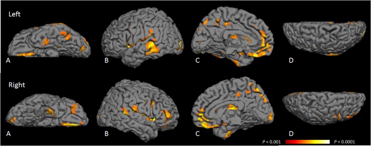
Results: T2DM was associated with more cerebral infarcts and lower total gray, white, and hippocampal volumes (all P < 0.05) but not with microbleeds or WMH. T2DM-related gray matter loss was distributed mainly in medial temporal, anterior cingulate, and medial frontal lobes, and white matter loss was distributed in frontal and temporal regions. T2DM was associated with poorer visuospatial construction, planning, visual memory, and speed (P ≤ 0.05) independent of age, sex, education, and vascular risk factors. The strength of these associations was attenuated by almost one-half when adjusted for hippocampal and total gray volumes but was unchanged by adjustment for cerebrovascular lesions or white matter volume.
Conclusions: Cortical atrophy in T2DM resembles patterns seen in preclinical Alzheimer disease. Neurodegeneration rather than cerebrovascular lesions may play a key role in T2DM-related cognitive impairment.
Figures
Comment in
-
Brain MRI correlates of cognitive dysfunction in type 2 diabetes: the needle recovered from the haystack?Diabetes Care. 2013 Dec;36(12):3855-6. doi: 10.2337/dc13-1501. Diabetes Care. 2013. PMID: 24265363 Free PMC article. No abstract available.
References
-
- Ott A, Stolk RP, van Harskamp F, Pols HA, Hofman A, Breteler MM. Diabetes mellitus and the risk of dementia: the Rotterdam Study. Neurology 1999;53:1937–1942 - PubMed
-
- Peila R, Rodriguez BL, Launer LJ, Honolulu-Asia Aging Study Type 2 diabetes, APOE gene, and the risk for dementia and related pathologies: the Honolulu-Asia Aging Study. Diabetes 2002;51:1256–1262 - PubMed
-
- Biessels GJ, Staekenborg S, Brunner E, Brayne C, Scheltens P. Risk of dementia in diabetes mellitus: a systematic review. Lancet Neurol 2006;5:64–74 - PubMed
-
- Longstreth WT, Jr, Bernick C, Manolio TA, Bryan N, Jungreis CA, Price TR. Lacunar infarcts defined by magnetic resonance imaging of 3660 elderly people: the Cardiovascular Health Study. Arch Neurol 1998;55:1217–1225 - PubMed
-
- Vermeer SE, Den Heijer T, Koudstaal PJ, Oudkerk M, Hofman A, Breteler MM, Rotterdam Scan Study Incidence and risk factors of silent brain infarcts in the population-based Rotterdam Scan Study. Stroke 2003;34:392–396 - PubMed
Publication types
MeSH terms
LinkOut - more resources
Full Text Sources
Other Literature Sources
Medical
