. 2013 Nov;41(20):9371-81.
doi: 10.1093/nar/gkt709. Epub 2013 Aug 11.

Interaction of bacterial fatty-acid-displaced regulators with DNA is interrupted by tyrosine phosphorylation in the helix-turn-helix domain

Affiliations

Interaction of bacterial fatty-acid-displaced regulators with DNA is interrupted by tyrosine phosphorylation in the helix-turn-helix domain

Abderahmane Derouiche et al. Nucleic Acids Res. 2013 Nov.

Abstract

Bacteria possess transcription regulators (of the TetR family) specifically dedicated to repressing genes for cytochrome P450, involved in oxidation of polyunsaturated fatty acids. Interaction of these repressors with operator sequences is disrupted in the presence of fatty acids, and they are therefore known as fatty-acid-displaced regulators. Here, we describe a novel mechanism of inactivating the interaction of these proteins with DNA, illustrated by the example of Bacillus subtilis regulator FatR. FatR was found to interact in a two-hybrid assay with TkmA, an activator of the protein-tyrosine kinase PtkA. We show that FatR is phosphorylated specifically at the residue tyrosine 45 in its helix-turn-helix domain by the kinase PtkA. Structural modelling reveals that the hydroxyl group of tyrosine 45 interacts with DNA, and we show that this phosphorylation reduces FatR DNA binding capacity. Point mutants mimicking phosphorylation of FatR in vivo lead to a strong derepression of the fatR operon, indicating that this regulatory mechanism works independently of derepression by polyunsaturated fatty acids. Tyrosine 45 is a highly conserved residue, and PtkA from B. subtilis can phosphorylate FatR homologues from other bacteria. This indicates that phosphorylation of tyrosine 45 may be a general mechanism of switching off bacterial fatty-acid-displaced regulators.


Figures
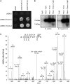
Figure 1.
Figure 1.
PtkA phosphorylates FatR at the residue tyrosine 45. (A) Specific interaction of the C-terminal part of FatR (residues 101–194) with TkmA in the yeast two-hybrid assay. Gene fatR 101–194 was cloned in the plasmids pGAD as a translational fusion with activating domain of the gal4 regulator, and tkmA was cloned in pGBDU, in fusion with the binding domain of gal4. Two clones for each construct were tested, designated (1) and (2). Vectors without fatR 101–194 and tkmA were used as negative controls. Eight days after the drop of yeast cells on the selective (−)LUH SD medium, the development of colonies was observed for tested strains expressing interacting proteins. (B) In vitro phosphorylation assay of FatR. Presence of purified proteins is indicated above each reaction lane. All reactions contained [γ-32P] ATP, MgCl2 and Tris-HCl, pH 7.5: concentrations and reaction conditions are given in Materials and Methods section. In the left, PtkA and FatR are from B. subtilis, and in the right, they are from B. megaterium. TkmA is from B. subtilis in all reactions. (C) After in vitro phosphorylation of B. subtilis FatR by PtkA, the sample was digested in solution with trypsine, and phosphopeptides were enriched by titanium dioxide chromatography and subjected to mass spectrometry. The spectrum shows the fragmentation pattern of the FatR phosphopeptide AHVGTGTIY(ph)R phosphorylated at the tyrosine 45.
Figure 2.
Figure 2.
Residue tyrosine 45 of FatR is conserved and involved in interaction with DNA. (A) Alignment of the helix-turn-helix domain of FatR with its homologues from Vibrio parahaemolyticus (3HE0), P. aeruginosa (2GEN), and S. aureus (1JT0). Position of tyrosine 45 of FatR is indicated by a red arrow. (B) Ribbon diagrams of structures of 3HE0 and 2GEN superimposed. The position of the residue tyrosine 45 is highlighted as a ball and stick model. 2GEN is in rainbow from blue to red, and 3HE0 is in green. (C) Zoom-in view of the interaction of Tyr40 in 1JT0 (conserved residue Tyr45 in FatR) with its HTH-DNA-binding domain. Tyr45 is highlighted as a ball and stick model in magenta, DNA is presented in ball and stick model, green and coral. Hydrogen bond is in blue dashes. (D) In vitro phosphorylation assay of FatR homologues from different bacteria. Presence of purified proteins is indicated above each reaction lane, and the species of origin below the lanes (no origin is indicated for B. subtilis proteins). All reactions contained [γ-32P] ATP, MgCl2 and Tris–HCl, pH 7.5: concentrations and reaction conditions are given in Materials and Methods section.
Figure 3.
Figure 3.
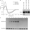
Hydroxyl group of FatR tyrosine 45 is essential for DNA binding. (A) CD spectra of FatR-WT (dashed line), FatR-Y45F (dotted line) and FatR-Y45E (solid line). Graphs are plotted in units of mean residue ellipticity (θ in 105 deg cm2 mol−1) at the respective wavelength (nm). (B) In vitro phosphorylation assay of different versions of FatR. Presence of purified proteins is indicated above each reaction lane. All reactions contained [γ-32P] ATP, MgCl2 and Tris-HCl, pH 7.5: concentrations and reaction conditions are given in Materials and Methods section. (C) Binding of different versions of FatR to its operator sequence analysed by electrophoretic mobility-shift assays. All reactions contained 30 pmol of DNA. Lane 1 is a control with no protein. Wild-type FatR is present in lanes 2–6, at final concentration of 120, 240, 480, 960 and 1920 pmol, respectively. This corresponds to molar ratios of protein to DNA of 1:4, 1:8, 1:16, 1:32 and 1:64, respectively. Lanes 7 and 8 contained 960 and 1920 pmol of FatR Y45F (protein:DNA ratios of 1:32 and 1:64), respectively. Lanes 9 and 10 contained 960 and 1920 pmol of FatR Y45E (protein:DNA ratios of 1:32 and 1:64), respectively. The signals corresponding to free DNA and FatR-DNA complex are indicated by arrows.
Figure 4.
Figure 4.
Phosphomimetic mutant fatR Y45E leads to a strong derepression of the fatR-cyp102A3 operon in vivo. (A) The unique FatR binding site detected by ChIP-on-chip on the B. subtilis genome in the WT and the ΔptkA strain. Genes in the B. subtilis genome are represented on top. The ChIP-on-chip profile of interaction with FatR-SPA is given for the WT and ΔptkA strain (replicates 1 and 2 coloured in black and grey, respectively). The peaks correspond to enrichment of a specific DNA sequence co-purified in complex with FatR-SPA. The vertical line indicates the position of the binding site. (B) Promoter activity of the operon fatR-cyp102A3 measured as Miller units of the β-galactosidase reporter gene. Experiments were performed using the following strains: WT, ΔptkA, ΔfatR, fatR Y45E and fatR Y45F. Noninduced strains were grown in LB with DMSO 0.1% (open circles), and 10 µM linoleate (in DMSO 0.1%) and 0.4 M NaCl were added to induce the promoter (filled circles). The results represent the maximal induction reached in each strain, at the end of exponential phase. Average of three independent measurements is shown, and standard deviations are indicated with error bars.

References

    1. Grangeasse C, Cozzone AJ, Deutscher J, Mijakovic I. Tyrosine phosphorylation: an emerging regulatory device of bacterial physiology. Trends Biochem. Sci. 2007;32:86–94. - PubMed
    1. Grangeasse C, Nessler S, Mijakovic I. Bacterial tyrosine kinases: evolution, biological function and structural insights. Philos. Trans. Roy. Soc. Lond. B Biol. Sci. 2012;367:2640–2655. - PMC - PubMed
    1. Jadeau F, Grangeasse C, Shi L, Mijakovic I, Deléage G, Combet C. BYKdb: the bacterial protein tYrosine Kinase database. Nucleic Acids Res. 2012;40:D321–D324. - PMC - PubMed
    1. Olivares-Illana V, Meyer P, Bechet E, Gueguen-Chaignon V, Soulat D, Lazereg-Riquier S, Mijakovic I, Deutscher J, Cozzone AJ, Laprévote O, et al. Structural basis for the regulation mechanism of the tyrosine kinase CapB from Staphylococcus aureus. PLoS Biol. 2008;6:e143. - PMC - PubMed
    1. Morona JK, Morona R, Paton JC. Attachment of capsular polysaccharide to the cell wall of Streptococcus pneumoniae type 2 is required for invasive disease. Proc. Natl Acad. Sci. USA. 2006;103:8505–8510. - PMC - PubMed

Publication types

MeSH terms

LinkOut - more resources