Comparative Study
. 2013 Oct 1;73(19):6036-45.
doi: 10.1158/0008-5472.CAN-13-0186. Epub 2013 Aug 12.

Double minute chromosomes in glioblastoma multiforme are revealed by precise reconstruction of oncogenic amplicons

Affiliations
Comparative Study

Double minute chromosomes in glioblastoma multiforme are revealed by precise reconstruction of oncogenic amplicons

J Zachary Sanborn et al. Cancer Res. .

Abstract

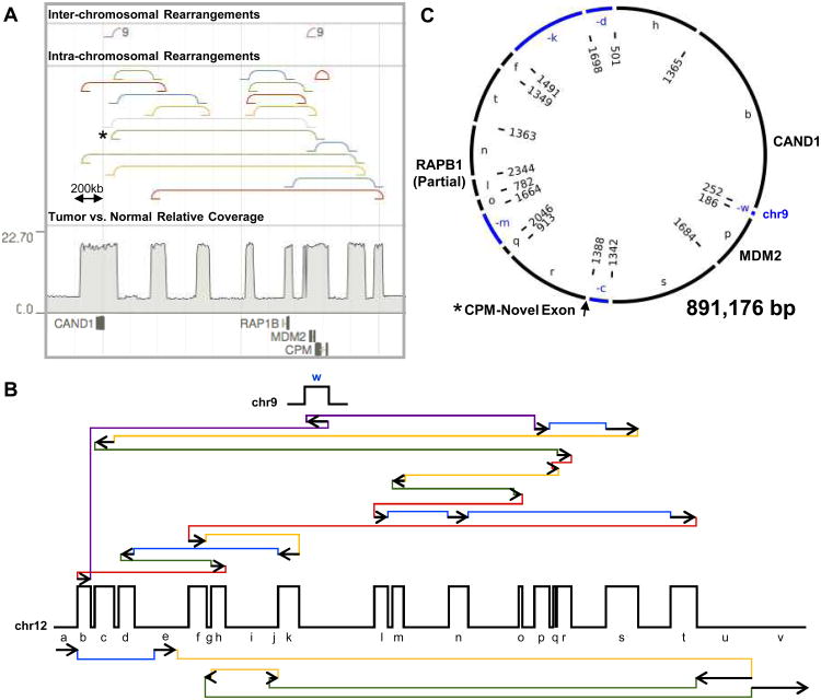
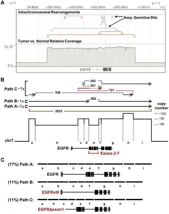
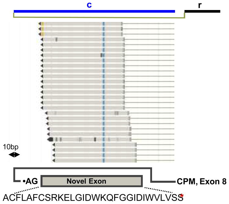
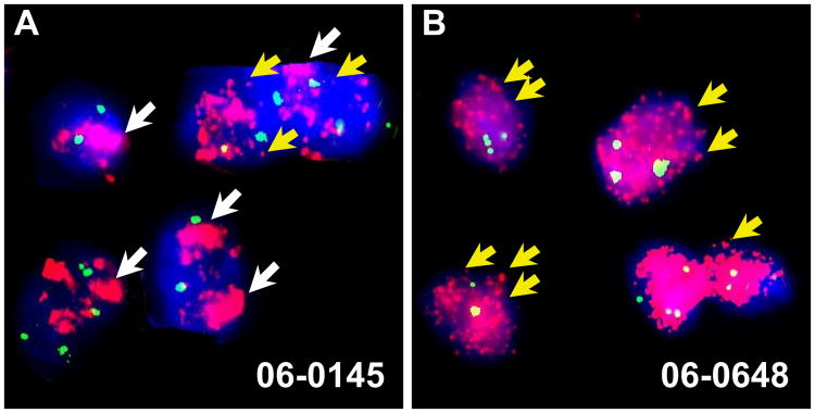
DNA sequencing offers a powerful tool in oncology based on the precise definition of structural rearrangements and copy number in tumor genomes. Here, we describe the development of methods to compute copy number and detect structural variants to locally reconstruct highly rearranged regions of the tumor genome with high precision from standard, short-read, paired-end sequencing datasets. We find that circular assemblies are the most parsimonious explanation for a set of highly amplified tumor regions in a subset of glioblastoma multiforme samples sequenced by The Cancer Genome Atlas (TCGA) consortium, revealing evidence for double minute chromosomes in these tumors. Further, we find that some samples harbor multiple circular amplicons and, in some cases, further rearrangements occurred after the initial amplicon-generating event. Fluorescence in situ hybridization analysis offered an initial confirmation of the presence of double minute chromosomes. Gene content in these assemblies helps identify likely driver oncogenes for these amplicons. RNA-seq data available for one double minute chromosome offered additional support for our local tumor genome assemblies, and identified the birth of a novel exon made possible through rearranged sequences present in the double minute chromosomes. Our method was also useful for analysis of a larger set of glioblastoma multiforme tumors for which exome sequencing data are available, finding evidence for oncogenic double minute chromosomes in more than 20% of clinical specimens examined, a frequency consistent with previous estimates.


Conflict of interest statement

Conflict of interest: J. Zachary Sanborn is a co-founder, equity holder and Chief Technology Officer, and David Haussler is a co-founder, equity holder and member of the Scientific Advisory Board of Five3 Genomics, LLC.

Figures

Figure 1
Figure 1. Reconstruction of TCGA-06-0648 double minutes
(a) Tumor browser view of a region of chromosome 12 from TCGA-06-0648. The bottom track shows protein-coding genes overlapping the amplified segments. The track above shows relative copy number of the tumor DNA compared to the normal, showing distinct blocks of elevated copy number with similar total copy number between blocks. The next two tracks show intra- and inter-chromosomal rearrangement breakpoints. All rearrangements shown are supported by at least 100 discordant reads. The type of rearrangement is indicated by the color of the line: duplication (red), deletion (blue), inversion (yellow and green), inter-chromosomal rearrangement (purple). (b) A diagram of the amplified segments and structural variants identified on chromosome 12 and 9 showing. Walking through this diagram results in a circular solution suggesting the DM diagrammed in (c) where segments inverted relative to their orientation in the reference genome are indicated by (–) and colored blue. The letters inside the circle correspond to the segments in (b). The numbers inside the circle indicate the number of sequencing reads supporting each breakpoint.
Figure 2
Figure 2. Reconstruction of 06-0145 amplicons
(a) Browser shot of the amplicon on chromosome 7. Tracks as described for figure 1. The two “Amp. Germline SVs” are known germline breakpoints present on the amplicon. (b) Diagram of the three paths that together form a potential solution of the breakpoint graph for sample TCGA-06-0145 that accounts for all observed, highly- supported breakpoints. The location of the gene EGFR is noted. Note that all paths, A, B and C, can create circular solutions by using the encompassing tandem duplication shown in red to connect the end of each path to the beginning. The tandem duplication may also connect path A to path B, path C to path B, etc. (c) The effects of each path on the EGFR gene, showing that path B creates an oncogenic form of EGFR, EGFRvIII, through specific deletion of exons 2-7.
Figure 3
Figure 3. Novel CPM C-terminal exon expressed from the TCGA-06-0648 DM
Top line shows the appropriate region from (Fig. 1c) with sequencing reads where the other pair maps to exon 8 of CPM below. The bottom panel shows the orientation of the new exon relative to CPM and its amino acid sequence.
Figure 4
Figure 4. Visualization of GBM tumor oncogenic amplicons
FISH analysis of formalin-fixed paraffin embedded (FFPE) blocks matching samples 06-0145 (a) and 06-0648 (b). Probes for the centromeric region of chromosome 7 (a) and chromosome 12 (b) shown in green gave 2-4 foci per cell. Probes for EGFR (a) and MDM2 (b) are shown in pink. White arrows point to broad, HSR-like staining patterns, and yellow arrows point to discrete extrachromosomal spots.

References

    1. Sindi S, Helman E, Bashir A, Raphael BJ. A geometric approach for classification and comparison of structural variants. Bioinformatics. 2009;25:i222–30. - PMC - PubMed
    1. Chen K, Wallis JW, McLellan MD, Larson DE, Kalicki JM, Pohl CS, et al. BreakDancer: an algorithm for high-resolution mapping of genomic structural variation. Nat Meth. 2009;6:677–81. - PMC - PubMed
    1. Bass AJ, Lawrence MS, Brace LE, Ramos AH, Drier Y, Cibulskis K, et al. Genomic sequencing of colorectal adenocarcinomas identifies a recurrent VTI1A-TCF7L2 fusion. Nature genetics. 2011;43:964–8. - PMC - PubMed
    1. Barker PE. Double minutes in human tumor cells. Cancer Genetics and Cytogenetics. 1982;5:81–94. - PubMed
    1. Lundberg G, Rosengren AH, Hakanson U, Stewenius H, Jin Y, Stewenius Y, et al. Binomial mitotic segregation of MYCN-carrying double minutes in neuroblastoma illustrates the role of randomness in oncogene amplification. PLoS ONE. 2008;3:e3099. - PMC - PubMed

Publication types