. 2013 Aug 27;110(35):14498-503.
doi: 10.1073/pnas.1308412110. Epub 2013 Aug 12.

Dirigent domain-containing protein is part of the machinery required for formation of the lignin-based Casparian strip in the root

Affiliations

Dirigent domain-containing protein is part of the machinery required for formation of the lignin-based Casparian strip in the root

Prashant S Hosmani et al. Proc Natl Acad Sci U S A. .

Erratum in

  • Proc Natl Acad Sci U S A. 2013 Oct 1;110(40):16283

Abstract

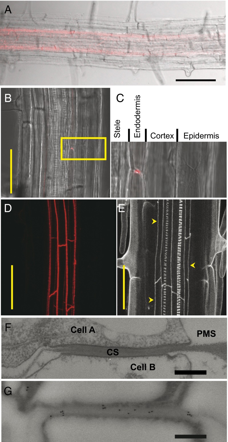
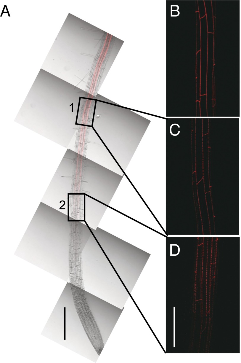
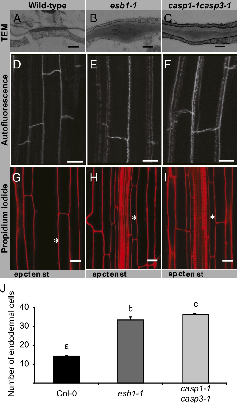
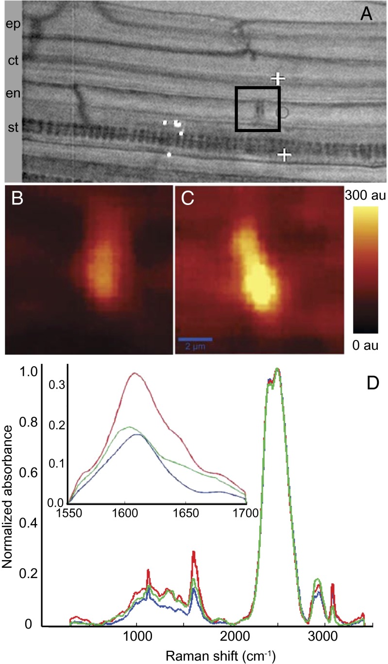
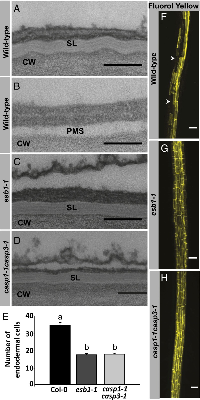
The endodermis acts as a "second skin" in plant roots by providing the cellular control necessary for the selective entry of water and solutes into the vascular system. To enable such control, Casparian strips span the cell wall of adjacent endodermal cells to form a tight junction that blocks extracellular diffusion across the endodermis. This junction is composed of lignin that is polymerized by oxidative coupling of monolignols through the action of a NADPH oxidase and peroxidases. Casparian strip domain proteins (CASPs) correctly position this biosynthetic machinery by forming a protein scaffold in the plasma membrane at the site where the Casparian strip forms. Here, we show that the dirigent-domain containing protein, enhanced suberin1 (ESB1), is part of this machinery, playing an essential role in the correct formation of Casparian strips. ESB1 is localized to Casparian strips in a CASP-dependent manner, and in the absence of ESB1, disordered and defective Casparian strips are formed. In addition, loss of ESB1 disrupts the localization of the CASP1 protein at the casparian strip domain, suggesting a reciprocal requirement for both ESB1 and CASPs in forming the casparian strip domain.


Conflict of interest statement

The authors declare no conflict of interest.

Figures

Fig. 1.
Fig. 1.
The dirigent domain containing protein ESB1 accumulates uniquely in the root endodermis at the Casparian strips. (AD) ESB1-mCherry expression from native ESB1 promoter in roots of esb1-1. Confocal image (red) merged with transmission image (AC). (C) Enlargement of area boxed in B. (D) Z-stack confocal image (red) in the same area as B. (EG) Casparian strips in wild-type roots. (E) Z-stack confocal image of lignin autofluorescence (arrows indicate Casparian strips). (F) Transmission electron micrograph of a representative Casparian strip. (G) Representative image of immunogold-electron micrograph at the anticlinal wall of an endodermal cell by using anti-ESB1 antibodies. CS, Casparian strip; PMS, space generated by plasmolysis. (Scale bars: A, 100 µm; B, D, and E, 50 µm; F and G, 500 nm.)
Fig. 2.
Fig. 2.
The dirigent domain containing protein ESB1 localized at the endodermis in equatorially located patches that coalesce into a continuous strip. (AD) ESB1-mCherry expression in esb1-1 transgenic plants were grown for 6 d, and mCherry fluorescence was observed by confocal microscope. (A) ESB1-mCherry observed in a 5-mm section of root. Confocal image (red) was merged with transmission image, and multiple merged images were tiled to form a combined image. Higher magnification Z-stack confocal images taken 6 mm from the root tip (B), in boxed area 1 (C), and in boxed area 2 (D). (Scale bars: A, 500 µm; BD, 100 µm.)
Fig. 3.
Fig. 3.
The dirigent-domain protein ESB1 is required for the normal morphology and function of the lignin Casparian strip. Transmission electron micrographs showing Casparian strip in wild-type (A), esb1-1 (B), and casp1-1casp3-1 (C). Autofluorescence visualization after clearing root in wild-type (D), esb1-1 (E), and casp1-1casp3-1 (F). PI penetration in wild-type (G), esb1-1 (H), and casp1-1casp3-1 (I). Asterisks mark the 30th endodermal cell after onset of elongation. Onset of elongation is defined as the zone where the length of an endodermal cell was observed to be more than twice its width. (J) Quantification of PI penetration into the stele quantified as number of endodermal cells from the first fully expanded cell in wild-type, esb1-1, and casp1-1casp3-1. Casparian strips in wild-type plants form a barrier to apoplastic diffusion of PI starting at the 13th endodermal cell from the onset of elongation, whereas in esb1-1, this barrier does not form until the 32nd endodermal cell. Different letters (a, b, and c) indicate statistically significant differences between means by one-way analysis of variance (ANOVA) with Tukey–Kramer separation of means (P < 0.05), n = 15 roots. (Scale bars: AC, 250 nm; DF, 10 µm; G–I, 20 µm.) ct, cortex; en, endodermis; ep, epidermis; st, stele.
Fig. 4.
Fig. 4.
Chemical imaging using Raman confocal microscope showing increased deposition of lignin at the disrupted Casparian strip in esb1-1 mutant. (A) Representative image showing an endodermal cell junction used for acquiring 2D Raman spectra (marked with a black square box signifying a 10 × 10 µm2 area). Raman images showing the modification of lignin deposition at the Casparian strip were obtained by integrating absorption intensities between 1,550 and 1,700 cm−1 in wild-type (B) and esb1-1 (C). (D) Raman spectra extracted from the wild-type Casparian strip (blue), wild-type xylem (green), and esb1-1 Casparian strip (red). D Insert shows the lignin spectra (1,550–1,700 cm−1). Intensities were normalized to the peak height of D2O at ∼2,500 cm−1, representing the O-D stretching band intensity. au, arbitrary units; ct, cortex; en, endodermis; ep, epidermis; st, stele.
Fig. 5.
Fig. 5.
Disruption of the Casparian strip through loss of ESB1 function leads to ecotopic deposition of suberin lamellae closer to the root tip and around passage cells in the endodermis. Transmission electron micrographs at periclinal wall of endodermal cells in wild-type mature region (A), wild-type young region (B), esb1-1 young region (C), and casp1-1casp3-1 young region (D). (FH) Suberin deposition in roots detected by Fluorol Yellow staining of wild-type (F), esb1-1 (G), and casp1-1casp3-1 (H). Counting of the number of endodermal cells from the first fully expanded cell shows the early appearance of suberin in both esb1-1 and casp1-1casp3-1 double mutant compared with the wild type, with Fluorol yellow staining starting at the 17th endodermal cell after onset of elongation in esb1-1, and casp1-1casp3-1 compared with the 33rd endodermal cell in wild-type (E). Different letters (a and b) indicate statistically significant differences between means by one-way analysis of variance (ANOVA) with Tukey–Kramer separation of means (P < 0.05), n = 15 roots. Arrowheads represent unsuberised passage cells. CW, cell wall; PMS, space generated by plasmolysis; SL, suberin lamellae. (Scale bars: AD, 200 nm; FH, 50 μm.)

References

    1. Steudle E. The cohesion-tension mechanism and the acquisition of water by plant roots. Annu Rev Plant Physiol Plant Mol Biol. 2001;52:847–875. - PubMed
    1. Naseer S, et al. Casparian strip diffusion barrier in Arabidopsis is made of a lignin polymer without suberin. Proc Natl Acad Sci USA. 2012;109(25):10101–10106. - PMC - PubMed
    1. Steudle E. Water uptake by plant roots: An integration of views. Plant Soil. 2000;226:45–56.
    1. White PJ. The pathways of calcium movement to the xylem. J Exp Bot. 2001;52(358):891–899. - PubMed
    1. Geldner N. The endodermis. Annu Rev Plant Biol. 2013;64:531–558. - PubMed

Publication types