. 2013 Aug 27;110(35):E3291-300.
doi: 10.1073/pnas.1302797110. Epub 2013 Aug 12.

let-7 and miR-140 microRNAs coordinately regulate skeletal development

Affiliations

let-7 and miR-140 microRNAs coordinately regulate skeletal development

Garyfallia Papaioannou et al. Proc Natl Acad Sci U S A. .

Abstract

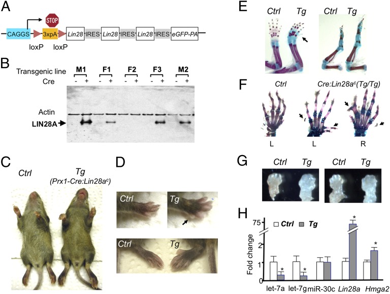
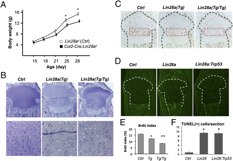
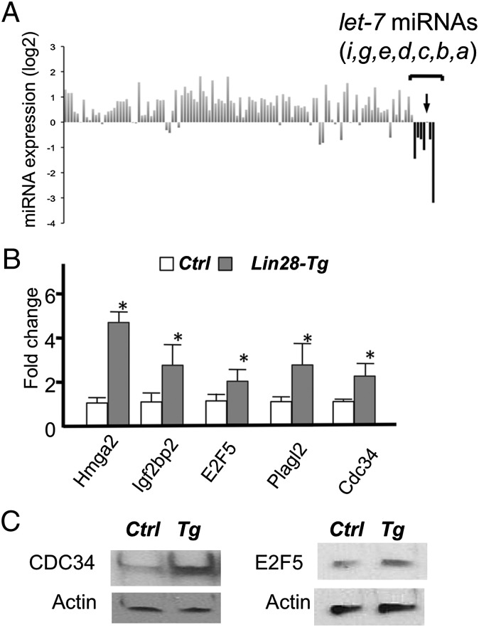
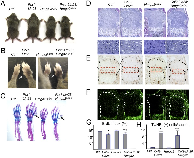
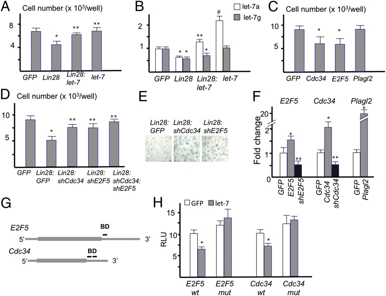
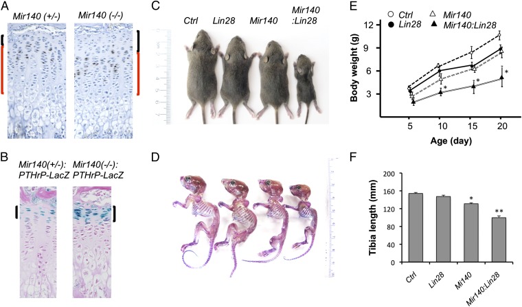
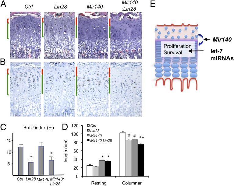
MicroRNAs (miRNAs) play critical roles in multiple processes of skeletal development. A global reduction of miRNAs in growth plate chondrocytes results in defects in both proliferation and differentiation; however, specific microRNAs responsible for these defects have not been identified. In this study, we provide evidence that let-7 miRNAs and microRNA-140 (miR-140), among other miRNAs expressed in chondrocytes, play major roles in endochondral bone development. We overexpressed lin-28 homolog A (Lin28a) to inhibit let-7 miRNA biogenesis in growth plate chondrocytes. Lin28a overexpression efficiently and specifically reduced let-7 miRNAs and up-regulated let-7 target genes. However, unlike the previous notion that let-7 miRNAs inhibit proliferation and growth, suppression of let-7 miRNAs via Lin28a overexpression decreased proliferation in growth plate chondrocytes, likely through up-regulation of the let-7 target cell cycle regulators cell division cycle 34 (Cdc34) and E2F transcription factor 5 (E2F5). Deficiency of the chondrocyte-specific miRNA, miR-140, causes a differentiation defect in growth plate chondrocytes. Although either Lin28a overexpression or miR-140 deficiency alone caused only mild growth impairment, mice with both miR-140 deficiency and Lin28a overexpression in chondrocytes showed a dramatic growth defect. Deregulation of distinct processes in the absence of these miRNAs synergistically decreased the proliferating chondrocyte mass; miR-140 deficiency reduced differentiation into proliferating chondrocytes, whereas Lin28a overexpression decreased proliferation per se.

Keywords: chondrocyte differentiation; chondrocyte proliferation; mouse.


Conflict of interest statement

The authors declare no conflict of interest.

Figures

Fig. 1.
Fig. 1.
Lin28a overexpression in limb mesenchymal cells causes overgrowth and polydactyly. (A) Cre-inducible Lin28a expression construct (Lin28ac). Three copies of the Lin28a coding sequence along with an EGFP cDNA, connected by IRES, were placed downstream of the ubiquitous promoter, CAGGS, followed by a loxP-flanked “stop cassette” composed of three copies of a poly(A) (3×pA) signal sequence. (B) LIN28A is induced in tail fibroblasts from four transgenic lines on adenovirus-mediated Cre expression. (C) Limb size is greater in 3-wk-old Prx1-Cre:Lin28ac (Tg) mice than in Cre-negative control (Ctrl) littermates. (D) Overgrowth of forelimbs (Upper) and hind limbs (Lower) in Tg mice. The arrow indicates an additional digit at the posterior side. Polydactyly is usually absent in hind limbs, but it can be observed in homozygous Tg mice. (E) Skeletal preparation of forelimbs (Left) and hind limbs (Right). The skeletal size is increased in Tg mice compared with Ctrl. The arrow indicates an additional digit. (F) Homozygous Tg mice show diverse digit abnormalities. Left (L) and right (R) forelimbs are shown. (G) Increased limb bud size of forelimbs (Left) and hind limbs (Right) of E12.5 Tg embryos. (H) Gene expression of E13.5 limbs. Prx1-Cre:Lin28ac transgenic limbs show decreased expression of let-7a and let-7g, and increased expression of the let-7 target gene, Hmga2. Expression levels are shown relative to Ctrl levels (n = 4; *P < 0.05 vs. Ctrl).
Fig. 2.
Fig. 2.
Lin28a overexpression decreases proliferation and increases cell death in chondrocytes. (A) Growth of Col2-Cre:Lin28ac mice (black squares), assessed by body weight, is mildly impaired compared with that of Cre-negative Ctrl (open circles) (n = 5; *P < 0.05 vs. Ctrl). Error bars depict mean ± SEM. (B) Growth plate morphology of the proximal tibia of postnatal 4.5-d-old mice. Col2-Cre:Lin28ac mice with one allele (heterozygote) and two alleles (homozygote) of the Lin28ac transgene are indicated by Lin28a (Tg) and Lin28a (Tg/Tg), respectively. (Lower) Cell density of the growth plate is decreased in the columnar proliferating zone of Lin28a transgenic mice. Transgenic mice show a somewhat disorganized columnar structure with the appearance of enlarged “hypertrophic-like” chondrocytes. (Original magnification, Top, ×40; Bottom, ×200.) (C) BrdU labeling index is reduced in postnatal 4.5-d-old heterozygous (Tg) and homozygous (Tg/Tg) Lin28a transgenic mice. (Magnification, ×40.) (D) Number of TUNEL-positive cells was increased in the growth plate of the transgenic tibia of postnatal 2.5-d-old Lin28a mice. Cell death was not rescued by simultaneous ablation of Trp53 (Col2-Cre:Lin28ac:TrP53fl/fl, labeled as Lin28a:Trp53). (Magnification, ×40.) (E) Quantification of the BrdU labeling index (n = 3; *P < 0.05 vs. Ctrl; **P < 0.05 vs. Tg). (F) Quantification of TUNEL-positive cells (*P < 0.05 vs. Ctrl). There is no significant difference between Lin28a transgenic mice and Lin28a:Trp53 doubly mutant mice.
Fig. 3.
Fig. 3.
Lin28a overexpression suppresses let-7 expression and derepresses let-7 target genes in chondrocytes. (A) miRNA expression profiles in primary rib chondrocytes of Cre-negative Ctrl and Col2-Cre:Lin28ac (Lin28-Tg) mice. Expression levels of 165 miRNAs detected in chondrocytes were normalized by that of the U6 level. Expression levels of miRNAs in Lin28-Tg chondrocytes are shown as fold differences relative to those of controls. The let-7 miRNAs were relatively specifically down-regulated in Lin28-Tg, except for let-7c (arrow). (B) Target let-7 genes were up-regulated in Lin28-Tg rib chondrocytes. Some of microarray results (Table S1) were verified by qRT-PCR (n = 3; *P < 0.05 vs. Ctrl). (C) Up-regulation of CDC34 and E2F5 proteins in primary rib chondrocytes of Lin28-Tg mice was verified.
Fig. 4.
Fig. 4.
Hmga2 up-regulation does not contribute to skeletal phenotypes of Lin28a transgenic mice. (A) Limb overgrowth of Prx1-Cre:Lin28ac (Prx1-Lin28) mice is present in the absence of Hmga2 . An Hmga2-null allele [pygmy (pg)] was bred into Prx1-Cre:Lin28ac mice to generate compound mutant mice, Prx1-Cre:Lin28ac:Hmga2pg/pg (Prx1-Lin28:Hmga2pg/pg). Three-wk-old littermates are shown. (B) Polydactyly and overgrowth of forelimbs are present in Lin28:Hmga2pg/pg mice. (C) Skeletal preparation demonstrates bone overgrowth and polydactyly in Lin28:Hmga2pg/pg mice. (D) Growth plate abnormalities [hypocellularity and appearance of large chondrocytes in Col2-Cre:Lin28ac (Col2-Lin28)] are present in the absence of Hmga2. (Original magnifications, Top, ×40; Bottom, ×200.) (E) BrdU labeling assay. The proliferation defect is still present in Lin28:Hmga2pg/pg mice. (Magnification, ×40.) (F) TUNEL assay. Chondrocyte cell death caused by Lin28a overexpression is not rescued by the absence of Hmga2. (Magnification, ×40.) (G) Quantification of E (n = 3; *P < 0.05 vs. Ctrl; **P < 0.05 vs. Ctrl; not significant vs. Lin28). (H) Quantification of F (n = 3; *P < 0.05 vs. Ctrl; **P < 0.05 vs. Ctrl; not significant vs. Col2-Lin28).
Fig. 5.
Fig. 5.
Overexpression of let-7 target genes decreases chondrocyte proliferation. (A) Primary rib chondrocytes are infected with MSCV-GFP viruses with no insert (GFP), Lin28a (Lin28), and/or let-71a-f1 (let-7). The cell number was counted 48 h after infection (n = 5; *P < 0.01 vs. GFP; **P < 0.05 vs. Lin28). (B) Expression levels of let-7a and let-7g. The let-7 virus contains let-7a and let-7f but not let-7g (n = 5; *P < 0.05 vs. GFP; **P < 0.05 vs. Lin28; #P < 0.01 vs. GFP). (C) Cell numbers of primary rib chondrocytes 48 h after infection of indicated viruses (n = 5; *P < 0.05 vs. GFP control). (D) Cell numbers of primary rib chondrocytes 72 h after infection with indicated viruses (n = 5; *P < vs. GFP; **P < 0.05 vs. Lin28). (E) Alcian blue staining of primary rib chondrocytes isolated from Col2-Cre:Lin28ac mice. Cartilaginous nodules were stained 4 d after infection with the indicated viruses. (Original magnification, ×40.) (F) Validation of gene knockdown and overexpression by qRT-PCR. E2F5, Cdc34, and Plagl2 levels were determined after infection with viruses with empty (GFP), overexpression (gray bars), or knockdown (black bars) constructs of indicated genes (n = 4; *P < 0.05 vs. GFP; **P < 0.05 vs. GFP). (G) Schematic representation of let-7 binding sites in the E2F5 and Cdc34 mRNAs. DNA containing the predicted let-7 binding site(s) (BD) is indicated by bars in 3′-untranslated regions. (H) Luciferase activities after transfection with luciferase reporter constructs carrying WT (wt) or mutated (mut) BD sequences with control (GFP) or a let-7a1-f1 expression construct (let-7) (n = 5; *P < 0.05 vs. control).
Fig. 6.
Fig. 6.
Simultaneous manipulation of Lin28a and Mir140 causes a substantial growth defect. (A) BrdU staining depicts an expansion of the resting zone and mild shortening of the proliferating columnar zone of the tibial growth plate of 3-wk-old Mir140-null mice. The resting zone, composed of mostly BrdU-negative cells, is indicated by black brackets. The columnar proliferating zone flanked by the resting zone and hypertrophic zone is indicated by red brackets. (Original magnification, ×200.) (B) Expansion of the resting zone is demonstrated by the increase in cells expressing LacZ from the endogenous PTHrP gene promoter (brackets). PTHrP is expressed exclusively by the resting zone chondrocytes in the growth plate. (Magnification, ×200.) (C) Doubly mutant mice, Col2-Cre:Lin28ac:Mir140−/− (Mir140:Lin28), are significantly smaller than single-mutant mice, Col2-Cre:Lin28a (Lin28) or Mir140−/− (Mir140), at 3 wk of age. (D) Skeletal preparation of C. (E) Growth of single- and double-mutant mice (n ≥ 5; *P < 0.05 vs. Lin28 or Mir140 single-mutant mice). (F) Tibia of double-mutant mice is significantly shorter than that of Ctrl or single-mutant mice (n ≥ 3, *P < 0.05 vs. Ctrl; **P < 0.05 vs. Lin28 or Mir140 single-mutant mice).
Fig. 7.
Fig. 7.
miR-140 and let-7 regulate distinct processes of growth plate development. (A) Histological appearances of Ctrl, Col2-Cre:Lin28ac (Lin28), Mir140−/− (Mir140), and Col2-Cre:Lin28ac:Mir140−/− (Mir140:Lin28) mice. The resting zone and columnar proliferating zone are indicated by red and green brackets, respectively. An expansion of the resting zone and shortening of the columnar proliferating zone are observed in Mir140:Lin28 mice. (Original magnification, ×100.) (B) BrdU staining shows an expansion of the resting zone and a reduction in the proliferation of columnar proliferating chondrocytes in Mir140:Lin28 mice. (Magnification, ×100.) (C) BrdU index in the columnar proliferating region is reduced in Lin28 and Mir140:Lin28 transgenic growth plates compared with Ctrl or Mir140 mice (n = 3 each group; *P < 0.05 vs. Ctrl or Mir140 mice). (D) Length of the resting and columnar regions of growth plates. The resting is zone is expanded in Mir140 and Mir140:Lin28 mice, whereas the columnar region is reduced in both Lin28 and Mir140 mice. Mir140:Lin28 mice show a further reduction in the length of the columnar region (n ≥ 3; *P < 0.05 vs. Ctrl or Lin28 mice; **P < 0.05 vs. Ctrl, Lin28, or Mir140 mice; #P < 0.05 vs. Ctrl). (E) Proposed model. Mir140 miRNAs are necessary for normal differentiation of resting zone chondrocytes into columnar proliferating chondrocytes. Proper regulation of this process is necessary to maintain an adequate number of proliferating chondrocytes, whereas let-7 miRNAs are necessary for normal proliferation and cell survival of columnar proliferating chondrocytes. Pathways regulated by Mir140 and let-7 miRNAs thus control the mass of proliferating chondrocytes through distinct mechanisms.

References

    1. Kronenberg HM. Developmental regulation of the growth plate. Nature. 2003;423(6937):332–336. - PubMed
    1. Ambros V, Chen X. The regulation of genes and genomes by small RNAs. Development. 2007;134(9):1635–1641. - PubMed
    1. Bartel DP. MicroRNAs: Target recognition and regulatory functions. Cell. 2009;136(2):215–233. - PMC - PubMed
    1. Park CY, Choi YS, McManus MT. Analysis of microRNA knockouts in mice. Hum Mol Genet. 2010;19(R2):R169–R175. - PMC - PubMed
    1. Kobayashi T, et al. Dicer-dependent pathways regulate chondrocyte proliferation and differentiation. Proc Natl Acad Sci USA. 2008;105(6):1949–1954. - PMC - PubMed

Publication types

LinkOut - more resources