. 2013 Oct 15;591(20):5125-39.
doi: 10.1113/jphysiol.2013.262733. Epub 2013 Aug 12.

Properties of myenteric neurones and mucosal functions in the distal colon of diet-induced obese mice

Affiliations

Properties of myenteric neurones and mucosal functions in the distal colon of diet-induced obese mice

François Reichardt et al. J Physiol. .

Abstract

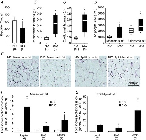
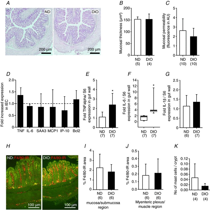
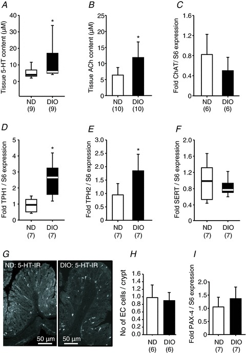
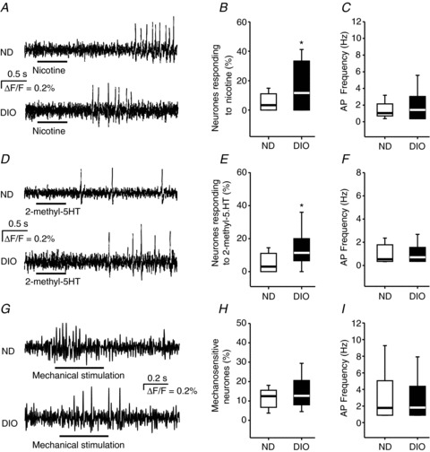
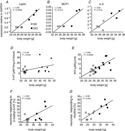
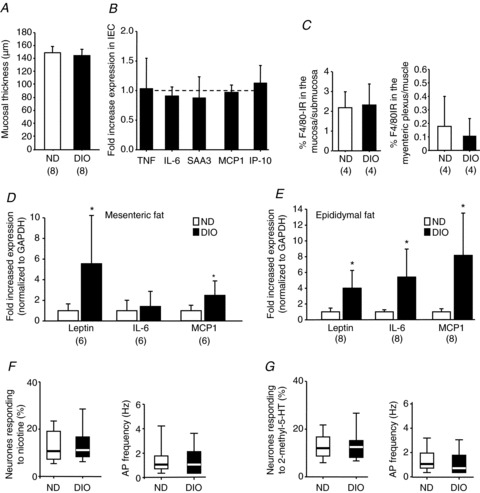
Colonic transit and mucosal integrity are believed to be impaired in obesity. However, a comprehensive assessment of altered colonic functions, inflammatory changes and neuronal signalling of obese animals is missing. In mice, we studied the impact of diet-induced obesity (DIO) on: (i) in vivo colonic transit; (ii) signalling in the myenteric plexus by recording responses to nicotine and 2-methyl-5-hydroxytryptamine (2-methyl-5-HT), together with the expression of tryptophan hydroxylase (TPH) 1 and 2, serotonin reuptake transporter, choline acetyltransferase and the paired box gene 4; and (iii) expression of proinflammatory cytokines, epithelial permeability and density of macrophages, mast cells and enterochromaffin cells. Compared with controls, colon transit and neuronal sensitivity to nicotine and 2-methyl-5-HT were enhanced in DIO mice fed for 12 weeks. This was associated with increased tissue acetylcholine and 5-hydroxytryptamine (5-HT) content, and increased expression of TPH1 and TPH2. In DIO mice, upregulation of proinflammatory cytokines was found in fat tissue, but not in the gut wall. Accordingly, mucosal permeability or integrity was unaltered without signs of immune cell infiltration in the gut wall. Body weight showed positive correlations with adipocyte markers, tissue levels of 5-HT and acetylcholine, and the degree of neuronal sensitization. DIO mice fed for 4 weeks showed no neuronal sensitization, had no signs of gut wall inflammation and showed a smaller increase in leptin, interleukin-6 and monocyte chemoattractant protein 1 expression in fat tissue. DIO is associated with faster colonic transit and impacts on acetylcholine and 5-HT metabolism with enhanced responsiveness of enteric neurones to both mediators after 12 weeks of feeding. Our study demonstrates neuronal plasticity in DIO prior to the development of a pathological histology or abnormal mucosal functions. This questions the common assumption that increased mucosal inflammation and permeability initiate functional disorders in obesity.


Figures

Figure 1
Figure 1. Diet-induced obesity accelerated colonic transit and increased mesenteric and epididymal adipose tissue mass, adipocyte size and expression of the adipokines leptin, interleukin-6 (IL-6) and monocyte chemoattractant protein 1 (MCP1) (12 weeks of feeding)
The shorter bead expulsion time indicated faster colonic transit in DIO mice (A). Mesenteric (B) and epididymal (C) fat mass was increased in DIO mice. Adipocyte sizes were also increased in both fat tissues (D). Images in (E) illustrate representative examples of adipocytes in mesenteric and epididymal fat revealed by haematoxylin and eosin staining; scale bar applies to all images. Expression of leptin, IL-6 and MCP1 was increased in DIO mice in both mesenteric (F) and epididymal (G) fat. Numbers in parentheses indicate number of animals. Asterisks mark significant changes, see text for P values. DIO, diet-induced obese mice; GAPDH, glyceraldehyde 3-phosphate dehydrogenase; ND, mice receiving normal diet.
Figure 2
Figure 2. Normal appearance, integrity and permeability of distal colon in DIO mice (12 weeks of feeding)
Haematoxylin and eosin staining revealed normal histology of the distal colon in DIO mice (A) and no increased mucosal thickness (B). Paracellular permeability was comparable between ND and DIO mice as mucosal to serosal translocation of fluorescein sulfonic acid was unchanged (C). The expression profile of markers for inflammation [tumour necrosis factor (TNF), interleukin-6 (IL-6), serum amyloid A 3 (SAA3)], chemotaxis [monocyte chemoattractant protein 1 (MCP1), interferon-inducible protein-10 (IP-10)] or apoptosis (B-cell lymphoma 2, Bcl2) was similar in ND (six animals) and DIO (four animals) mice (D). However, in extracts of the entire colonic wall (including fat tissue), the expression of TNF-α (E) and IL-6 (F), but not interleukin-1β (IL-1β) (G), was increased in DIO mice. Images in (H) are representative examples of F4/80 immunoreactive macrophages in cross-sections of the distal colon in ND and DIO mice. Analysis of macrophage densities in the mucosa/submucosa region (I) and myenteric plexus/muscle layer region (J) revealed no increased macrophage density in either region in DIO mice. Likewise, the number of CD117-positive mast cells was not altered significantly (K). Numbers in parentheses indicate number of animals. Asterisks mark significant changes, see text for P values. DIO, diet-induced obese mice; IEC, isolated epithelial cell; ND, mice receiving normal diet.
Figure 3
Figure 3. Tissue acetylcholine (ACh) and 5-hydroxytryptamine (5-HT) contents are increased in DIO mice, together with increased expression of tryptophan hydroxylase 1 (TPH1) and tryptophan hydroxylase 2 (TPH2), but not acetylcholine transferase (ChAT) or serotonin reuptake transporter (SERT) (12 weeks of feeding)
Colonic tissue content of 5-HT (A) and ACh (B) is significantly enhanced in DIO mice. These changes occurred without altered expression in ChAT (C, rate-limiting enzyme in the synthesis of ACh), but was associated with increased expression of TPH1 (D) and TPH2 (E), which are involved in the synthesis of 5-HT. SERT (F) expression was not changed in the colon of DIO mice. Images in (G) are representative examples of 5-HT immunoreactive enterochromaffin (EC) cells in the colonic mucosa of ND and DIO mice. Number of mucosal EC cells was similar in ND and DIO mice (H); note that 5-HT-positive mast cells in the lamina propria were not counted. Similar expression of paired box gene 4 (PAX4) in the colon of ND and DIO mice (I). Numbers in parentheses indicate number of animals. Asterisks mark significant changes, see text for P values. DIO, diet-induced obese mice; ND, mice receiving normal diet.
Figure 4
Figure 4. Nicotine and 2-methyl-5-hydroxytryptamine (2-methyl-5-HT) activated more colonic myenteric neurones in DIO mice (12 weeks of feeding)
Myenteric neurones in ND and DIO mice fired action potentials (AP) in response to 500 ms pressure pulse application (indicated by the bars below the traces) of nicotine (A) and 2-methyl-5-HT (D). In DIO mice, more neurones responded to nicotine (B) and 2-methyl-5-HT (E), whereas the AP frequencies of the neurones responding to nicotine (C) or 2-methyl-5-HT (F) were similar (for nicotine: 41 of 235 neurones from 28 ganglia of six DIO mice and 18 of 277 neurones from 30 ganglia of six ND mice; for 2-methyl-5-HT: 33 of 223 neurones from 18 ganglia of six DIO mice and 14 of 209 neurones from 18 ganglia of six ND mice). Intraganglionic volume injection (indicated by the bar below the traces) caused a rapidly adapting spike discharge in colonic myenteric neurones of ND and DIO mice (G). The proportion of mechanosensitive neurones (H) as well as their spike discharge frequency (I) were similar in ND (six mice, 32 ganglia, 89 neurones) and DIO (six mice, 28 ganglia, 80 neurones) mice. Asterisks mark significant changes, see text for P values. DIO, diet-induced obese mice; ND, mice receiving normal diet.
Figure 5
Figure 5. Increased body weight correlated with neuronal sensitization, with leptin, monocyte chemoattractant protein 1 (MCP1) and interleukin-6 (IL-6) expression in mesenteric fat tissue and with 5-hydroxytryptamine (5-HT) and acetylcholine (ACh) tissue levels (12 weeks of feeding)
Leptin (A), MCP1 (B) and IL-6 (C) levels in fat tissue highly correlated with body weight. Body weight was also positively correlated with 5-HT (D) and ACh (E) tissue concentrations in the gut wall, as well as with the numbers of myenteric neurones responding to 2-methyl-5-hydroxytryptamine (2-methyl-5-HT) (F) and nicotine (G). Symbol key in (A) applies to all panels. DIO, diet-induced obese mice; ND, mice receiving normal diet.
Figure 6
Figure 6. Lack of inflammation and neuronal sensitization in the colon of DIO mice fed for 4 weeks
DIO mice fed for 4 weeks had normal mucosal thickness (A), normal expression of epithelial proinflammatory cytokines (B) and normal macrophage density (C). The expression levels of leptin, interleukin-6 (IL-6) and monocyte chemoattractant protein 1 (MCP1) in different fat tissues were increased (D, E); however, the fold increase was less than after 12 weeks of feeding (compare with Fig. 1F and G). The number of myenteric neurones responding to nicotine (F) or 2-methyl-5-hydroxytryptamine (2-methyl-5-HT) (G), as well as the spike discharge, did not differ between ND and DIO mice fed for 4 weeks (nicotine: eight DIO mice, 28 ganglia, 80 neurones and eight ND mice, 27 ganglia, 78 neurones; 2-methyl-5-HT: seven DIO mice, 29 ganglia, 63 neurones and seven ND mice, 19 ganglia, 53 neurones). Numbers in parentheses indicate number of animals. Asterisks mark significant changes, see text for P values. AP, action potential; DIO, diet-induced obese mice; GAPDH, glyceraldehyde 3-phosphate dehydrogenase; IEC, isolated epithelial cell; IP-10. interferon-inducible protein-10; ND, mice receiving normal diet; SAA3, serum amyloid A 3; TNF, tumour necrosis factor.

Comment in

Similar articles

Cited by

References

    1. Baudry C, Reichardt F, Marchix J, Bado A, Schemann M, Bruley des Varranes S, Neunlist M, Moriez R. Diet-induced obesity has neuroprotective effects in murine gastric enteric nervous system: involvement of leptin and GDNF. J Physiol. 2012;590:533–544. - PMC - PubMed
    1. Bertrand R, Senadheera S, Markus I, Liu L, Howitt L, Chen H, Murphy TV, Sandow SL, Bertrand PP. A Western Diet increases serotonin availability in rat small intestine. Endocrinology. 2011;152:36–47. - PubMed
    1. Bertrand RL, Senadheera S, Tanoto A, Tan KL, Howitt L, Chen H, Murphy TV, Sandow SL, Liu L, Bertrand PP. Serotonin availability in rat colon is reduced during a Western diet model of obesity. Am J Physiol Gastrointest Liver Physiol. 2012;303:G424–G434. - PubMed
    1. Bogumil R, Koal T, Weinberger KM, Dammeier S. Massenspektrometrische Analyse von Blutplasma im Kitformat. Laborwelt. 2008;2:17–23.
    1. Delgado-Aros S, Camilleri M, Garcia MA, Burton D, Busciglio I. High body mass alters colonic sensory-motor function and transit in humans. Am J Physiol Gastrointest Liver Physiol. 2008;295:382–388. - PMC - PubMed

Publication types

MeSH terms