. 2013:8:2719-32.
doi: 10.2147/IJN.S45174. Epub 2013 Jul 31.

Green chemistry approach for the synthesis of biocompatible graphene

Affiliations

Green chemistry approach for the synthesis of biocompatible graphene

Sangiliyandi Gurunathan et al. Int J Nanomedicine. 2013.

Abstract

Background: Graphene is a single-atom thick, two-dimensional sheet of hexagonally arranged carbon atoms isolated from its three-dimensional parent material, graphite. One of the most common methods for preparation of graphene is chemical exfoliation of graphite using powerful oxidizing agents. Generally, graphene is synthesized through deoxygenation of graphene oxide (GO) by using hydrazine, which is one of the most widespread and strongest reducing agents. Due to the high toxicity of hydrazine, it is not a promising reducing agent in large-scale production of graphene; therefore, this study focused on a green or sustainable synthesis of graphene and the biocompatibility of graphene in primary mouse embryonic fibroblast cells (PMEFs).

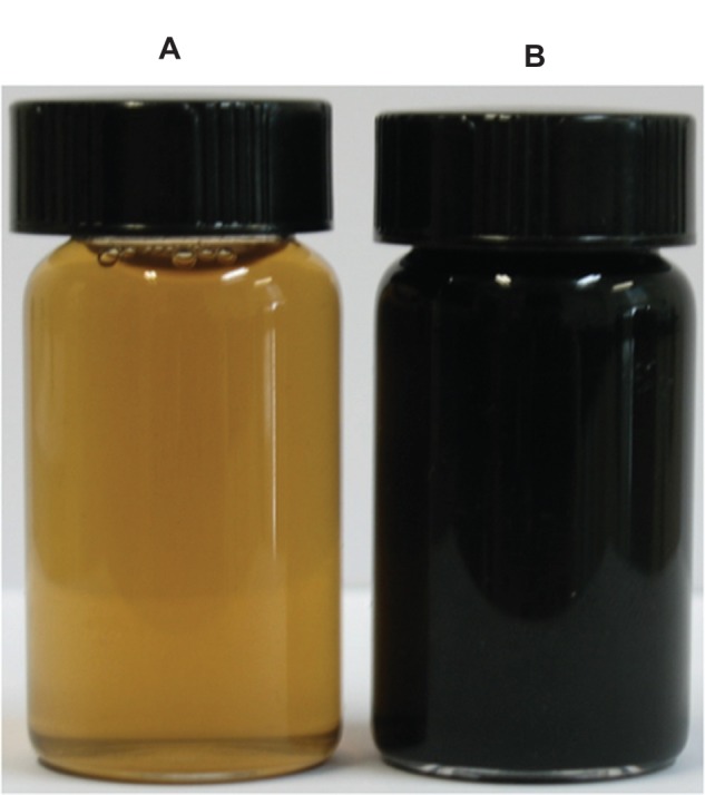
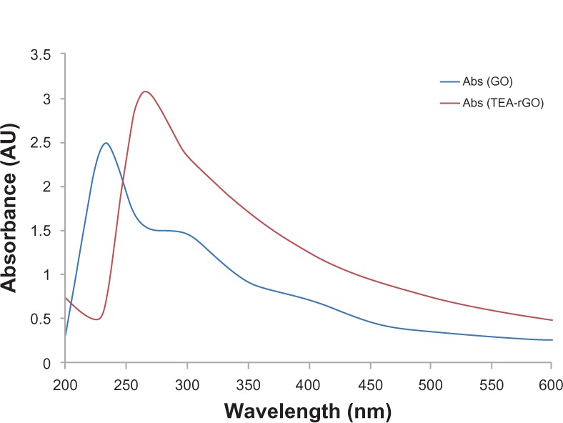
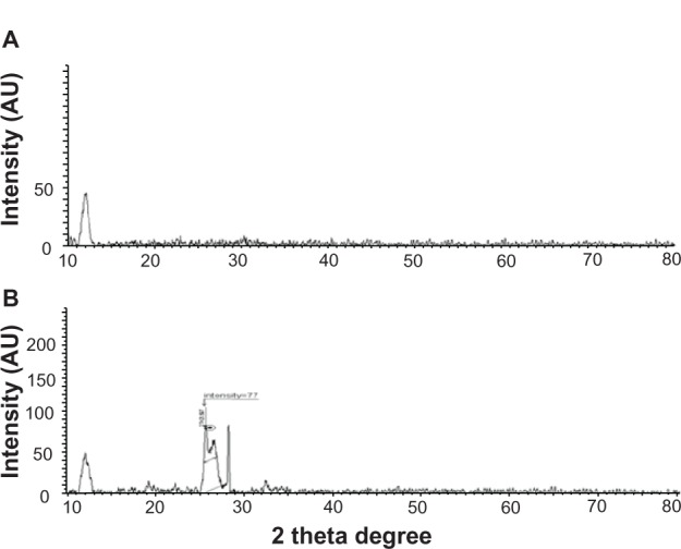
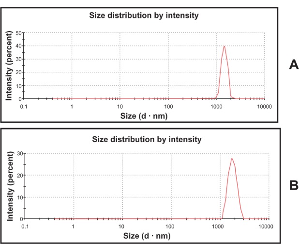
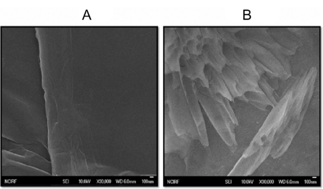
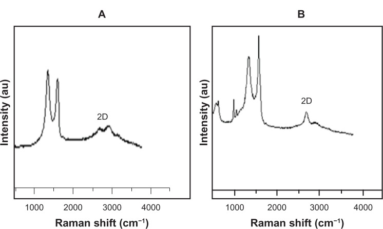
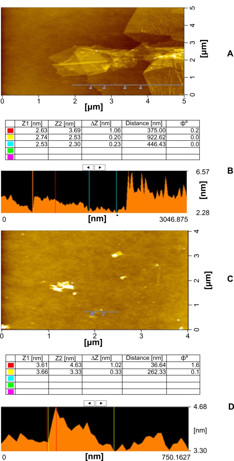
Methods: Here, we demonstrated a simple, rapid, and green chemistry approach for the synthesis of reduced GO (rGO) from GO using triethylamine (TEA) as a reducing agent and stabilizing agent. The obtained TEA reduced GO (TEA-rGO) was characterized by ultraviolet (UV)-visible absorption spectroscopy, X-ray diffraction (XRD), particle size dynamic light scattering (DLS), scanning electron microscopy (SEM), Raman spectroscopy, and atomic force microscopy (AFM).

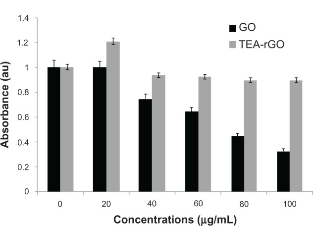
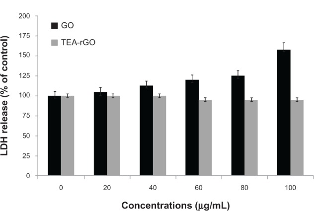
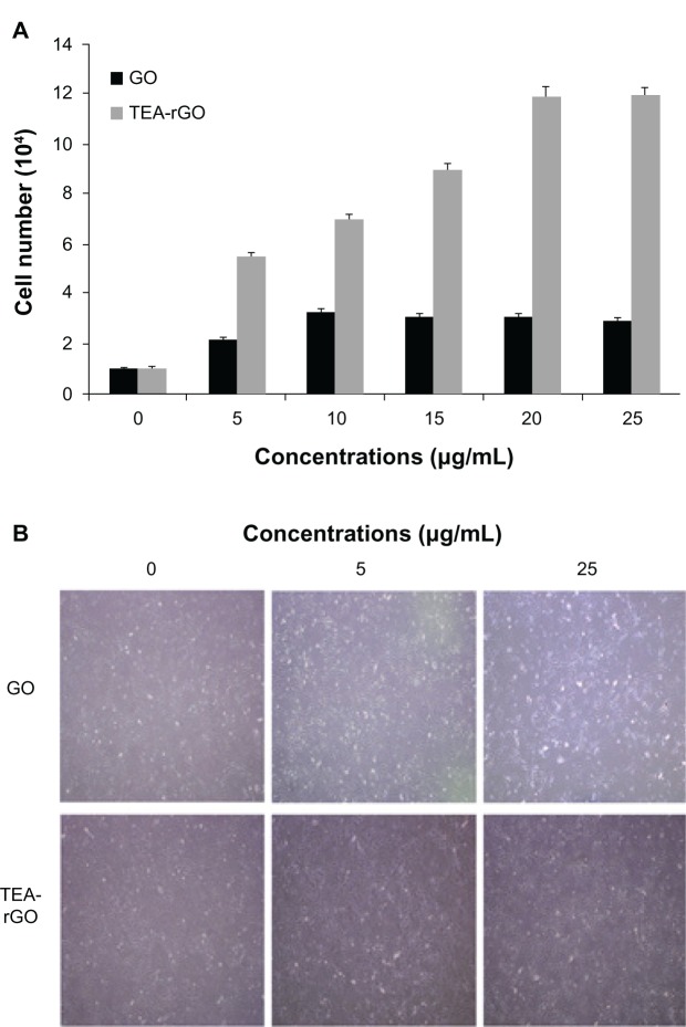
Results: The transition of graphene oxide to graphene was confirmed by UV-visible spectroscopy. XRD and SEM were used to investigate the crystallinity of graphene and the surface morphologies of prepared graphene respectively. The formation of defects further supports the functionalization of graphene as indicated in the Raman spectrum of TEA-rGO. Surface morphology and the thickness of the GO and TEA-rGO were analyzed using AFM. The presented results suggest that TEA-rGO shows significantly more biocompatibility with PMEFs cells than GO.

Conclusion: This is the first report about using TEA as a reducing as well as a stabilizing agent for the preparation of biocompatible graphene. The proposed safe and green method offers substitute routes for large-scale production of graphene for several biomedical applications.

Keywords: Raman spectroscopy; atomic force microscopy; graphene; graphene oxide; triethylamine; ultraviolet; visible spectroscopy.


Figures

Figure 1
Figure 1
Digital photographs of aqueous dispersions of GO before and after reduction with TEA. Notes: Digital photographs of aqueous dispersions (0.5 mg/mL) of GO before (A) and after (B) the reduction with TEA, which were kept at 30°C for 60 minutes. Abbreviations: GO, graphene oxide; TEA, triethylamine.
Figure 2
Figure 2
UV–visible absorption spectra of GO and the TEA-rGO suspension in water. Abbreviations: UV, ultraviolet; GO, graphene oxide; TEA, triethylamine; Abs, absorbance.
Figure 3
Figure 3
XRD pattern of GO and TEA-rGO. Notes: X-ray diffraction (A) GO and (B) TEA-rGO. Abbreviations: XRD, X-ray diffraction; GO, graphene oxide; TEA-rGO, triethylamine-reduced graphene oxide.
Figure 4
Figure 4
Hydrodynamic size distribution of GO and TEA-rGO. Notes: Hydrodynamic size distribution of (A) GO and (B) TEA-rGO (500 μg/mL) measured by DLS at room temperature in DI water. Abbreviations: GO, graphene oxide; TEA-rGO, triethylamine-reduced graphene oxide; DLS, dynamic light scattering; DI, deionized.
Figure 5
Figure 5
SEM images of GO and TEA-rGO. Notes: SEM images of (A) GO and (B) TEA-rGO. Abbreviations: SEM, scanning electron microscopy; GO, graphene oxide; TEA-rGO, triethylamine-reduced graphene oxide.
Figure 6
Figure 6
Raman spectra of GO and TEA-rGO. Notes: Raman spectra of (A) GO and (B) TEA-rGO. Abbreviations: GO, graphene oxide; TEA-rGO, triethylamine-reduced graphene oxide; 2D, 2-dimensional; au, absorbance units.
Figure 7
Figure 7
AFM images and height profiles of GO and TEA-rGO. Notes: (A) AFM images and (B) height profile of GO; (C) AFM images and (D) height profile of TEA-rGO. Abbreviations: AFM, atomic force microscopy; GO, graphene oxide; TEA-rGO, triethylamine-reduced graphene oxide.
Figure 8
Figure 8
Effect of GO and TEA-rGO on cell viability of PMEFs cells. Notes: Cell viability of PMEFs cells was determined using WST-8 assay after 24 hours exposure to different concentrations of GO or TEA-rGO. The results represent the means of three separate experiments, and error bars represent the standard error of the mean. GO treated groups showed statistically significant differences from the control group by the Student’s t-test (P < 0.05). Abbreviations: GO, graphene oxide; TEA-rGO, triethylamine-reduced graphene oxide; PMEFs, primary mouse embryonic fibroblast cells; au, absorbance units; WST, water-soluble tetrazolium salt.
Figure 9
Figure 9
Effect of GO and TEA-rGO on LDH activity in PMEFs cells. Notes: LDH activity was measured by changes in optical densities due to NAD+ reduction, monitored at 490 nm, as described in Materials and methods, using the Cytotoxicity Detection Lactate Dehydrogenase kit. The results represent the means of three separate experiments, and error bars represent the standard error of the mean. GO treated groups showed statistically significant differences from the control group by the Student’s t-test (P < 0.05). Abbreviations: GO, graphene oxide; TEA-rGO, triethylamine-reduced graphene oxide; LDH, lactate dehydrogenase; PMEFs, primary mouse embryonic fibroblast cells; NAD+, nicotinamide adenine dinucleotide.
Figure 10
Figure 10
The effect of lower concentration of TEA-rGO on proliferation of PMEFs cells. Notes: (A) PMEFs cells were treated with various concentrations of GO and TEA-rGO for 24 hours, cells were harvested with 0.05% trypsin and 0.02% EDTA in PBS, and counted. Data are expressed as number of cells. The results represent the means of three separate experiments and error bars represent the standard error of the mean. GO and TEA-rGO treated groups showed statistically significant differences from the control group by the Student’s t-test (P < 0.05). (B) Representative microscopic images of GO and TEA-rGO treated cells (0.5 and 25 μg/mL). Abbreviations: GO, graphene oxide; TEA-rGO, triethylamine-reduced graphene oxide; EDTA, ethylenediaminetetraacetic acid; PBS, phosphate buffered saline; PMEFs, primary mouse embryonic fibroblast cells.
Figure 11
Figure 11
Effect of GO and TEA-rGO on attachment of PMEFs cells. Notes: (A) The cells were grown in coated dishes for 24 hours. The numbers of cells attached to coated dishes were counted after 24 hours. Triplicate cell counts were performed in each experiment, and the experiment was repeated three times. The results represent the means of three separate experiments and error bars represent the standard error of the mean. GO and TEA-rGO (5 μg/mL) treated groups showed statistically significant differences from the control group by the Student’s t-test (P < 0.05). Data are expressed as percentage of attached cells compared to initial cells. (B) Representative microscopic images of GO and TEA-rGO treated cells. Abbreviations: GO, graphene oxide; TEA-rGO, triethylamine-reduced graphene oxide; PMEFs, primary mouse embryonic fibroblast cells; CON, control.

Similar articles

Cited by

References

    1. Novoselov KS, Geim AK, Morozov SV, et al. Electric field effect in atomically thin carbon films. Science. 2004;306(5696):666–669. - PubMed
    1. Geim AK, Novoselov KS. The rise of graphene. Nat Mater. 2007;6(3):183–191. - PubMed
    1. Rao CN, Sood AK, Subrahmanyam KS, Govindaraj A. Graphene: the new two-dimensional nanomaterial. Angew Chem Int Ed Engl. 2009;48(42):7752–7777. - PubMed
    1. Dikin DA, Stankovich S, Zimney EJ, et al. Preparation and characterization of graphene oxide paper. Nature. 2007;448(7152):457–460. - PubMed
    1. Dreyer DR, Park S, Bielawski C, Ruoff RS. The chemistry of graphene oxide. Chem Soc Rev. 2009;39(1):228–240. - PubMed

Publication types

LinkOut - more resources