. 2013 Spring;18(2):129-38.

Pulmonary embolism, part I: Epidemiology, risk factors and risk stratification, pathophysiology, clinical presentation, diagnosis and nonthrombotic pulmonary embolism

Affiliations

Pulmonary embolism, part I: Epidemiology, risk factors and risk stratification, pathophysiology, clinical presentation, diagnosis and nonthrombotic pulmonary embolism

Jan Bĕlohlávek et al. Exp Clin Cardiol. 2013 Spring.

Abstract

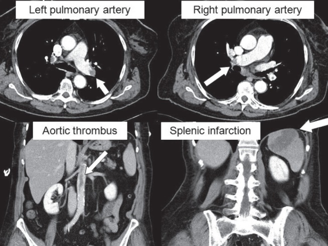
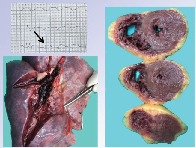
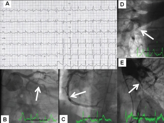
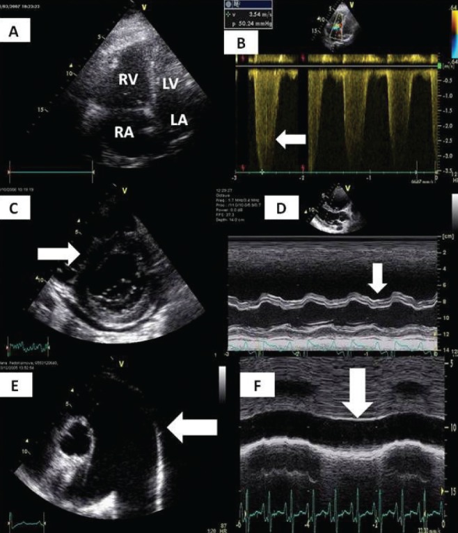
Pulmonary embolism is an important clinical entity with considerable mortality despite advances in diagnosis and treatment. In the present article, the authors offer a comprehensive review focused mainly on epidemiology, risk factors, risk stratification, pathophysiological considerations and clinical presentation. Diagnosis based on assessment of clinical likelihood, electrocardiography, chest x-ray, D-dimer levels, markers of myocardial injury and overload, and blood gases is discussed in detail. Special attention is devoted to the clinical use of computed tomography, pulmonary angiography and echocardiography in the setting of pulmonary embolism.

Keywords: Diagnosis; Epidemiology; Pulmonary embolism; Risk stratification.


Figures

Figure 1)
Figure 1)
A female patient with clinically small (ie, hemodynamically not significant), yet morphologically extensive, pulmonary embolism (arrows point to large emboli in the left and right pulmonary artery branches on computed tomography angiography). This clinical case is additionally complicated by embolization across a patent foramen ovale to the aorta and spleen. Patient’s clinical course was, however, favourable. Images were reproduced with permission from archives of the Department of Diagnostic Radiology, General University Hospital and 1st Medical School, Charles University in Prague, Prague, Czech Republic
Figure 2)
Figure 2)
Massive pulmonary embolism in a 35-year-old woman presenting with cardiac arrest and asystole. The patient was brought in by emergency medical services under continuous resuscitation; transient resumption of electrical activity was followed by clear ST-segment elevations in lead III (upper left panel). The patient was then transferred to the catheter laboratory and connected to veno-arterial extracorporeal membrane oxygenation, where pulmonary embolism diagnosis was confirmed by pulmonary angiography detecting virtually complete pulmonary bed obstruction. Thrombolytic therapy and catheter-based mechanical thrombus fragmentation failed to restore adequate flow through the pulmonary bed. The lower panel clearly shows the macroscopic autopsy finding of the pulmonary bed filled up with fresh thrombi. The right panel displays a myocardial section (documenting massive right ventricular wall effusion) indicating critical right-heart overload and possible mimicking of the above electocardiographic finding. Intraresuscitation selective coronary angiography ruled out coronary artery obstruction. (Images reproduced with permission from archives of the Department of Pathology, General University Hospital and 1st Medical School, Charles University in Prague, Prague, Czech Republic)
Figure 3)
Figure 3)
Computed tomography image of a minor pulmonary infarction (arrow) in a patient with submassive pulmonary embolism. (Image reproduced with permission from archives of the Department of Diagnostic Radiology, General University Hospital and Charles University Medical School 1, Prague, Czech Republic)
Figure 4)
Figure 4)
Patient referred as a case of acute coronary syndrome with ST-segment elevation and cardiogenic shock (electrocardiography [panel A – typical picture of right bundle branch block with ST-segment elevation]). Coronary angiography failed to explain the severity of status (panel B) with a normal finding on the left main coronary artery and an (at most) borderline stenosis (arrow) on the right coronary artery (panel C). Pulmonary artery angiography documented an extensive pulmonary embolism (filling defects indicated with arrows – right and left pulmonary arteries in panels D and E, respectively). The procedure continued with local thrombolytic delivery into the pulmonary artery and catheter-based fragmentation with prompt resolution of complaints and hemodynamic stabilization
Figure 5)
Figure 5)
Classical echocardiographic signs of massive and submassive pulmonary embolisms. A Dilation of the right atrium (RA) and right ventricle (RV). B Jet of tricuspid regurgitation corresponding with severe pulmonary hypertension, gradient >50 mmHg. C ‘D-Shape’ of left ventricle (LV). D Abnormal motion of interventricular septum. E Dilation of the pulmonary artery. F Dilation and absence of respiratory variability of inferior vena cava corresponding with increased RA pressure. LA Left atrium

References

    1. Oger E. Incidence of venous thromboembolism in a community-based study in western France. Thromb Haemost. 2000;83:657–60. - PubMed
    1. Widimský J, Malý J, Eliáš P, et al. Doporučení pro diagnostiku a léčbu akutní plicní embolie. Vnitř. Lék. 2008;54:1S25–1S72.
    1. Torbicki A, Perrier A, Konstantidines S, et al. Guidelines on the diagnosis and management of acute pulmonary embolism. Eur Heart J. 2008;29:2276–315. - PubMed
    1. Meignan M, Rosso J, Gauthier H, et al. Systematic lung scans reveal a high frequency of silent pulmonary embolism in patients with proximal deep venous thrombosis. Arch Intern Med. 2000;160:159–64. - PubMed
    1. Pineda LA, Hathwar VS, Grant BJ. Clinical suspicion of fatal pulmonary embolism. Chest. 2001;120:791–5. - PubMed

LinkOut - more resources