. 2013 Aug 5;8(8):e70194.
doi: 10.1371/journal.pone.0070194. Print 2013.

Enhancement of natural killer cell cytotoxicity by sodium/iodide symporter gene-mediated radioiodine pretreatment in breast cancer cells

Affiliations

Enhancement of natural killer cell cytotoxicity by sodium/iodide symporter gene-mediated radioiodine pretreatment in breast cancer cells

Hae Won Kim et al. PLoS One. .

Abstract

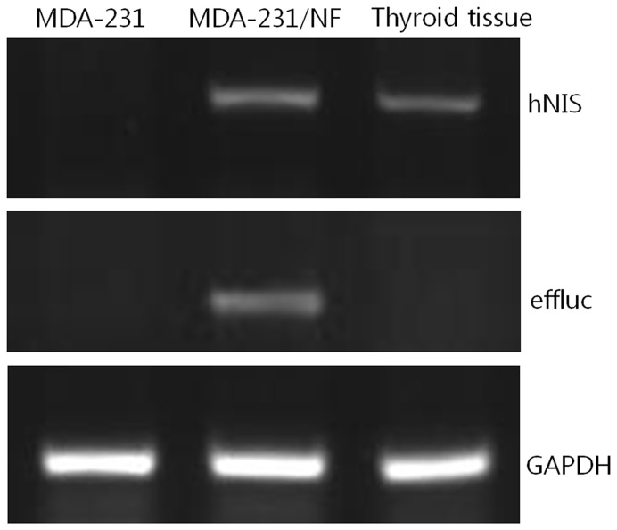
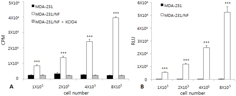
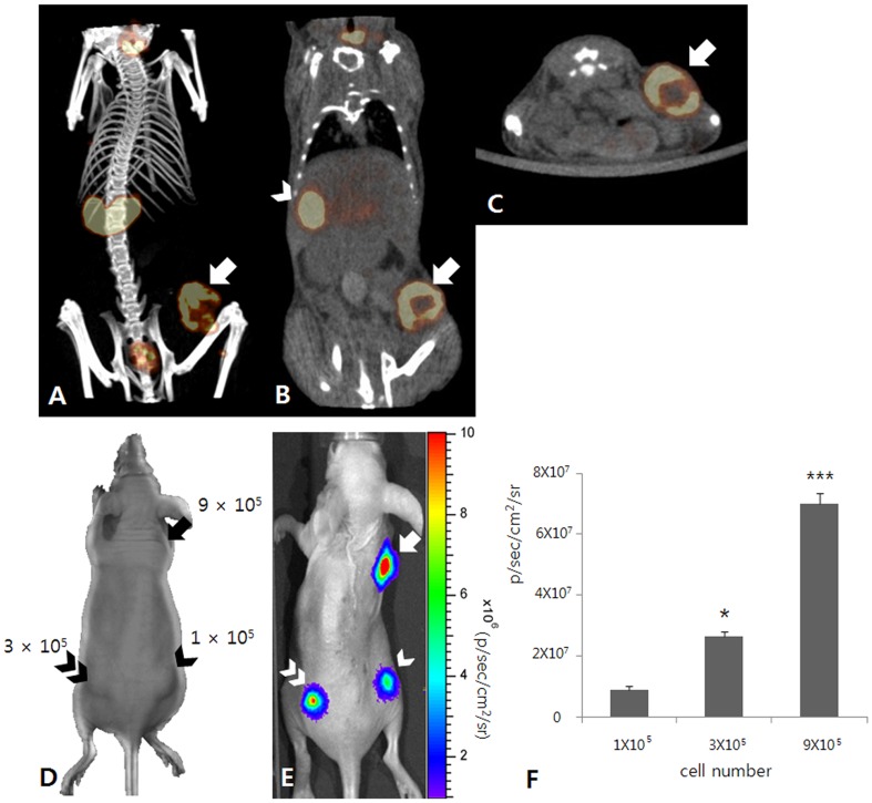
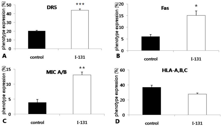
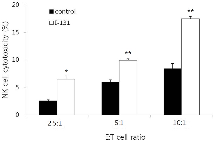
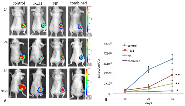
A phase II study of NK cell therapy in treatment of patients with recurrent breast cancer has recently been reported. However, because of the complexities of tumor microenvironments, effective therapeutic effects have not been achieved in NK cell therapy. Radioiodine (I-131) therapy inhibits cancer growth by inducing the apoptosis and necrosis of cancer cells. Furthermore, it can modify cancer cell phenotypes and enhance the effect of immunotherapy against cancer cells. The present study showed that I-131 therapy can modulate microenvironment of breast cancer and improve the therapeutic effect by enhancing NK cell cytotoxicity to the tumor cells. The susceptibility of breast cancer cells to NK cell was increased by precedent I-131 treatment in vitro. Tumor burden in mice treated with I-131 plus NK cell was significantly lower than that in mice treated with NK cell or I-131 alone. The up-regulation of Fas, DR5 and MIC A/B on irradiated tumor cells could be the explanation for the enhancement of NK cell cytotoxicity to tumor cells. It can be applied to breast cancer patients with iodine avid metastatic lesions that are non-responsive to conventional treatments.


Conflict of interest statement

Competing Interests: The authors have declared that no competing interests exist.

Figures

Figure 1
Figure 1. RT-PCR analysis of human sodium/iodide symporter (hNIS) and enhanced firefly luciferase (effluc) gene expression in MDA-231, MDA-231/NF cells and human thyroid tissue.
RT-PCR revealed hNIS mRNA expression in MDA-231/NF cells and human thyroid tissue, and effluc mRNA expression in MDA-231/NF cells. RT-PCR fragments have lengths of 583 bp and 316 bp for hNIS and effluc in MDA-231/NF cells; however, these bands do not appear in MDA-231 cells.
Figure 2
Figure 2. In vitro I-125 uptake assay and luciferase assay in MDA-231 and MDA-231/NF cells.
(A) I-125 uptake by MDA-231/NF cells increased according to cell number. I-125 uptake by MDA-231 cells remained at the basal level. *** p<0.001 compared with MDA-231 and MDA-231/NF cells blocked by KClO4. (B) Bioluminescence signals of MDA-231/NF cells increased according to cell number. Bioluminescence signal of MDA-231 cells remained at the basal level. CPM: count per minute, RLU: Relative Light Units, *** p<0.001 compared with MDA-231 cells.
Figure 3
Figure 3. Characterization image of MDA-231/NF cells.
(A–C) Tc-99m pertechnetate SPECT/CT images show focal tracer uptake (arrow) in the right flank of the MDA-231/NF tumor xenograft. There is also noted tracer uptake at the stomach (arrow head). (D–F) In vivo bioluminescence imaging for a mouse with MDA-231/NF cell implantation. (D) MDA-231/NF cells were implanted subcutaneously into the right hind-flank (1×10⁵; arrow head), left hind-flank (3×10⁵; double arrow head), and right fore-flank (9×10⁵; arrow) of the mouse. (E) Bioluminescence signals from implanted MDA-231/NF cells are clearly visualized in the right hind-flank (arrow head), left hind-flank (double arrow head), and right fore-flank (arrow). (F) The signal intensity from tumor cells increased with increasing cell number. * p<0.05 compared with 1×10⁵ cells, *** p<0.001 compared with 1×10⁵ and 3×10⁵ cells.
Figure 4
Figure 4. Phenotype analysis in MDA-231/NF cells by flow cytometry.
(A, B and C) The levels of DR5, Fas, and MIC A/B expression in tumor cells that received I-131 were significantly higher than in tumor cells received. (D) I-131 therapy resulted in decreased expression of HLA-A,B,C, but without statistical significance. * p<0.05, ** p<0.005, *** p<0.001 compared with control.
Figure 5
Figure 5. In vitro Cytotoxicity assay using Calcein-AM.
NK cell cytotoxocity is significantly increased according to effector : target cell (E : T) ratio in the control and I-131 groups. NK cell cytotoxicities were significantly higher in irradiated cells than in non-irradiated cells at all E : T ratios of 2.5 : 1, 5 : 1, and 10 : 1. * p<0.05, ** p<0.005 compared with control.
Figure 6
Figure 6. Tumor burdens monitored by bioluminescence imaging 14, 24, and 34 days after tumor inoculation.
(A and B) The tumor signals in the I-131 and NK groups were significantly lower than those in the control group at 34 days. The combined group showed the lowest stationary tumor signal over time, which was significantly lower than that in the I-131 and NK groups at 34 days. * p<0.05 compared with I-131 and NK groups,** p<0.005 compared with control group.

References

    1. DeSantis C, Siegel R, Bandi P, Jemal A (2011) Breast cancer statistics, 2011. CA Cancer J Clin 61: 409–418. - PubMed
    1. Ugnat AM, Xie L, Morriss J, Semenciw R, Mao Y (2004) Survival of women with breast cancer in Ottawa, Canada: variation with age, stage, histology, grade and treatment. Br J Cancer 90: 1138–1143. - PMC - PubMed
    1. Nieto Y (2003) The verdict is not in yet. Analysis of the randomized trials of high-dose chemotherapy for breast cancer. Haematologica 88: 201–211. - PubMed
    1. Dent R, Trudeau M, Pritchard KI, Hanna WM, Kahn HK, et al. (2007) Triple-negative breast cancer: clinical features and patterns of recurrence. Clin Cancer Res 13: 4429–4434. - PubMed
    1. Dewan MZ, Terunuma H, Takada M, Tanaka Y, Abe H, et al. (2007) Role of natural killer cells in hormone-independent rapid tumor formation and spontaneous metastasis of breast cancer cells in vivo. Breast Cancer Res Treat 104: 267–275. - PubMed

Publication types