. 2013 Aug 5;8(8):e70419.
doi: 10.1371/journal.pone.0070419. Print 2013.

A biodegradable, sustained-released, prednisolone acetate microfilm drug delivery system effectively prolongs corneal allograft survival in the rat keratoplasty model

Affiliations

A biodegradable, sustained-released, prednisolone acetate microfilm drug delivery system effectively prolongs corneal allograft survival in the rat keratoplasty model

Yu-Chi Liu et al. PLoS One. .

Abstract

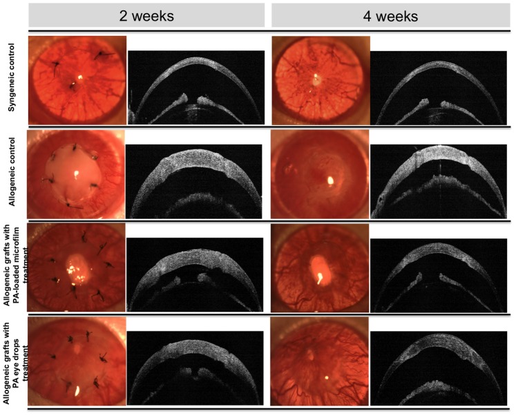
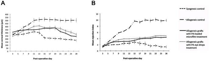
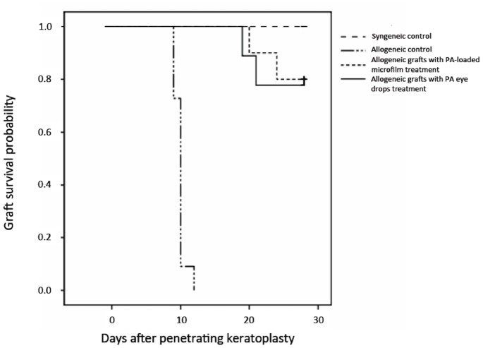
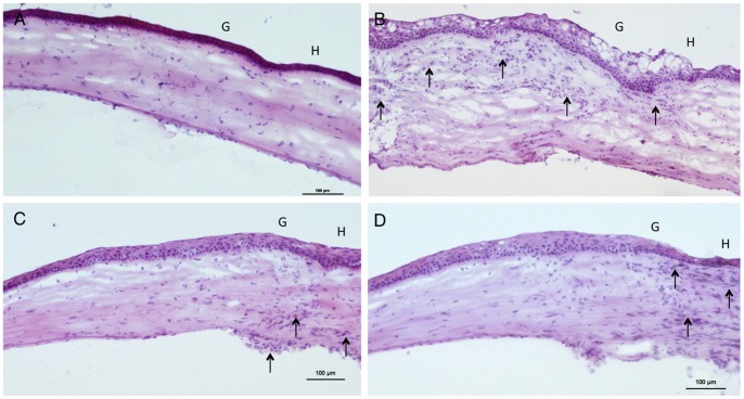
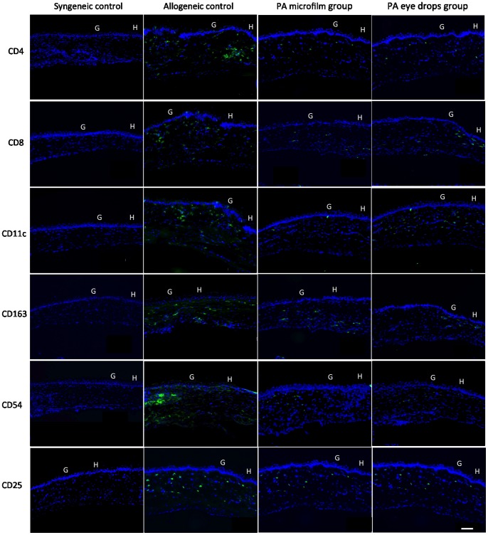
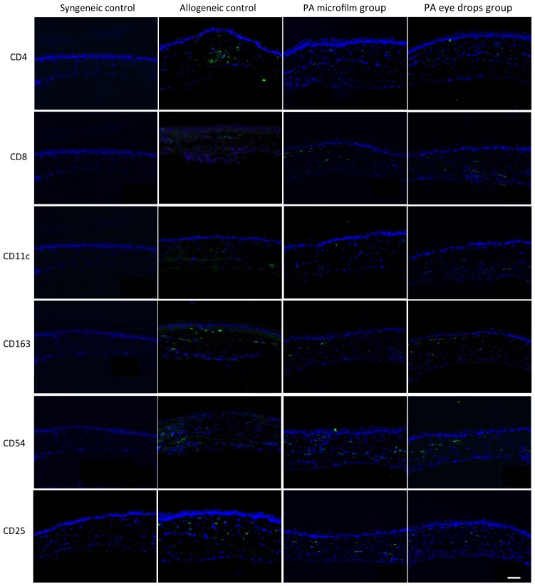
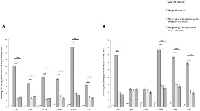
Frequent and long-term use of topical corticosteroids after corneal transplantation is necessary to prevent graft rejection. However, it relies heavily on patient compliance, and sustained therapeutic drug levels are often not achieved with administration of topical eye drops. A biodegradable drug delivery system with a controlled and sustained drug release may circumvent these limitations. In this study, we investigated the efficacy of a prednisolone acetate (PA)-loaded poly (d,l-lactide-co-ε-caprolactone) (PLC) microfilm drug delivery system on promoting the survival of allogeneic grafts after penetrating keratoplasty (PK) using a rat model. The drug release profiles of the microfilms were characterized (group 1). Subsequently, forty-eight PK were performed in four experimental groups: syngeneic control grafts (group 2), allogeneic control grafts (group 3), allogeneic grafts with subconjunctivally-implanted PA microfilm (group 4), and allogeneic grafts with PA eye drops (group 5; n = 12 in each). PA-loaded microfilm achieved a sustained and steady release at a rate of 0.006-0.009 mg/day, with a consistent aqueous drug concentration of 207-209 ng/ml. The mean survival days was >28 days in group 2, 9.9±0.8 days in group 3, 26.8±2.7 days in group 4, and 26.4±3.4 days in group 5 (P = 0.023 and P = 0.027 compared with group 3). Statistically significant decrease in CD4+, CD163+, CD 25+, and CD54+ cell infiltration was observed in group 4 and group 5 compared with group 3 (P<0.001). There was no significant difference in the mean survival and immunohistochemical analysis between group 4 and group 5. These results showed that sustained PA-loaded microfilm effectively prolongs corneal allograft survival. It is as effective as conventional PA eye drops, providing a promising clinically applicable alternative for patients undergoing corneal transplantation.


Conflict of interest statement

Competing Interests: The authors have read the journal's policy and have the following conflicts: Tina T. Wong and Jodhbir S. Mehta are listed as co-inventors for the patent filed on prednisolone microfilm PCT/SG2011/000282. This does not alter the authors' adherence to all the PLOS ONE policies on sharing data and materials.

Figures

Figure 1
Figure 1. Clinical evaluation of corneal grafts by slit lamp biomicroscopy and ASOCT at 2 and 4 weeks.
At 2 weeks, all allogeneic control grafts exhibited rejection episodes with severe graft edema and opacity, whereas grafts from the PA microfilm and PA eye drop groups had minimal graft edema and opacity. At 4 weeks, all allogeneic control grafts progressed to complete opacification and pronounced edema with neovascularization. The grafts from the PA microfilm and PA eye drop groups appeared clear with visible pupillary margin. All syngeneic control grafts remained clear during the follow-up period. On ASOCT, all grafted corneas exhibited good anatomic position without graft-host dehiscence or anterior chamber collapse.
Figure 2
Figure 2. Changes of graft thickness and mean rejection scores with time for different groups.
(A) The mean central graft thickness measured by ASOCT per time point for different groups. (B) The mean of the graft RI per time point for different groups.
Figure 3
Figure 3. Clinical and histological evaluation of PA microfilm implantation.
(A) Slit lamp photo showing the subconjunctivally-implanted microfilm at 4 weeks. Arrows indicated the implanted microfilm. (B) Histological section with H&E staining at 4 weeks. Arrows indicated the implanted microfilm. Original magnification×100. Scale bar: 100 µm.
Figure 4
Figure 4. Kaplan-Meier analysis of the rejection-free graft survival.
The survival probability at 28 days was 100%, 0%, 80.0% and 76.7% in the syngeneic control, allogeneic control, PA microfilm, and PA eye drop groups, respectively. The PA microfilm and PA eye drop groups had significantly longer survivals as compared to the allogeneic control group (P = 0.012 and P = 0.014, log-rank test). There was no significant difference between the PA microfilm and PA eye drop groups (P = 0.58, log-rank test).
Figure 5
Figure 5. Histological sections with H&E staining for different groups.
(A) Syngeneic control group. The graft presented normal architecture and thickness with very scant cell infiltrate in the stroma. (B) Allogeneic control group. There was marked graft edema and diffuse infiltration of mononuclear cells in the stroma (arrow). (C) Allogeneic grafts treated with PA microfilms (D) Allogeneic grafts treated with PA eye drops. Thickening of stroma and cellular infiltration (arrow) were appreciably less in the grafts treated with PA microfilms or PA eye drops. G: Graft. H: Host. Original magnification×100. Scale bar: 100 µm.
Figure 6
Figure 6. Immunohistochemistry staining for CD4+, CD8+, CD11c+, CD163+, CD54+, and CD25+ cells at graft-host junction for different groups.
The grafts treated with the PA microfilms or PA eye drops had less dense and diffuse cell infiltration in comparison with the allogeneic control grafts. The syngeneic grafts were nearly free of immunologic cell infiltration. G: Graft. H: Host. Original magnification ×100. Scale bar: 100 µm.
Figure 7
Figure 7. Immunohistochemistry staining for CD4+, CD8+, CD11c+, CD163+, CD54+, and CD25+ cells at central grafts for different groups.
The syngeneic grafts were nearly free of immunologic cell infiltration. The grafts treated with the PA microfilms or PA eye drops had less dense and diffuse cell infiltration in comparison with the allogeneic control grafts. G: Graft. H: Host. Original magnification ×100. Scale bar: 100 µm.
Figure 8
Figure 8. Quantification of CD4+, CD8+, CD11c+, CD163+, CD54+, and CD25+ positively stained cells for different groups.
(A) Quantification of positively stained cells at graft-host junction. The grafts from the PA microfilm and PA eye drop groups showed a statistically significantly decreased infiltration of CD4+, CD8+, CD25+, CD54+, CD11c, and CD163+ cells compared to the allogeneic control grafts. (B) Quantification of positively stained cells at central grafts. The grafts from the PA microfilm and PA eye drop groups showed a statistically significantly decreased infiltration of CD4+, CD25+, CD54+, and CD163+ cells compared to the allogeneic control grafts. Error bars represent SE and asterisks indicate P<0.001.

References

    1. Tan DT, Dart JK, Holland EJ, Kinoshita S (2012) Corneal transplantation. Lancet 379: 1749–1761. - PubMed
    1. Eye Banking Statistical Report Eye Bank Association of America 2011. Available: http://www.restoresight.org. Accessed 2012 Nov 12.
    1. Tan DT, Janardhanan P, Zhou H, Chan YH, Htoon HM, et al. (2008) Penetrating keratoplasty in Asian eyes: the Singapore Corneal Transplant Study. Ophthalmology 115: 975–982.e1. - PubMed
    1. Shimazaki J, Iseda A, Satake Y, Shimazaki-Den S (2012) Efficacy and safety of long-term corticosteroid eye drops after penetrating keratoplasty: a prospective, randomized, clinical trial. Ophthalmology 119: 668–673. - PubMed
    1. Panda A, Vanathi M, Kumar A, Dash Y, Priya S (2007) Corneal graft rejection. Surv Ophthalmol 52: 375–396. - PubMed

Publication types