. 2013 Aug 5;8(8):e70575.
doi: 10.1371/journal.pone.0070575. Print 2013.

Mito-tempol and dexrazoxane exhibit cardioprotective and chemotherapeutic effects through specific protein oxidation and autophagy in a syngeneic breast tumor preclinical model

Affiliations

Mito-tempol and dexrazoxane exhibit cardioprotective and chemotherapeutic effects through specific protein oxidation and autophagy in a syngeneic breast tumor preclinical model

Jennifer S Dickey et al. PLoS One. .

Abstract

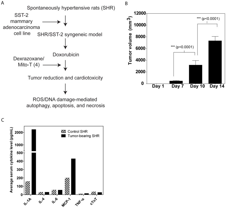
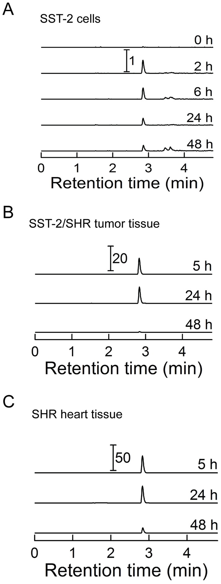
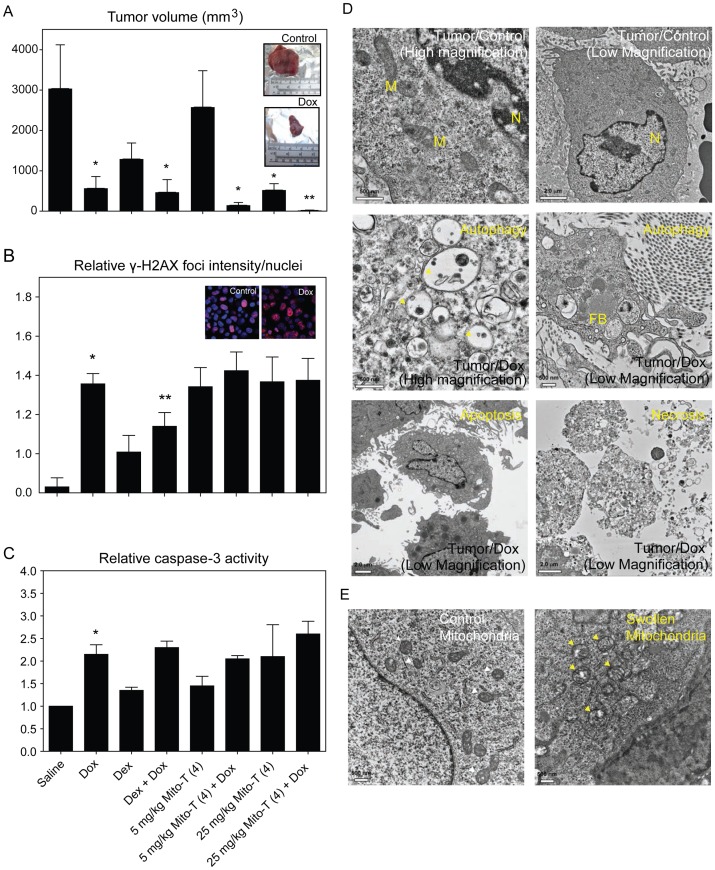
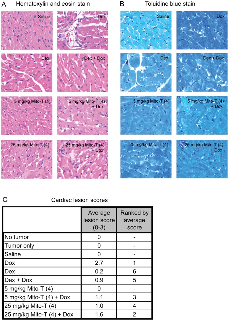
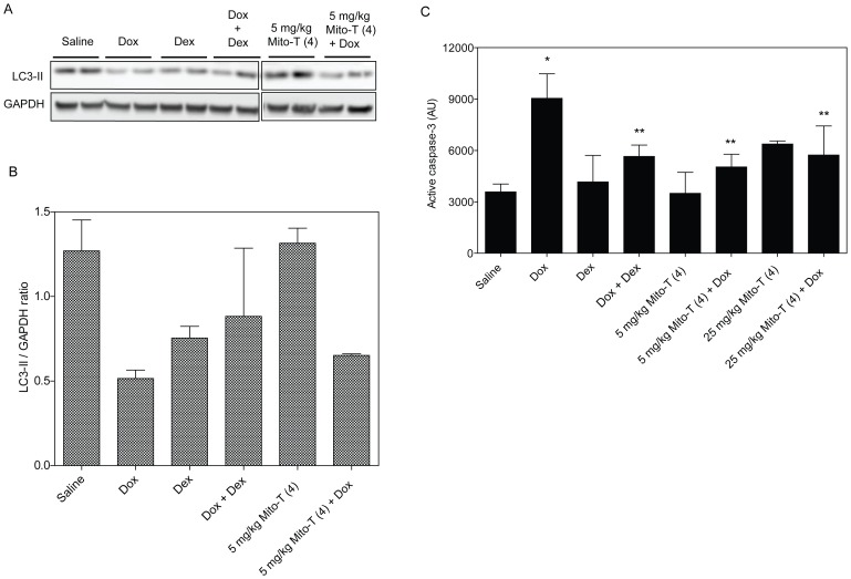
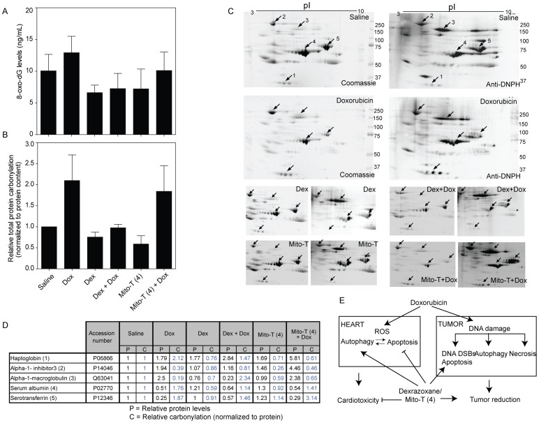
Several front-line chemotherapeutics cause mitochondria-derived, oxidative stress-mediated cardiotoxicity. Iron chelators and other antioxidants have not completely succeeded in mitigating this effect. One hindrance to the development of cardioprotectants is the lack of physiologically-relevant animal models to simultaneously study antitumor activity and cardioprotection. Therefore, we optimized a syngeneic rat model and examined the mechanisms by which oxidative stress affects outcome. Immune-competent spontaneously hypertensive rats (SHRs) were implanted with passaged, SHR-derived, breast tumor cell line, SST-2. Tumor growth and cytokine responses (IL-1A, MCP-1, TNF-α) were observed for two weeks post-implantation. To demonstrate the utility of the SHR/SST-2 model for monitoring both anticancer efficacy and cardiotoxicity, we tested cardiotoxic doxorubicin alone and in combination with an established cardioprotectant, dexrazoxane, or a nitroxide conjugated to a triphenylphosphonium cation, Mito-Tempol (4) [Mito-T (4)]. As predicted, tumor reduction and cardiomyopathy were demonstrated by doxorubicin. We confirmed mitochondrial accumulation of Mito-T (4) in tumor and cardiac tissue. Dexrazoxane and Mito-T (4) ameliorated doxorubicin-induced cardiomyopathy without altering the antitumor activity. Both agents increased the pro-survival autophagy marker LC3-II and decreased the apoptosis marker caspase-3 in the heart, independently and in combination with doxorubicin. Histopathology and transmission electron microscopy demonstrated apoptosis, autophagy, and necrosis corresponding to cytotoxicity in the tumor and cardioprotection in the heart. Changes in serum levels of 8-oxo-dG-modified DNA and total protein carbonylation corresponded to cardioprotective activity. Finally, 2D-electrophoresis/mass spectrometry identified specific serum proteins oxidized under cardiotoxic conditions. Our results demonstrate the utility of the SHR/SST-2 model and the potential of mitochondrially-directed agents to mitigate oxidative stress-induced cardiotoxicity. Our findings also emphasize the novel role of specific protein oxidation markers and autophagic mechanisms for cardioprotection.


Conflict of interest statement

Competing Interests: Eisai Co., Ltd. donated the SST-2 cells used in this study. The authors have no patents, products in development, marketed products or competing interests to declare. This does not alter the authors' adherence to all the PLOS ONE policies on sharing data and materials. The views expressed in this article are those of the authors and do not necessarily reflect the official policy or position of the United States Food and Drug Administration and the Department of Health and Human Services, nor does mention of trade names, commercial products, or organizations imply endorsement by the United States Government.

Figures

Figure 1
Figure 1. Optimization of the syngeneic SHR/SST-2 model.
A, Study design for the optimization and application of the syngeneic SHR/SST-2 model. SHRs were subcutaneously implanted with exponentially-growing SST-2 breast cancer cells in their right mammary fat pads. Doxorubicin and dexrazoxane were used as control chemotherapeutic and chemoprotective agents, respectively. Mito-T (4) was tested as a chemoprotective agent. In addition to overall tumor reduction and cardiac lesions, protein oxidation, DNA oxidation, autophagy, apoptosis, and necrosis were measured as mechanistic endpoints. B, Tumors were measured at days 1, 7, 10, and 14. Results are expressed as tumor volume. C, Serum from control and tumor-bearing SHRs was compared for levels of inflammatory cytokines, IL-1A, IL-4, IL-6, MCP-1, and TNF-α. Cardiac troponin T (cTnT) was compared as an indicator of cardiac toxicity. Mean values from 30 animals per group are shown.
Figure 2
Figure 2. Accumulation of Mito-T (4) in mitochondria of heart and tumor cells and tissue.
A, SST-2 cells were incubated with 10 μM Mito-T (4) for the indicated period of time and collected. Mito-T (4) was detected in isolated mitochondrial fractions by LC-MS/MS as described in the Materials and Methods section. B, SHR rats were implanted with SST-2 cells. Upon tumor growth of 1 cm3 over approximately 6 days, animals were treated with 25 mg/kg Mito-T (4). Necropsy was performed at 5, 24, and 48 h following administration of Mito-T (4) and tissue was harvested and frozen. Tumor tissue was homogenized and mitochondrial fraction assayed for Mito-T (4) content by LC-MS-MS. C, Same as panel B, but mitochondria from heart tissue were analyzed. All LC traces represent the MRM transition 489.0 → 474.2 and were scaled to the same protein level. The numbers with the scale bars indicate differences in signal intensities between the panels and relative to the signals from SST-2 cells (panel A).
Figure 3
Figure 3. Tumor growth and response to chemotherapeutic and chemoprotective agents in SHR/SST-2 animals.
A, SHRs were implanted with SST-2 cells and 24 h later were administered either doxorubicin (10 mg/kg), dexrazoxane (50 mg/kg), Mito-T (4) (5 or 25 mg/kg), a combination of doxorubicin and dexrazoxane, or a combination of doxorubicin and Mito-T (4). Each treatment group consisted of 10 animals. The mean tumor volumes (mm3) measured 14 days after drug treatment are shown for each treatment group. The two inset images show representative excised tumors from saline and doxorubicin-treated SHR/SST-2 animals. B, DNA damage by doxorubicin and chemoprotective agents was measured in tumor cells by confocal microscopic detection of γ-H2AX foci. Average foci intensity was measured in at least 100 cells per drug treatment observed from at least 10 representative fields. The data are represented as fold increase over no treatment tumor cells (Control). C, Active caspase-3 levels were assessed as a marker of apoptosis induction. Average fold increase is shown over saline control tumor tissue samples. * =  statistically significant compared to saline and ** = statistically significant compared to doxorubicin (A–C). D, Transmission electron microscopy analysis of tumor samples from rats exposed to doxorubicin showing the formation of autophagic vacuoles (Autophagy panel), or nuclear condensation and membrane blebbing (Apoptosis panel), and membrane breakdown (Necrosis panel). Saline treated (Tumor/Control panels) are shown as controls. Representative low- and high-magnification images for the control and autophagic samples are shown for clarity. Mitochondria (labeled M), nucleus (N), and fat bodies (FB) are labeled within the images. E, Swollen mitochondria following doxorubicin treatment (right) are indicated with yellow arrows in comparison to mitochondria from saline-treated animals (left) indicated with white arrows. Scale bars and magnification are indicated on each panel. Quantification of the mitochondrial cross-section areas is provided in Figure S2.
Figure 4
Figure 4. Cardiotoxicity and cardioprotection in SHR/SST-2 animals.
A–B, Representative photomicrographs of cardiac histological sections are shown following either hematoxylin and eosin staining (panel A) or toluidine blue staining (panel B). C, Numerical cardiomyopathy scores (maximum severity score  = 3) are shown following analysis of each treatment group (n = 5). Please refer to the methods and results sections for descriptions of the scoring method and treatment-induced cardiomyopathy, respectively.
Figure 5
Figure 5. Autophagy and apoptosis in cardiac tissue after cardiotoxic and cardioprotective treatment.
A, Western blotting of autophagy-indicative lipidated LC-3 II protein in cardiac tissue samples. Protein extracts analyzed for LC3-II from two representative SHR/SST-2 animals per treatment group are shown. B, The mean LC3-II/GAPDH ratio in each treatment group by Western blotting analysis is shown. Quantitation was performed by densitometric analysis of the LC3-II bands from panel A. GAPDH protein levels were used as a loading control. C, Paraffin-embedded cardiac tissue were stained with anti-active caspase-3 antibody. The intensity of HRP-tagged secondary antibody was quantified as an indication of active caspase-3 using the ScanScope software. Mean intensities are shown in the graph and derived from at least 10 images per animal (5 animals per group). * =  statistically significant compared to saline and ** = statistically significant compared to doxorubicin.
Figure 6
Figure 6. Oxidative damage to DNA and proteins in SHR serum.
A, DNA oxidative modification as measured by 8-hydroxy-2′-deoxyguanosine levels in SHR serum using an ELISA assay. Average from at least 4 animals per group is shown B, Protein carbonylation of serum was measured as an indication of protein oxidation. Serum samples (5 µg) from drug treated groups of animals were derivatized with DNP and electrophoresed on two parallel gels for each experiment. One gel was stained with coomassie blue to determine total protein and the other gel was used for Western blotting. Carbonylated proteins were detected using anti-DNPH antibody. Total serum protein carbonylation levels shown are relative values compared to total protein carbonylation of the saline treated samples normalized to 1. Quantitation was performed by densitometric analysis of the whole lanes for gel staining and Western blot. C, Representative 2D-gels from serum samples from drug treated, tumor bearing SST-2 rats are shown. Left panels for each sample represent the coomassie gel staining and right panels represent the Western blot analysis using anti-DNP antibody. Spots labeled 1–5 were the major serum proteins that exhibited significant changes in carbonylation and concentration comparing saline and doxorubicin. The top four panels show the complete gel data for saline and doxorubicin samples, while the bottom eight panels show the significant spots from other drug treated samples. The carbonylated proteins with the most appreciable changes were identified by LC-MS/MS analysis as haptoglobin (1), alpha-1-inhibitor 3 (2), alpha-1-macroglobulin (3), serum albumin (4), and serotransferrin (5). D, Table shows relative protein content and specific protein carbonylation in drug treated SHRs compared to saline treated rats determined by 2D-gel electrophoresis from panel C. Data are representative of three animals analyzed per treatment group. E, Proposed model for pro-autophagic and anti-apoptotic mechanism of action for dexrazoxane and Mito-T (4) in combination with doxorubicin.

Similar articles

Cited by

References

    1. Albini A, Pennesi G, Donatelli F, Cammarota R, De Flora S, et al. (2010) Cardiotoxicity of anticancer drugs: the need for cardio-oncology and cardio-oncological prevention. J Natl Cancer Inst 102: 14–25. - PMC - PubMed
    1. Cardinale D, Colombo A, Torrisi R, Sandri MT, Civelli M, et al. (2010) Trastuzumab-induced cardiotoxicity: clinical and prognostic implications of troponin I evaluation. J Clin Oncol 28: 3910–3916. - PubMed
    1. Mellor HR, Bell AR, Valentin JP, Roberts RR (2011) Cardiotoxicity associated with targeting kinase pathways in cancer. Toxicol Sci: 14–32. - PubMed
    1. Ferrans VJ, Clark JR, Zhang J, Yu ZX, Herman EH (1997) Pathogenesis and prevention of doxorubicin cardiomyopathy. Tsitologiia 39: 928–937. - PubMed
    1. Levine RL, Stadtman ER (2001) Oxidative modification of proteins during aging. Exp Gerontol 36: 1495–1502. - PubMed

Publication types

MeSH terms