. 2013 Aug 5;8(8):e71057.
doi: 10.1371/journal.pone.0071057. Print 2013.

A human in vitro whole blood assay to predict the systemic cytokine response to therapeutic oligonucleotides including siRNA

Affiliations

A human in vitro whole blood assay to predict the systemic cytokine response to therapeutic oligonucleotides including siRNA

Christoph Coch et al. PLoS One. .

Abstract

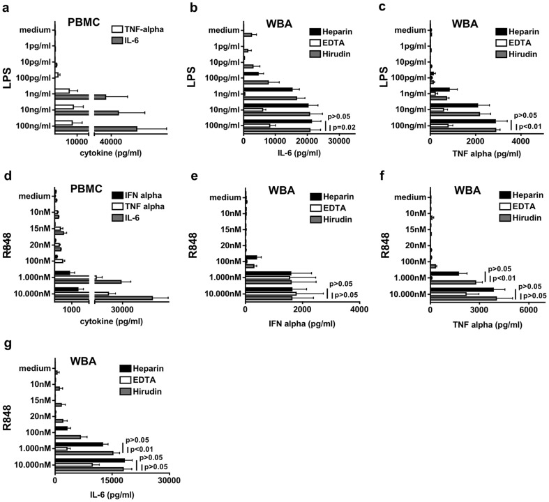
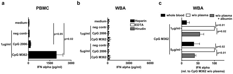
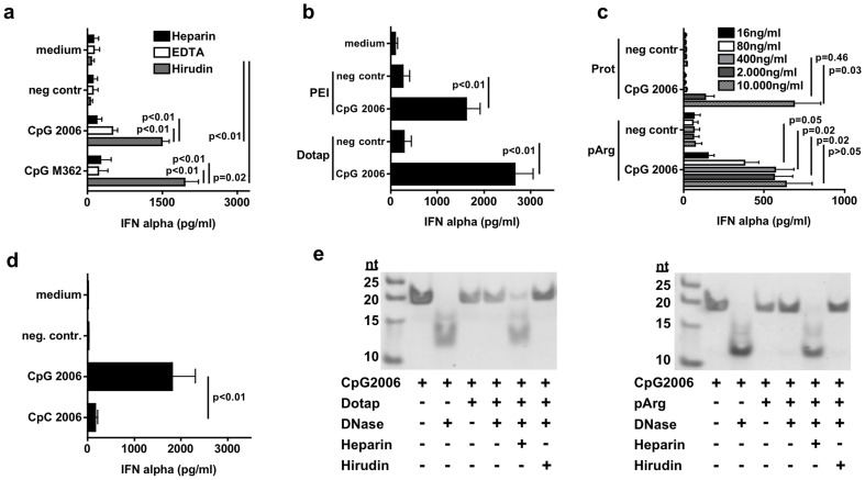
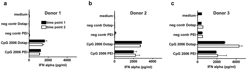
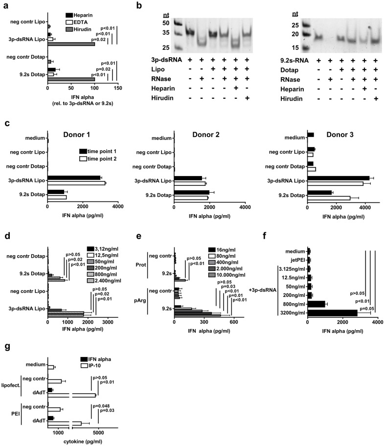
Therapeutic oligonucleotides including siRNA and immunostimulatory ligands of Toll-like receptors (TLR) or RIG-I like helicases (RLH) are a promising novel class of drugs. They are in clinical development for a broad spectrum of applications, e.g. as adjuvants in vaccines and for the immunotherapy of cancer. Species-specific immune activation leading to cytokine release is characteristic for therapeutic oligonucleotides either as an unwanted side effect or intended pharmacology. Reliable in vitro tests designed for therapeutic oligonucleotides are therefore urgently needed in order to predict clinical efficacy and to prevent unexpected harmful effects in clinical development. To serve this purpose, we here established a human whole blood assay (WBA) that is fast and easy to perform. Its response to synthetic TLR ligands (R848: TLR7/8, LPS: TLR4) was on a comparable threshold to the more time consuming peripheral blood mononuclear cell (PBMC) based assay. By contrast, the type I IFN profile provoked by intravenous CpG-DNA (TLR9 ligand) in humans in vivo was more precisely replicated in the WBA than in stimulated PBMC. Since Heparin and EDTA, but not Hirudin, displaced oligonucleotides from their delivery agent, only Hirudin qualified as the anticoagulant to be used in the WBA. The Hirudin WBA exhibited a similar capacity as the PBMC assay to distinguish between TLR7-activating and modified non-stimulatory siRNA sequences. RNA-based immunoactivating TLR7/8- and RIG-I-ligands induced substantial amounts of IFN-α in the Hirudin-WBA dependent on delivery agent used. In conclusion, we present a human Hirudin WBA to determine therapeutic oligonucleotide-induced cytokine release during preclinical development that can readily be performed and offers a close reflection of human cytokine response in vivo.


Conflict of interest statement

Competing Interests: The authors have declared that no competing interests exist.

Figures

Figure 1
Figure 1. Comparison of cytokines induced by LPS and R848 in PBMC and in a human WBA.
a – PBMC were incubated in 96 well with increasing concentrations of LPS. After 48 h supernatant was analyzed for TNF-α and IL-6 using ELISA. Results of eight donors are shown as mean +/− SD. b–c – Whole blood anticoagulated either with Heparin, EDTA or Hirudin was incubated in 96 wells with increasing concentrations of LPS and after 48 h supernatant was checked for IL-6 (b), TNF-α (c). Mean of six donors is shown +/− SD. d – Done as described in (a) but R848 was used for stimulation and supernatant was analyzed for IFN-α, TNF-α and IL-6. Mean of eight donors (three for IFN-α) is shown +/− SD. e–g – Done as described for (b) but R848 was used for stimulation and IFN-α (e), TNF-α (f) and IL-6 (g) were measured in supernatant. Results of four (IFN-α) and six donors are shown as mean +/− SD.
Figure 2
Figure 2. The WBA simulates IFN-α induction by CpG-ODN in vivo more accurately than the PBMC assay.
a – PBMC were stimulated with B-type (CpG 2006) and C-type (M362) CpG-oligonucleotides as indicated. C20-DNA served as a negative control (neg contr). After 48 h induction of IFN-α was analyzed using ELISA. b – Done as described for (a) but whole blood anticoagulated with Heparin, EDTA or Hirudin was used. c – Complete whole blood or whole blood in which plasma was replaced by medium or medium+albumin was anticoagulated with Hirudin and stimulated with B-type (CpG 2006) and C-type (CpG M362) CpG-oligonucleotides or C20-DNA (neg contr) as indicated. After 48 h supernatant was analyzed for IFN-α concentration. Graph shows percentage of induced IFN-α compared to CpG M362 (induced IFN-α concentrations: 1 µg/ml CpG M362: 1697 pg/ml; 5 µg/ml CpG M362: 3627 pg/ml). Results of at least six donors are shown as mean +/− SD in (a–c).
Figure 3
Figure 3. Amplification by transfection tolerates anticoagulation with Hirudin but not Heparin or EDTA in the WBA.
a – Whole blood was anticoagulated with Heparin, EDTA or Hirudin and stimulated with 0.8 µg/ml B-type (2006) or C-type (M362) CpG-oligonucleotide or C20-DNA (negative control) in complex with DOTAP. After 48 h supernatant was analyzed for IFN-α induction by ELISA. Results of at least six donors are shown as mean +/− SD. b – Done as described for (a) but additional polyethylenimine (PEI) was used as delivery agent. Results of at least four donors are shown as mean +/− SD. c – Done as described for (a) but Protamine (Prot) and poly-L-Arginine (pArg) were used as delivery agents in complex with increasing concentrations of DNA as indicated. Results of at least eight donors are shown as mean +/− SD. d – Whole blood was anticoagulated with Hirudin and stimulated with 0.8 µg/ml CpG 2006 or CpC 2006 (CpG replaced by CpC sequences), both delivered with DOTAP. C20-DNA served as negative control. After 48 h supernatant was analyzed for IFN-α by ELISA. Results of eight donors are shown as mean +/− SD. e – CpG 2006 was incubated with DOTAP or pArg with or without addition of Heparin or Hirudin and digested with DNase before gel electrophoresis.
Figure 4
Figure 4. The Hirudin anticoagulated WBA exhibits high reliability upon repeated testing of the same donor.
Whole blood of three different donors (a–c) anticoagulated with Hirudin was stimulated twice with one week inbetween with 0.8 µg/ml CpG 2006 delivered with polyethylenimine (PEI) or DOTAP. C20-DNA served as negative control. After 48 h supernatant was analyzed for IFN-α by ELISA. Mean of duplicates +/− SD is shown for each timepoint.
Figure 5
Figure 5. Hirudin-WBA – application as fast screening method to determine immunostimulatory side effects of siRNA therapeutics.
a–b – PBMC were stimulated with siRNA without (no meth) or with three 2′-O ribose methylated nucleotides (3x meth) delivered with p-L-Arginine (pArg) in increasing concentrations as indicated. After 48 h concentrations of IFN-α (a) and TNF-α (b) in supernatant was analyzed by ELISA. c–d – Done as described for (a) and (b) but whole blood assay anticoagulated with Hirudin was used. Results of eight donors are shown as mean +/− SD in (a–d). For sequence details of siRNA used see lower right paragraph.
Figure 6
Figure 6. RNA ligands for TLR7 and RIG-I induce IFN-α in Hirudin-whole blood assay.
a – Whole blood was anticoagulated with Heparin, EDTA or Hirudin and stimulated with 0.8 µg/ml 3p-dsRNA, 9.2s-RNA or A20-RNA (negative control) delivered with Lipofectamine2000 or DOTAP. After 48 h supernatant was analyzed for IFN-α induction by ELISA. Graph shows percentage of induced IFN-α compared to 3p-dsRNA and 9.2s-RNA respectively (induced IFN-α concentrations: 3p-dsRNA: 2641 pg/ml; 9.2s-RNA: 1438 pg/ml). Results of four donors are shown as mean +/− SD. b – 3p-dsRNA was incubated with Lipofectamine2000 and 9.2s-RNA with DOTAP with or without addition of Heparin or Hirudin. Complexes were digested by RNase and subsequently analyzed by gel electrophoresis. c – Whole blood of three different donors anticoagulated with Hirudin was stimulated at two different time points with 0.8 µg/ml 3p-dsRNA, 9.2s-RNA or A20-RNA (negative control). After 48 h supernatant was analyzed for IFN-α using ELISA. Mean of duplicates +/− SD are shown for each timepoint. d – Whole blood was anticoagulated with Hirudin and stimulated with increasing concentrations of 3p-dsRNA and 9.2s-RNA. Results of eight donors are shown as mean +/− SD. e – Done as described for (d) but Protamine (Prot) and poly-L-Arginine (pArg) were used as delivery agents for increasing concentrations of 9.2s-RNA. Results of eight donors are shown as mean +/− SD. f – Done as described for (d) but polyethylenimine (PEI) was used to deliver increasing concentrations of 3p-dsRNA. Results of six donors are shown as mean +/− SD. g – Done as described for (d) but 0.8 µg/ml of dAdT was used to stimulate the cells. Results show mean of IFN-α and IP-10 induction of six donors +/− SD.

References

    1. Vaishnaw AK, Gollob J, Gamba-Vitalo C, Hutabarat R, Sah D, et al. (2010) A status report on RNAi therapeutics. Silence 1: 14 doi:10.1186/1758-907X-1-14 - DOI - PMC - PubMed
    1. Krieg AM (2006) Therapeutic potential of Toll-like receptor 9 activation. Nat Rev Drug Discov 5: 471–484 doi:10.1038/nrd2059 - DOI - PubMed
    1. Barchet W, Wimmenauer V, Schlee M, Hartmann G (2008) Accessing the therapeutic potential of immunostimulatory nucleic acids. Curr Opin Immunol 20: 389–395 doi:10.1016/j.coi.2008.07.007 - DOI - PubMed
    1. Hornung V, Guenthner-Biller M, Bourquin C, Ablasser A, Schlee M, et al. (2005) Sequence-specific potent induction of IFN-alpha by short interfering RNA in plasmacytoid dendritic cells through TLR7. Nat Med 11: 263–270 doi:10.1038/nm1191 - DOI - PubMed
    1. Krieg AM, Yi AK, Matson S, Waldschmidt TJ, Bishop GA, et al. (1995) CpG motifs in bacterial DNA trigger direct B-cell activation. Nature 374: 546–549 doi:10.1038/374546a0 - DOI - PubMed

Publication types

MeSH terms