. 2013 Oct;170(4):883-92.
doi: 10.1111/bph.12334.

UDP induces intestinal epithelial migration via the P2Y6 receptor

Affiliations

UDP induces intestinal epithelial migration via the P2Y6 receptor

Tatsuro Nakamura et al. Br J Pharmacol. 2013 Oct.

Abstract

Background and purpose: Extracellular nucleotides are released at high concentrations from damaged cells and function through P2 receptor activation. Intestinal epithelial restitution, which is defined as cell migration independent of cell proliferation, is an important initial step in the process of wound healing. In this study, we investigated the role of extracellular nucleotides in intestinal epithelial migratory responses.

Experimental approach: Wound-healing and trans-well migration assays were performed with a rat intestinal epithelial cell line (IEC-6). The concentrations of extracellular nucleotides released from injured IEC-6 cells were measured by HPLC. TGF-β expression was assessed by RT-PCR and elisa.

Key results: Scratching the monolayer of IEC-6 cells induced cell migration. Pretreatment with apyrase or MRS2578, a selective P2Y6 antagonist, inhibited the wound-induced cell migration. Among the cellular nucleotides, only ATP and uridine 5'-diphosphate (UDP) were detected in the culture medium after cell wounding. Exogenously applied UDP dose-dependently enhanced the migration more effectively than ATP but did not induce proliferation. In addition, cell wounding and UDP increased the expression of TGF-β, and both the wound-induced and UDP-enhanced migration were inhibited by MRS2578 or ALK5Inhibitor (ALK5i), a TGF-β receptor blocker. Furthermore, cell wounding and UDP stimulation up-regulated the expression of P2Y6 receptor mRNA, and this effect was suppressed by MRS2578 or ALK5i.

Conclusion and implications: Wound-induced UDP evokes intestinal epithelial restitution by activation of P2Y6 receptors, which mediates de novo synthesis of TGF-β. In addition, the expression of P2Y6 receptors is increased by cell wounding and UDP, which constitutes a positive-feedback loop for mucosal repair.

Keywords: P2Y6 receptor; TGF-β; UDP; extracellular nucleotides; intestinal epithelial migration.


Figures

Figure 1
Figure 1
Wound stimulation enhances intestinal epithelial migration through P2Y6 receptors. (A) The confluent monolayer of IEC-6 cells was pretreated with apyrase (30 U·mL−1), MRS2578 (1 μmol·L−1), suramin (100 μmol·L−1) or PPADS (100 μmol·L−1) for 30 min before being scratched with a pipette tip. The monolayer was photographed immediately (0 h) and 8 h after the scratch formation. (B) After 8 h, the number of cells that crossed the wounded area was counted with the ImageJ software at three different locations in each experiment. The values are expressed as a percentage compared with the control. Data are means ± SEM from four independent experiments. **P < 0.01 as compared with the control.
Figure 2
Figure 2
UDP is released into culture medium after wounding. (A) Typical reverse-phase HPLC graph showing the presence of standard nucleotides (ATP, ADP, UTP and UDP: 5 μmol·L−1) (upper panel). Lower panel shows the HPLC pattern of the medium of injured cells. The signal peaks of UDP and ATP were detected at 1.5 and 3.0–3.5 min after injection of the nucleotides respectively. (B) The concentration of the nucleotides in the medium after cell scratch formation at the indicated time. The data are expressed as means ± SEM obtained from four independent experiments. *P < 0.05 as compared with ATP.
Figure 3
Figure 3
UDP enhances cell migration in both the trans-well and wound-healing assays. (A) A confluent monolayer of IEC-6 cells was scratched with a pipette tip. After being washed three times, each nucleotide (ATP, ADP, UTP and UDP: 100 μmol·L−1) was added to the wounded monolayer. The monolayer was photographed immediately after scratch formation (0 h) (upper panel) and 8 h after scratch formation (lower panel). (B) The number of cells that crossed the wounded area at 8 h (left panel) or 24 h (right panel) was counted at three different places in each experiment. (C) UDP (1–100 μmol·L−1) was added to the scratched monolayer of IEC-6 cells. After 8 h, the number of cells that crossed the wounded area was counted at three different places in each experiment. (D) After incubation with MRS2578 (a P2Y6 antagonist: 0.1–1 μmol·L−1) for 30 min, UDP (100 μmol·L−1) was added to the scratched cell monolayer. Migration was evaluated as a percentage compared with the control. Values are expressed as means ± SEM from four independent experiments. *P < 0.05, **P < 0.01 as compared with the control.#P < 0.05, ##P < 0.01 as compared with 100 μmol·L−1 UDP.
Figure 4
Figure 4
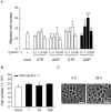
UDP dose-dependently induces intestinal cell migration, but not proliferation, via P2Y6 receptors. (A) A trans-well migration assay was performed by using a Boyden chamber. Serum-starved IEC-6 cells (5 × 10⁴) were seeded on the insert wells, and the indicated concentrations of nucleotides were added to the upper and lower wells. After 8 h, the number of cells that migrated from the upper to the lower wells was counted with the ImageJ software. (B) 1 × 10⁵ cells were seeded onto the 12-well plates and then serum-starved for 24 h after becoming adherent. After 24 h of incubation with 1–100 μmol·L−1 UDP, the cell number was counted with a cytometer. (C) Cell morphology before (left) and 24 h after stimulation with UDP (100 μmol·L−1) (right). The data are expressed as means ± SEM from at least four independent experiments. *P < 0.05, **P < 0.01 as compared with the control.
Figure 5
Figure 5
UDP did not increase [Ca2+]i in IEC-6 cells. (A) Representative tracing of the change in fluorescence ratio (F340/F380) in IEC-6 cells treated with UDP (10 μmol·L−1) followed by ATP (100 μmol·L−1). (B) Analytical data of changes in [Ca2+]i (AUC: 0–1 min) treated with UDP (10, 100 μmol·L−1) and ATP (100 μmol·L−1). Data are expressed as means ± SEM from three independent experiments.
Figure 6
Figure 6
UDP/P2Y6-induced cell migration is mediated by a TGF-β-dependent pathway. (A) Confluent IEC-6 cells were incubated with an ALK5Inhibitor (ALK5i) (a TGF-β receptor blocker: 10 μmol·L−1) or AG1478 (an EGF receptor blocker: 100 nmol·L−1) for 30 min. The monolayer was photographed immediately after scratch formation (0 h) and 8 h after scratch formation, with or without 100 μmol·L−1 UDP or TGF-β (10 ng·mL−1). Migration was evaluated as a percentage by counting the number of cells across the wounded area. Values are expressed as means ± SEM from four independent experiments. **P < 0.01 as compared with the control. ##P < 0.01 as compared with 100 nmol·L−1 AG1478. ##P < 0.01 as compared with 100 μmol·L−1 UDP. $$P < 0.01 as compared with 10 ng·mL−1 TGF-β. (B) Monolayer of IEC-6 cells was stimulated by 100 μmol·L−1 UDP or cell wounding for 4 h. TGF-β or TGF-α mRNA expression levels are expressed as ratios to GAPDH mRNA. Values are expressed as mean ± SEM from four independent experiments. **P < 0.01 as compared with resting cells. (C) A monolayer of IEC-6 cells was stimulated by 100 μmol·L−1 UDP or cell wounding, and the samples were collected after 6 h. TGF-β levels were measured with elisa. Values are expressed as mean ± SEM from four independent experiments. **P < 0.01 as compared with resting cells. #P < 0.05 as compared with UDP (100 μmol·L−1).
Figure 7
Figure 7
P2Y6 receptor expression is increased by UDP/TGF-β signalling. Confluent IEC-6 cells were incubated with or without ALK5i (a TGF-β receptor blocker: 10 μmol·L−1) or MRS2578 (a P2Y6 antagonist: 1 μmol·L−1) for 30 min. Cells were then stimulated with UDP (100 μmol·L−1), TGF-β (10 ng·mL−1) or scratching. After 4 h, the mRNA of the cells was extracted and translated into cDNA. The mRNA expression level is expressed as a percentage of the GAPDH mRNA level. Values are expressed as means ± SEM from four independent experiments. **P < 0.01 as compared with resting cells. ##P < 0.01 as compared with wounded cells. §§P < 0.01 as compared with UDP (100 μmol·L−1).
Figure 8
Figure 8
Proposed pathway for the cell restitution induced by UDP/TGF-β signalling. UDP released from wounded cells affects neighbouring cells to induce de novo synthesis of TGF-β through activation of the P2Y6 receptor. Up-regulated P2Y6 receptors promote UDP-induced responses (positive-feedback loop). UDP and TGF-β cooperate with each other to induce migration and contribute to the intestinal epithelial wound healing.

Similar articles

Cited by

References

    1. Abbracchio MP, Burnstock G, Boeynaems JM, Barnard EA, Boyer JL, Kennedy C, et al. International Union of Pharmacology LVIII: update on the P2Y G protein-coupled nucleotide receptors: from molecular mechanisms and pathophysiology to therapy. Pharmacol Rev. 2006;58:281–341. - PMC - PubMed
    1. Alexander SPH, Mathie A, Peters JA. Guide to receptors and channels (GRAC) Br J Pharmacol. (5th edn) 2011;164(Suppl 1):S1–324. - PMC - PubMed
    1. Bar I, Guns PJ, Metallo J, Cammarata D, Wilkin F, Boeynams JM, et al. Knockout mice reveal a role for P2Y6 receptor in macrophages, endothelial cells, and vascular smooth muscle cells. Mol Pharmacol. 2008;74:777–784. - PubMed
    1. Blikslager AT, Moeser AJ, Gookin JL, Jones SL, Odle J. Restoration of barrier function in injured intestinal mucosa. Physiol Rev. 2007;87:545–564. - PubMed
    1. Brinson AE, Harden TK. Differential regulation of the uridine nucleotide-activated P2Y4 and P2Y6 receptors. SER-333 and SER-334 in the carboxyl terminus are involved in agonist-dependent phosphorylation desensitization and internalization of the P2Y4 receptor. J Biol Chem. 2001;276:11939–11948. - PubMed

Publication types

MeSH terms

Substances

LinkOut - more resources