. 2013 Dec;33(12):1897-908.
doi: 10.1038/jcbfm.2013.144. Epub 2013 Aug 14.

Neuroprotective effects of geranylgeranylacetone in experimental traumatic brain injury

Affiliations

Neuroprotective effects of geranylgeranylacetone in experimental traumatic brain injury

Zaorui Zhao et al. J Cereb Blood Flow Metab. 2013 Dec.

Abstract

Geranylgeranylacetone (GGA) is an inducer of heat-shock protein 70 (HSP70) that has been used clinically for many years as an antiulcer treatment. It is centrally active after oral administration and is neuroprotective in experimental brain ischemia/stroke models. We examined the effects of single oral GGA before treatment (800 mg/kg, 48 hours before trauma) or after treatment (800 mg/kg, 3 hours after trauma) on long-term functional recovery and histologic outcomes after moderate-level controlled cortical impact, an experimental traumatic brain injury (TBI) model in mice. The GGA pretreatment increased the number of HSP70(+) cells and attenuated posttraumatic α-fodrin cleavage, a marker of apoptotic cell death. It also improved sensorimotor performance on a beam walk task; enhanced recovery of cognitive/affective function in the Morris water maze, novel object recognition, and tail-suspension tests; and improved outcomes using a composite neuroscore. Furthermore, GGA pretreatment reduced the lesion size and neuronal loss in the hippocampus, cortex, and thalamus, and decreased microglial activation in the cortex when compared with vehicle-treated TBI controls. Notably, GGA was also effective in a posttreatment paradigm, showing significant improvements in sensorimotor function, and reducing cortical neuronal loss. Given these neuroprotective actions and considering its longstanding clinical use, GGA should be considered for the clinical treatment of TBI.


Figures
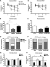
Figure 1
Figure 1
Geranylgeranylacetone (GGA) pretreatment improves cognitive function assessed by the standard Morris water maze (sMWM) and novel object recognition tests. (A) Escape latency. A significant difference was detected between the sham-injured and vehicle (Veh) traumatic brain injury (TBI) groups at day 2 (*P<0.05), day 3 (*P<0.05), and day 4 (***P<0.001). The GGA pretreatment attenuated spatial learning and memory deficit caused by TBI, which was shown by a significant decreased latency at day 4 as compared with vehicle group (#P<0.05). CCI, controlled cortical impact. (B) Probe test. The GGA-pretreated mice spent significant more time in the target quadrant than vehicle TBI mice (#P<0.05). Significant differences were also observed between sham-injured groups and vehicle TBI mice (**P<0.01). (C) Search strategy was examined on each of the four trials on acquisition day 4. Search strategy in the sMWM showed good separation between sham-injured, vehicle TBI, and GGA-pretreated group (χ2=265.3; P<0.001). (D) Reversal MWM (rMWM) escape latency. Significant differences were not detected between sham-injured, vehicle TBI, and GGA-pretreated groups across all training days. (E) Reversal probe test. Significant differences were not observed among these three groups. (F) Search strategy in the rMWM showed strong tendency of memory improvement in GGA-pretreated group compared with vehicle TBI group (χ2=120.96; P<0.01). (G, H) Novel object recognition test. Sham-injured, vehicle TBI, and GGA-pretreated mice spent equal time with the two identical objects during the sample phase on postinjury day (PID) 25 (dashed line at 15 seconds). At 1 hour after the sample phase, the time spent with the novel and familiar objects during the choice phase was recorded. The GGA-pretreated mice spent significantly more time with the novel object when compared with the vehicle TBI group (##P<0.05). Significant differences were also observed between sham-injured groups and vehicle TBI mice (*P<0.05). Analysis by repeated measures one-way analysis of variance (ANOVA; injury severity × day) in panels A and D and by one-way ANOVA in panels B, E, G and H) followed by post hoc adjustments using Student–Newman–Keuls test, mean±s.e.m. (C, F) were analyzed by χ2 analysis, n=6 to 10.
Figure 2
Figure 2
Geranylgeranylacetone (GGA) pretreatment improves motor function in beam walk test, reverses depressive-like behavior in the tail-suspension (TS) test, and improves overall behavioral outcomes in a composite score. (A) All animals had <10 foot faults before controlled cortical impact (CCI). Significant differences were observed between the vehicle (Veh) traumatic brain injury (TBI) and GGA-pretreated group on postinjury day (PID) 21 (#P<0.05). Significant differences were also observed between the sham-injured and vehicle TBI groups on day 1 (***P<0.001), day 3 (***P<0.001), day 7 (***P<0.001), day 14 (***P<0.001), day 21 (**P<0.01), and day 28 (**P<0.01). (B) The TS test was performed on PID 24. Significantly increased immobility times were observed in vehicle TBI group (*P<0.05) when compared with sham-injured group. The immobility time was significantly reduced in the GGA-pretreated group as compared with vehicle TBI group (##P<0.05). (C) Four behavioral indexes were converted to a composite score, using individual scales ranging from 0 (severe) to 5 (sham). The composite score was obtained by the sum of scores for the four indexes. The composite score was significantly decreased in the vehicle TBI groups (***P<0.001) as compared with sham-injured group. The GGA-pretreated group showed a significantly improved composite score as compared with vehicle group (###P<0.001). Analysis by repeated measures one-way analysis of variance (ANOVA; injury severity × day) in (A) and by one-way ANOVA in (B, C) followed by post hoc adjustments using Student–Newman–Keuls test, mean±s.e.m., n=6 to 10.
Figure 3
Figure 3
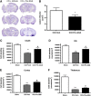
Geranylgeranylacetone (GGA) pretreatment reduces lesion size and attenuates neuronal cell loss in the Cornu Ammonis-2/3 (CA2/3) and dentate gyrus (DG) subregions of hippocampus, cortex, and thalamus after traumatic brain injury (TBI). Lesion volume was quantified using the Cavalieri method. Unbiased stereological assessment of lesion volume at 28 days after TBI was performed on cresyl violet–stained brain section. (A) Representative images from each group are shown. CCI, controlled cortical impact. Veh, vehicle. (B) Significant reduction of lesion volume was observed in pre-GGA-treated group (*P<0.05) when compared with the sham-injured group. Stereological assessment of neuronal cell on postinjury day (PID) 28 was performed on cresyl violet–stained sections in the CA1, CA2/3, and DG subregions of the hippocampus, cortex, and thalamus. (C, D, E, F). Significant differences of neuronal density were observed between sham-injured and vehicle TBI groups in the CA2/3 (**P<0.01) and DG (*P<0.01) subregions of the hippocampus, as well as the cortex (***P<0.001) and thalamus (***P<0.001). The GGA pretreatment significantly increased neuronal density in the CA2/3 (#P<0.05), DG (#P<0.05), cortex (#P<0.05), and thalamus (#P<0.05) compared with vehicle TBI group. Analysis by one-tailed paired Student's t-test in panel B and by one-way analysis of variance (ANOVA) in panels C, D, E and F followed by Student–Newman–Keuls test, mean±s.e.m., n=6 to 8.
Figure 4
Figure 4
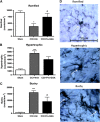
Geranylgeranylacetone (GGA) pretreatment attenuates microglia activation after traumatic brain injury (TBI). Unbiased stereological quantification of microglial cell number and activation status in the cortex at 28 days after TBI. Resting ramified (A) as well as activating hypertrophic (B) and bushy (C) microglia phenotypes were analyzed. Vehicle (Veh) TBI significantly reduced the densities of ramified microglia (*P<0.05) and increased the densities of hypertrophic (***P<0.001) and bushy microglia (***P<0.001) compared with sham-injured group. The GGA pretreatment significantly increased the densities of ramified microglia (#P<0.05) and attenuated activation of bushy microglia (#P<0.05) compared with vehicle TBI group. (D) Representative immunohistochemical images of ramified, hypertrophic, and bushy microglia illustrate the different morphologic features of each microglial phenotype. Analysis by one-way analysis of variance (ANOVA) followed by Student–Newman–Keuls test, mean±s.e.m., n=6.
Figure 5
Figure 5
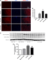
Geranylgeranylacetone (GGA) pretreatment upregulates heat-shock protein 70 (HSP70) expression and inhibits fodrin cleavage after traumatic brain injury (TBI). (A) Representative photomicrographs of ipsilateral cortex 6 hours after TBI with or without pre-GGA/post-GGA treatment, immunolabeled for HSP70 (red) and 4,6-diamino-2-phenylindole (DAPI, chromatin stain) (blue). Mice subjected to TBI showed increased expression of HSP70 compared with sham-injured mice. Mice subjected to TBI after GGA pretreatment showed higher level of expression of HSP70 compared with injured mice that received vehicle treatment. (B) The percentage of HSP70+ cells was significantly increased in the vehicle (Veh) TBI group as compared with the sham-injured mice (**P<0.01). The GGA pretreatment further increased expression of HSP70+ cells as compared with vehicle TBI group (#P<0.05). The number of HSP70+ cell was not significantly different in the GGA posttreatment group as compared with vehicle TBI group. CCI, controlled cortical impact. (C, D) Vehicle TBI significantly increased the 145/150 kDa fodrin cleavage products (***P<0.001) in the ipsilateral cortical tissue measured by western blot. The GGA pretreatment significantly attenuated expressions of 145/150 kDa fodrin cleavage products (##P<0.01) compared with vehicle TBI group. Analysis by one-way analysis of variance (ANOVA) followed by Student–Newman–Keuls test, mean±s.e.m., n=4 to 5 for immunohistochemistry and n=6 for western blot.
Figure 6
Figure 6
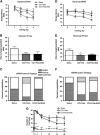
Geranylgeranylacetone (GGA) posttreatment improves motor function (beam walk test) after traumatic brain injury (TBI). (A) A significant difference was detected between the sham and vehicle groups at day 3 (*P<0.05) and day 4 (*P<0.05). The GGA-posttreated group failed to attenuate spatial learning and memory deficit caused by TBI compared with the vehicle (Veh) TBI group, as shown by no significant differences in escape latency (P>0.05). CCI, controlled cortical impact. (B) Probe test. Significant differences were not observed among these three groups. (C) Search strategy was examined on each of the four trials on acquisition day 4. Search strategy in the standard Morris water maze (sMWM) showed good separation between GGA posttreatment and vehicle TBI group (χ2=292.3; P<0.001). (D) Reversal MWM escape latency. Significant differences were not detected between sham-injured, vehicle TBI, and GGA-pretreated groups across all training days. (E) Reversal probe test. Significant differences were not observed between GGA posttreatment group and vehicle TBI groups. (F) Search strategy in the reversal MWM (rMWM) did not show memory improvement in the GGA-pretreated group compared with vehicle TBI group. (G) All animals had <10 foot faults before CCI. Significant differences were observed between the vehicle TBI and GGA posttreatment group on postinjury day (PID) 21 (#P<0.05). Significant differences were also observed between the sham-injured and vehicle TBI groups on day 1 (***P<0.001), day 3 (***P<0.001), day 7 (***P<0.001), day 14 (***P<0.001), day 21 (***P<0.001), and day 28 (***P<0.001). Analysis by repeated measures one-way analysis of variance (ANOVA; injury severity × day) in panels A, D and G and by one-way ANOVA in panels B and E followed by post hoc adjustments using Student–Newman–Keuls test, mean±s.e.m. (C, F) was analyzed by χ2 analysis; n=6 to 7.
Figure 7
Figure 7
Geranylgeranylacetone (GGA) posttreatment reduces cortical lesion size and attenuates neuronal cell loss in the cortex after traumatic brain injury (TBI). Unbiased stereological assessment of lesion volume at 28 days after TBI was performed on cresyl violet–stained brain sections. (A) Representative images from each group are shown. CCI, controlled cortical impact. (B) A significant reduction of lesion volume was observed in GGA posttreatment group (*P<0.05) when compared with vehicle (Veh) TBI group. Stereological assessment of neuronal densities on postinjury day (PID) 28 was performed on cresyl violet–stained sections. (C) Neuronal densities in the cortex were significantly reduced in vehicle TBI group compared with sham-injured group (***P<0.001). The GGA posttreatment attenuated neuronal cell loss in the cortex compared with vehicle TBI group (#P<0.05). Analysis by one-tailed paired Student's t-test in panel B and by one-way analysis of variance (ANOVA) followed by Student–Newman–Keuls test in panel C, mean±s.e.m., n=6.

Similar articles

Cited by

References

    1. Faul MXL, Wald MM, Coronado VG.Traumatic brain injury in the United States: emergency department visits, hospitalizations, and deathsIn: (ed). Centers for Disease Control and Prevention National Center for Injury Prevention and Control Atlanta; 201013
    1. Bramlett HM, Dietrich WD. Progressive damage after brain and spinal cord injury: pathomechanisms and treatment strategies. Prog Brain Res. 2007;161:125–141. - PubMed
    1. Loane DJ, Faden AI. Neuroprotection for traumatic brain injury: translational challenges and emerging therapeutic strategies. Trends Pharmacol Sci. 2010;31:596–604. - PMC - PubMed
    1. Evans CG, Chang L, Gestwicki JE. Heat shock protein 70 (hsp70) as an emerging drug target. J Med Chem. 2010;53:4585–4602. - PMC - PubMed
    1. Yenari MA, Fink SL, Sun GH, Chang LK, Patel MK, Kunis DM, et al. Gene therapy with HSP72 is neuroprotective in rat models of stroke and epilepsy. Ann Neurol. 1998;44:584–591. - PubMed

Publication types

MeSH terms