

Review
doi: 10.1038/nrm3643.
Epub 2013 Aug 14.
Restarting life: fertilization and the transition from meiosis to mitosis
Affiliations
- PMID: 23942453
- PMCID: PMC4021448
- DOI: 10.1038/nrm3643
Item in Clipboard
Review
Restarting life: fertilization and the transition from meiosis to mitosis
Nat Rev Mol Cell Biol.
2013 Sep.
Abstract
Fertilization triggers a complex cellular programme that transforms two highly specialized meiotic germ cells, the oocyte and the sperm, into a totipotent mitotic embryo. Linkages between sister chromatids are remodelled to support the switch from reductional meiotic to equational mitotic divisions; the centrosome, which is absent from the egg, is reintroduced; cell division shifts from being extremely asymmetric to symmetric; genomic imprinting is selectively erased and re-established; and protein expression shifts from translational control to transcriptional control. Recent work has started to reveal how this remarkable transition from meiosis to mitosis is achieved.
Figures
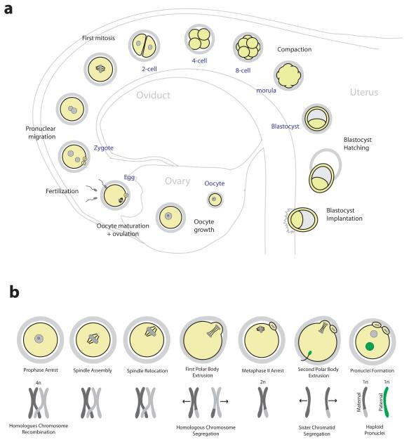
a | An overview of preimplantation development. Following ovulation, eggs are fertilized in the oviduct to form the zygote. After several mitotic divisions, the embryo undergoes compaction to form the morula. A fluid-filled cavity develops inside the embryo forming the blastocyst, which hatches from the zona pellucida to implant into the uterine wall. b | Stages of oocyte maturation and corresponding chromosome configuration. Following nuclear envelope breakdown the spindle assembles, relocates to the oocyte surface and segregates half the homologous chromosomes into a polar body. A spindle assembles around the remaining chromosomes and the egg arrests in metaphase II awaiting fertilization. Upon sperm binding, the egg segregates half of the sister chromatids into a second polar body. The resulting zygote contains haploid pronuclei from the mother and the father.
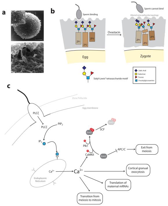
a | Scanning electron microscopy of a human egg surrounded by the zona pellucida (top). A human sperm can be seen approaching the surface of the zona pellucida (bottom). b | Schematic of sperm-egg binding. The zona pellucida (grey) consists of glycoproteins that are modified with oligosaccharide chains. Sperm can bind to the zona pellucida of unfertilized eggs. Following fertilization, cleavage of ZP2 by ovastacin renders the zona pellucida non-permissive for sperm binding. c | Following sperm-egg membrane fusion, sperm-derived PLCζ promotes the production of IP3, which binds to receptors on the endoplasmic reticulum causing release of Ca2+. Ca2+ activates CaMKII which, together with Plk1, phosphorylates Emi2 promoting its targeting for destruction by the SCF ubiquitin ligase. This releases Emi2-mediated inhibition of the APC/C, allowing the APC/C to promote exit from meiosis. The rise in intracellular Ca2+ also triggers cortical granule exocytosis, translation of maternal mRNAs and ultimately the transition from meiosis to mitosis. Images in part a are reproduced, with permission, from REF. © (1998) Oxford Journals.
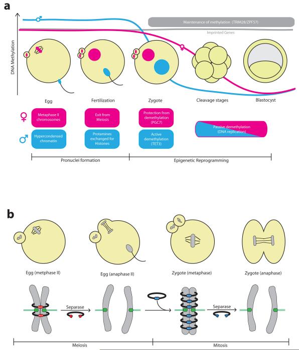
a | Schematic of chromatin remodelling during fertilization and early embryo development. Following fertilization, the egg exits from meiosis and assembles a haploid pronucleus. At the same time the sperm genome, which enters the egg in a highly compact state, undergoes decompaction. Sperm protamines are replaced by histones and the male haploid pronucleus is formed. Maternal and paternal genomes are methylated during gametogenesis, including parental-specific methylation at imprinted genes. The sperm pronucleus undergoes rapid active demethylation in the zygote mediated by Tet3. The maternal pronucleus is largely protected from this active demethylation by the action of PGC7. Following pronuclear fusion, the zygotic genome undergoes passive demethylation during the blastomeres cleavage stages, reaching a minimum in the blastocyst. The maintenance of methylation at imprinted regions depends on the ZPF57/TRIM28 DNA-binding complex. b | Sister chromatids in the egg are held together by Rec8-containing cohesin complex. Rec8 cleavage by separase triggers second polar body extrusion following fertilization. Scc1-containing cohesin complexes are loaded onto chromosomes immediately in the zygote, and Scc1 cleavage by separase triggers sister chromatid segregation during the first mitotic division.
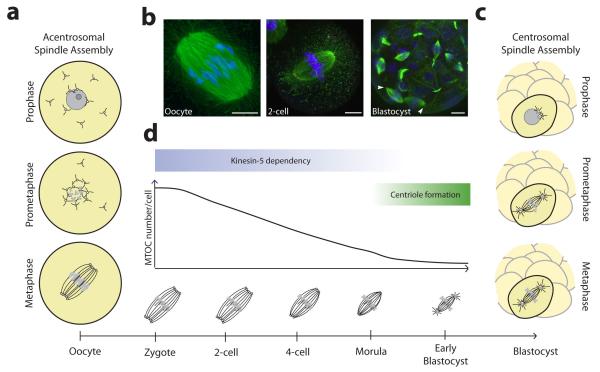
a | Acentrosomal spindle assembly in mouse oocytes is driven by multiple acentriolar microtubules organising centres (aMTOCs) that self-assemble a bipolar spindle. b | An acentrosomal spindle with unfocused poles in the mouse oocyte (left) and 2-cell embryo (middle). Cells in the blastocyst assemble from centrosomes and have focused spindle poles as indicated by arrow heads (right). Scale bars: 10 μm. c | Centrosomal spindle assembly involves the separation of two centrosomes that act as MTOCs and form the spindle poles. d | The transition from acentrosomal to centrosomal spindle assembly during mouse development is a gradual process that is completed by the blastocyst stage. Image of oocyte in part b is courtesy of the author D.Clift. Images of 2-cell embryo and blastocyst in part b are reproduced, with permission, from REF. © (2012) Rockefeller University Press.
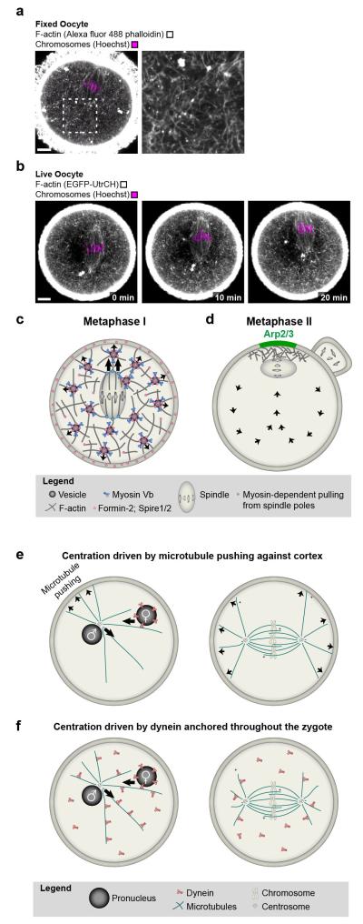
a | A cytoplasmic F-actin network mediates asymmetric spindle positioning during meiosis I in mouse oocytes. F-actin (grey; Alexa fluor 488 phalloidin) and chromosomes (magenta; Hoechst) in fixed mouse oocyte. Scale bar: 10 μm. b | Autofluorescence (green) around spindle region and chromosomes (magenta; Hoechst) in fixed mouse oocyte. Scale bar: 10 μm. c | F-actin (grey; EGFP-UtrCH) and chromosomes (magenta; Hoechst) in live oocyte during asymmetric spindle positioning. Scale bar: 10 μm. d | Model for vesicle-actin network mediated asymmetric spindle positioning during the first meiotic division e | Model for how the Arp2/3-complex helps to maintain the metaphase II spindle in cortical proximity while the egg awaits fertilization. f | Model for centration of pronuclei and first mitotic spindle by pushing of astral microtubules against the cortex. g | Model for centration of pronuclei and first mitotic spindle driven by dynein that is anchored throughout the cytoplasm.
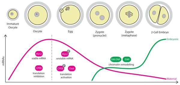
During the growing phase, oocytes accumulate large amounts of mRNA necessary to drive the transcriptionally silent phase of oocyte maturation and fertilization. Global mRNA stability in the oocyte is ensured by the RNA-binding protein Mys2 and many mRNAs are held in a translationally-repressed state by binding of CPEB to their 3′UTRs. Maternal mRNAs begin to be translated and/or degraded during oocyte maturation, which involves the phosphorylation of Mys2 and CPEB, and mRNA levels reach a minimum at the 2-cell stage. Transcription from the embryonic genome is initiated firstly in the zygote followed by a robust activation of transcription in the 2-cell embryo, and involves extensive chromatin remodelling.
References
-
- Johnson J, Canning J, Kaneko T, Pru JK, Tilly JL. Germline stem cells and follicular renewal in the postnatal mammalian ovary. Nature. 2004;428:145–150. - PubMed
-
- Li R, Albertini DF. The road to maturation: somatic cell interaction and self-organization of the mammalian oocyte. Nat Rev Mol Cell Biol. 2013;14:141–152. - PubMed
-
- Wassarman PM, Litscher ES. Mammalian fertilization: the egg’s multifunctional zona pellucida. Int J Dev Biol. 2008;52:665–76. - PubMed
-
- Bleil JD, Wassarman PM. Mammalian sperm-egg interaction: identification of a glycoprotein in mouse egg zonae pellucidae possessing receptor activity for sperm. Cell. (1980;20:873–82. - PubMed
Publication types
MeSH terms
Grants and funding
LinkOut - more resources
Full Text Sources
Other Literature Sources
