Review
. 2013 Sep;14(9):609-25.
doi: 10.1038/nrn3381. Epub 2013 Aug 14.

The brain reward circuitry in mood disorders

Affiliations
Review

The brain reward circuitry in mood disorders

Scott J Russo et al. Nat Rev Neurosci. 2013 Sep.

Erratum in

  • Nat Rev Neurosci. 2013 Oct;14(10):736

Abstract

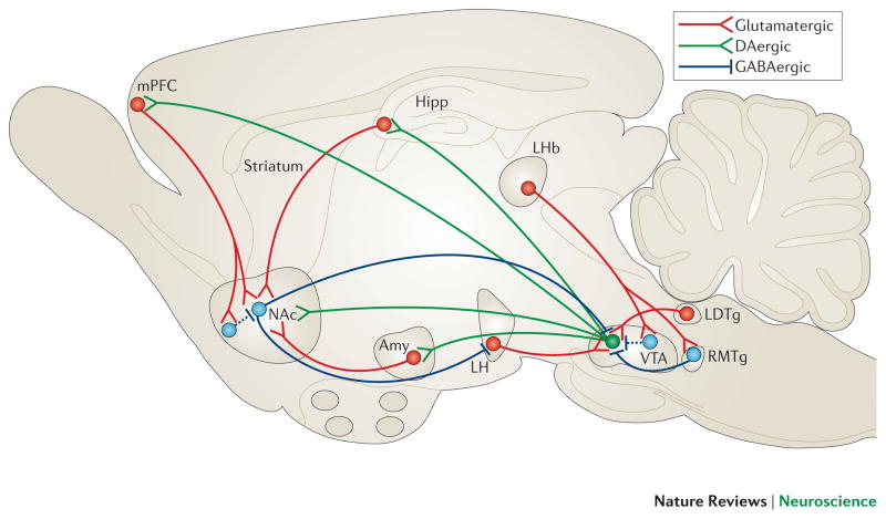
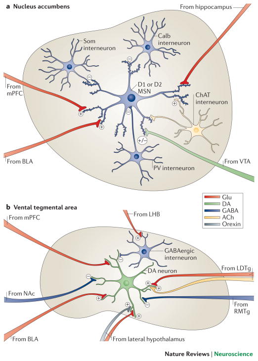
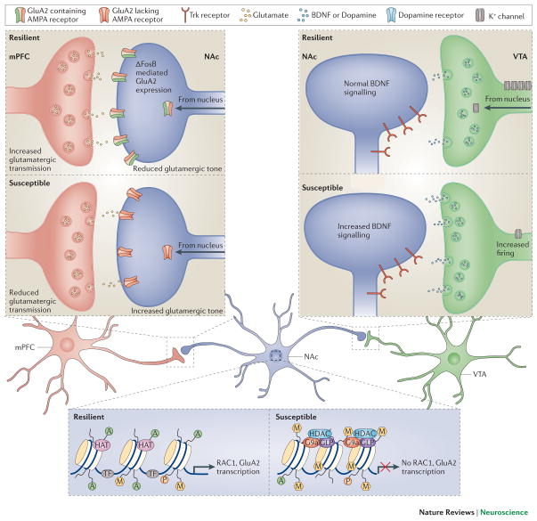
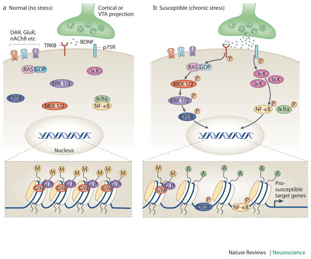
Mood disorders are common and debilitating conditions characterized in part by profound deficits in reward-related behavioural domains. A recent literature has identified important structural and functional alterations within the brain's reward circuitry--particularly in the ventral tegmental area-nucleus accumbens pathway--that are associated with symptoms such as anhedonia and aberrant reward-associated perception and memory. This Review synthesizes recent data from human and rodent studies from which emerges a circuit-level framework for understanding reward deficits in depression. We also discuss some of the molecular and cellular underpinnings of this framework, ranging from adaptations in glutamatergic synapses and neurotrophic factors to transcriptional and epigenetic mechanisms.


Figures

Figure 1
Figure 1. VTA-NAc reward circuit
A simplified schematic of the major dopaminergic, glutamatergic and GABAergic connections to and from the ventral tegmental area (VTA) and nucleus accumbens (NAc) (dopaminergic=green; glutamatergic=red; and GABAergic=blue) in the rodent brain. The primary reward circuit includes dopaminergic projections from the VTA to the NAc, which release dopamine in response to reward- (and in some cases aversion-) related stimuli. There are also GABAergic projections from the NAc to the VTA; projections through the direct pathway (mediated by D1-type medium spiny neurons [MSNs]) directly innervate the VTA, whereas projections through the indirect pathway (mediated by D2-type MSNs) innervate the VTA via intervening GABAergic neurons in ventral pallidum (not shown). The NAc also contains numerous types of interneurons (Figure 2). The NAc receives dense innervation from glutamatergic monosynaptic circuits from the medial prefrontal cortex (mPFC), hippocampus (Hipp) and amygdala (Amy), among other regions. The VTA receives such inputs from amygdala, lateral dorsal tegmentum (LDTg), lateral habenula (LHb) and lateral hypothalamus (LH), among others. These various glutamatergic inputs control aspects of reward-related perception and memory. The glutamatergic circuit from LH to VTA is also mediated by orexin (not shown). Greater details of these monosynaptic circuits for NAc and VTA are shown in Figure 2.
Figure 2
Figure 2. Local microcircuitry of the NAc and VTA
a | A close-up view detailing the presynaptic inputs onto D1- and D2-type GABAergic medium spiny neurons (MSNs) and onto several types of interneurons within the nucleus accumbens (NAc). The latter include GABAergic interneurons that express calretinin, (Calr), parvalbumin (PV), somatostatin (SOM) or calbindin (Calb), and large cholinergic interneurons that express choline acetyltransferase (ChAT). Glutamatergic neurons (Glu) from the medial prefrontal cortex (mPFC), hippocampus (Hipp) and basolateral amygdala (BLA) release glutamate onto spine synapses to provide excitatory signals to GABAergic MSN projection neurons. These excitatory inputs also synapse directly onto the GABAergic and cholinergic interneurons that modulate MSNs (not shown). D1-and D2-type MSNs also receive signals from dopamine though shaft or spine neck synapses (brown). The figure does not depict possible differences in glutamatergic innervation of D1-type versus D2-type MSNs, which are only now beginning to be explored. b | A close-up view detailing the presynaptic inputs onto ventral tegmental area (VTA) dopamine neurons and local GABAergic interneurons in the VTA. Glutamatergic neurons (Glu) from the amygdala (BLA), medial prefrontal cortex (mPFC) and lateral dorsal tegmentum (LDTg) synapse directly onto VTA dopamine neurons. In contrast, glutamatergic neurons from lateral habenula (LHb) synapse directly onto inhibitory GABAergic neurons in the rostromedial tegmentum (RMTg) or VTA proper, which then inhibit dopamine neurons and promote aversion. Dopamine neurons receive direct excitatory inputs from peptidergic (e.g., orexinergic) or glutamatergic neurons in lateral hypothalamus (LH), which increase dopamine release and promote reward. Although GABAergic projections from the NAc are shown innervating VTA dopamine neurons, much of this innervation is on VTA GABAergic neurons, whereas much of the GABAergic innervation of the dopamine neurons is indirect, via GABAergic neurons in ventral pallidum (not shown). Color code: dopaminergic=green; glutamatergic=red; GABAergic=blue; peptidergic=yellow; and cholinergic=orange.
Figure 3
Figure 3. Molecular mechanisms controlling depression-related circuit plasticity
Examples of maladaptive molecular processes that confer susceptibility to chronic social defeat stress are shown. a. In the mPFC (infralimbic and paralimbic cortex), susceptible animals display molecular evidence of decreased neural activity, which can be targeted through optogenetic stimulation with ChR2 to promote resilience. Using a glutamate cell specific promoter, stimulation of ChR2 in glutamatergic terminals in the NAc reverses stress-induced social avoidance behavior. b. VTA dopamine neurons of susceptible animals show a stress-induced increase in firing rate that drives maladaptive responses to stress. The result is greater release of BDNF and dopamine from terminals in the NAc to promote depression-like behavior. The increased excitability of VTA dopamine neurons of susceptible animals results from increased cationic current, which is completely compensated for in resilient animals via induction of K+ channels, driving neuronal firing back to normal levels. c. In the NAc, susceptibility is associated with increased glutamatergic responses on medium spiny neurons. The presynaptic input responsible for the increased glutamate tone is not known. Susceptible mice exhibit reduced Rac1 gene transcription in the NAc, which is associated with reduced histone pan-acetylation (acH3) and increased lysine 27 methylation (me3K27), leads to reorganization of the actin cytoskeleton and increased excitatory synapse number and function. In addition, there appears to be ΔFosB-mediated transcriptional events (not shown), which control the types of glutamate receptors functioning at these synapses among many other changes. Susceptible animals have lower levels of GluA2 (also known as Gria2), a Ca2+-impermeable AMPA glutamate receptor subunit, which in resilient mice limits overactive glutamate tone in NAc.
Figure 4
Figure 4. Enhanced vulnerability to stress via priming of BDNF signaling in the NAc
Increased vulnerability to the depression-like effects of chronic social defeat stress occurs in part via priming of BDNF signaling in the nucleus accumbens (NAc). Under control conditions (left), BDNF activation of TrkB signaling is limited. However, after some prior stimulus that increases susceptibility (e.g., repeated stress, chronic cocaine exposure; right), BDNF–TrkB signaling is increased in the NAc, causing enhanced phosphorylation and activity of several downstream-signaling mediators, including cAMP response element binding protein (CREB). This maladaptive response occurs not only through increased BDNF release into the NAc from the VTA, but also through epigenetic modifications that further prime BDNF signaling cascades. For example, chronic stress increases Ras expression in NAc of susceptible animals by decreasing G9a binding at the H-Ras1 gene promoter, causing reduced levels of repressive H3K9me2 (dimethyl-Lys9-histone H3, a major form of repressive histone methylation). Ras also appears to be a target for CREB, creating a pathological feed-forward loop that promotes CREB activation and Ras expression as well as depression-like behavior. The figure also shows the induction of another pathway downstream of BDNF, including inhibitor of kappa kinase (IKK) and nuclear factor kappa B (NFκB) — possibly downstream of the p75 BDNF receptor — in the NAc after chronic social defeat stress in susceptible animals.

References

    1. Kessler RC, Chiu WT, Demler O, Merikangas KR, Walters EE. Prevalence, severity, and comorbidity of 12-month DSM-IV disorders in the National Comorbidity Survey Replication. Arch Gen Psychiatry. 2005;62:617–27. - PMC - PubMed
    1. Culpepper L. Why do you need to move beyond first-line therapy for major depression? J Clin Psychiatry. 71(Suppl 1):4–9. - PubMed
    1. Conway KP, Compton W, Stinson FS, Grant BF. Lifetime comorbidity of DSM-IV mood and anxiety disorders and specific drug use disorders: results from the National Epidemiologic Survey on Alcohol and Related Conditions. J Clin Psychiatry. 2006;67:247–57. - PubMed
    1. Nestler EJ, Carlezon WA., Jr The mesolimbic dopamine reward circuit in depression. Biol Psychiatry. 2006;59:1151–9. - PubMed
    1. Koob GF, Le Moal M. Addiction and the brain antireward system. Annu Rev Psychol. 2008;59:29–53. - PubMed

Publication types