. 2013 Sep;140(17):3635-44.
doi: 10.1242/dev.094599.

CAF-1 promotes Notch signaling through epigenetic control of target gene expression during Drosophila development

Affiliations

CAF-1 promotes Notch signaling through epigenetic control of target gene expression during Drosophila development

Zhongsheng Yu et al. Development. 2013 Sep.

Abstract

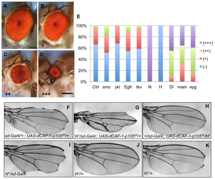
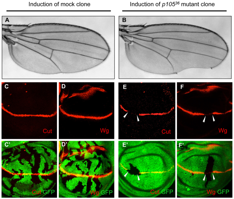
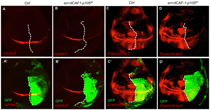
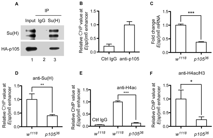
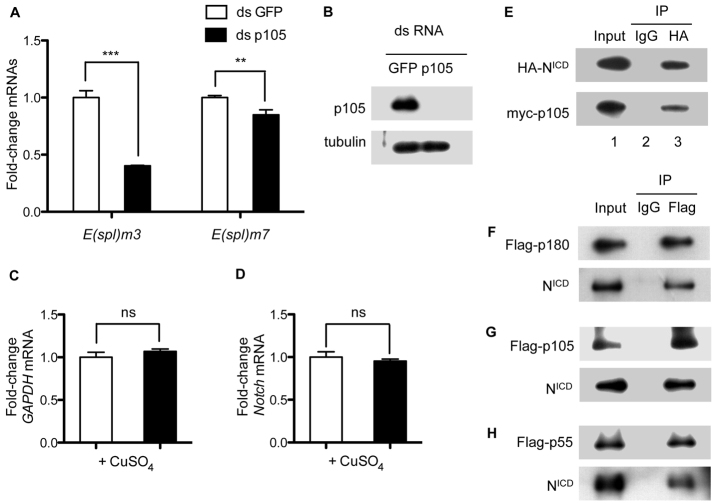
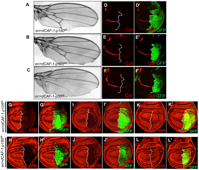
The histone chaperone CAF-1 is known for its role in DNA replication-coupled histone deposition. However, loss of function causes lethality only in higher multicellular organisms such as mice and flies, but not in unicellular organisms such as yeasts, suggesting that CAF-1 has other important functions than histone deposition during animal development. Emerging evidence indicates that CAF-1 also has a role in higher order chromatin organization and heterochromatin-mediated gene expression; it remains unclear whether CAF-1 has a role in specific signaling cascades to promote gene expression during development. Here, we report that knockdown of one of the subunits of Drosophila CAF-1, dCAF-1-p105 (Caf1-105), results in phenotypes that resemble those of, and are augmented synergistically by, mutations of Notch positive regulatory pathway components. Depletion of dCAF-1-p105 leads to abrogation of cut expression and to downregulation of other Notch target genes in wing imaginal discs. dCAF-1-p105 is associated with Suppressor of Hairless [Su(H)] and regulates its binding to the enhancer region of E(spl)mβ. The association of dCAF-1-p105 with Su(H) on chromatin establishes an active local chromatin status for transcription by maintaining a high level of histone H4 acetylation. In response to induced Notch activation, dCAF-1 associates with the Notch intracellular domain to activate the expression of Notch target genes in cultured S2 cells, manifesting the role of dCAF-1 in Notch signaling. Together, our results reveal a novel epigenetic function of dCAF-1 in promoting Notch pathway activity that regulates normal Drosophila development.

Keywords: CAF-1; Drosophila; Epigenetic regulation; H4 acetylation; Notch.


Figures

Fig. 1.
Fig. 1.
dCAF-1-p105 genetically interacts with the Notch pathway in the eye and wing. (A-E) Genetic interaction of dCAF-1-p105 and the Notch pathway in the eye. Eyes of wild-type adult flies (A) and flies that express dCAF-1-p105IR in a wild-type (B,C) or heterozygous N1 background (D) are classified into four classes according to eye size (E). Scale bar: 200 μm. (E) The penetrance and expressivity of small-eye phenotypes for flies of the different genotypes. Ctrl represents ey>dCAF-1-p105IR. smo, yki, Egfr, tkv, N, H, Dl, mam and eyg represent flies of ey>dCAF-1-p105IR in the background of one copy of the mutation for each indicated gene. (F-K) Genetic interaction of dCAF-1-p105 and the Notch pathway in the wing. (F) dCAF-1-p105 knockdown under the control of sd-Gal4 causes loss of wing margins (100%, n=50). (G) The loss of wing margin phenotype of sd>dCAF-1-p105IR is enhanced in the presence of one copy of N1 (100%, n=50; removing nearly all of the wing margin). (H) The loss of wing margin phenotype of sd>dCAF-1-p105IR is suppressed by a copy of the H1 mutation (66.1%, n=62). (J) H1/+ shows a mild Notch gain-of-function phenotype with an interruption of longitudinal vein 5 (LV5) in the distal part (100%, n=50). (I,K) Note that the notched wings of N1/+ can apparently also be enhanced in combination with sd-Gal4 (greater loss of wing margins), but is further enhanced when dCAF-1-p105IR is introduced (G). For I, the penetrance is 100%, referring to a phenotype that, on average, removes nearly 50% of the total wing margin (n=50), and for K the penetrance is 11.8%, referring to a weak phenotype that usually exhibits a single distal notch of the wing (N=110; supplementary material Fig. S1B).
Fig. 2.
Fig. 2.
Generation and molecular identification of the dCAF-1-p105 mutant. (A) Genomic organization of the dCAF-1-p105 locus and one of its neighboring genes, with the p[EP]Caf1-105G2212 P-element insertion site (white triangle) and the fragment deleted in p10536 (dashed line) indicated. Black bars indicate the coding regions of dCAF-1-p105 and CG11777; white bars indicate the 5′ and 3′ UTRs. PCR primers used to amplify fragments containing the deletions are indicated (forward and reverse arrows). (B) PCR analysis of genomic DNA of p10536 heterozygous animals shows a short, 638 bp fragment that is not present in the wild type in addition to the 3118 bp wild-type fragment. Sequencing (not shown) indicated that the deletion encompasses two regions of 2408 bp and 72 bp (see A). (C) RT-PCR illustrating that no transcripts of dCAF-1-p105 can be detected. Rp49 (RpL32) provides an internal control. (D) Comparison of the larvae of w1118 and p10536 at 24, 48 and 72 hours after egg deposition (AED), showing a developmental delay in the dCAF-1-p105 mutant. (E) Ubiquitous expression of the transgene UAS-HA-dCAF-1-p105 under the control of da-Gal4 rescues the lethality of p10536 mutants. The rescued flies did not exhibit any detectable defects compared with the wild type.
Fig. 3.
Fig. 3.
dCAF-1-p105 is required for the proper expression of Notch target genes cut and wg. (A,B) Induction of dCAF-1-p105 (p10536) mutant clones leads to a wing notch, whereas induction of mock clones leads to wild-type wings. Mock clones and p10536 mutant clones were induced at the second instar larval stage at 38°C for 1 hour. The genotype of flies with mock clones is hs-Flp/+; FRT42D, Minute, ubi-GFP/FRT42D. The genotype of flies with p10536 mutant clones is hs-Flp/+; FRT42D, Minute, ubi-GFP/FRT42D, p10536. (C-F′) In dCAF-1-p105 mutant clones, the expression of Cut (E,E′, GFP-negative area, arrowheads) and Wg (F,F′, GFP-negative area, arrowheads) is abolished in a cell-autonomous manner, whereas in the mock clones the expression of both Cut (C,C′) and Wg (D,D′) is unaffected. Wing discs were dissected for immunostaining 3 days after clonal induction.
Fig. 4.
Fig. 4.
Notch target genes are regulated by dCAF-1-p105 at the transcription level. (A-B′) The expression of cut-lacZ is downregulated upon depletion of dCAF-1-p105. In control discs (A,A′) cut-lacZ is expressed normally along the D/V boundary (dashed line) in both the anterior and posterior regions. Knocking down dCAF-1-p105 under the control of en-Gal4 (B,B′) substantially reduces cut-lacZ expression in the posterior region. Genotypes: (A,A′) UAS-Dicer2/+; en-Gal4, UAS-GFP/+; cut-lacZ/+; (B,B′) UAS-Dicer2/+; en-Gal4, UAS-GFP/UAS-dCAF-1-p105IR; cut-lacZ/+. UAS-Dicer2 was used to enhance RNAi efficiency. (C-D′) The expression of E(spl)mβ-lacZ is downregulated in the dCAF-1-p105-depleted area. In control discs (C,C′) E(spl)mβ-lacZ is expressed normally in the entire wing disc. Knocking down of dCAF-1-p105 under the control of en-Gal4 (D,D′) substantially reduces E(spl)mβ-lacZ expression. The genotype of Ctrl is UAS-Dicer2/+; en-Gal4, UAS-GFP/E(spl)mβ-lacZ, and en>dCAF-1-p105IR is UAS-Dicer2/+; en-Gal4, UAS-GFP/E(spl)mβ-lacZ, UAS-dCAF-1-p105IR.
Fig. 5.
Fig. 5.
dCAF-1-p105 associates with Su(H) and maintains the local histone H4 acetylation level. (A) Su(H) associates with HA-tagged dCAF-1-p105 in vivo. Total protein extracts were prepared from Drosophila embryos (0-12 hours AED) with ubiquitous expression of HA-tagged dCAF-1-p105 under the control of da-Gal4. Input (lane 1) represents 5% of the extracts that were used for immunoprecipitation (IP) with IgG (control) or Su(H) antibody. Antibodies used for western blot detection are indicated on the left. (B) ChIP assay shows that dCAF-1-p105 preferentially occupies the enhancer of the Notch target gene E(spl)mβ. The y-axis indicates relative protein occupancy values at the enhancer region as detected by qPCR after ChIP. (C) qPCR shows that the level of E(spl)mβ mRNA in p10536 mutants is significantly lower than in w1118 animals. (D) ChIP assay showing a decrease in Su(H) abundance at the E(spl)mβ enhancer in the absence of dCAF-1-p105. (E,F) ChIP assay shows a decrease in acetylated histone H4 (H4ac) at the E(spl)mβ enhancer in the absence of dCAF-1-p105. H4ac at the E(spl)mβ enhancer region is maintained at a significantly higher level in the wild type than in p10536 mutants. The abundance of H4ac relative to total histone H3 is higher in wild type than in p10536 mutants. The mean of three independent experiments after normalization is shown; error bars indicate s.e.m. *P<0.05, **P<0.001, ***P<0.0001; Student’s t-test.
Fig. 6.
Fig. 6.
dCAF-1 physically interacts with NICD and regulates Notch target gene expression in S2 cells upon artificial Notch signal induction. (A) qPCR to monitor mRNA levels of E(spl)m3 and E(spl)m7 when dCAF-1-p105 was knocked down in S2 cells with artificial induction of Notch signaling. E(spl)m3 and E(spl)m7 are highly expressed when Notch signaling is induced by CuSO4 in normal S2 cells (supplementary material Fig. S4B,C). A reduction in E(spl)m3 and E(spl)m7 transcription was observed upon dCAF-1-p105 RNAi manipulation (black bars in A). GFP dsRNA (white bars in A) was used to control for RNAi specificity. (B) dCAF-1-p105 mRNA was abolished by dsp105 treatment but not in the dsGFP treatment control. (C,D) In both groups of dsRNA treatment, Notch and Gapdh mRNA levels were not significantly altered. Error bars indicate s.e.m. ns, not significant; **P<0.001, ***P<0.0001; Student’s t-test. (E) HA-tagged NICD is associated with Myc-tagged dCAF-1-p105. HA-tagged NICD and Myc-tagged dCAF-1-p105 were co-transfected into cultured S2 cells and total cellular extracts prepared for the co-IP assay with IgG (control) or anti-HA antibody. Proteins detected by western blot are indicated to the left. (F-H) All three subunits of dCAF-1 are associated with NICD in response to the induction of Notch signaling in S2 cells. Each subunit of dCAF-1 and pMT-Notch were co-transfected into cultured S2 cells and CuSO4 was added to induce full-length Notch expression. (B,E-H) Antibodies used for IP are indicated at the top, proteins detected by western blot after IP are indicated to the left. Input lanes represent 5% of the S2 cell extracts that were used for IP.
Fig. 7.
Fig. 7.
The function of dCAF-1 in regulating Notch signaling is dependent on its integrity as a complex. (A-C) Depletion of any subunit of the dCAF-1 complex by RNAi under the control of en-Gal4 is sufficient to generate the notched wing phenotype at the wing posteriors. (D-F′) Depletion of any subunit of the dCAF-1 complex under the control of en-Gal4 is sufficient to downregulate cut expression (red). Complete genotypes: en>dCAF-1-p180IR is en-Gal4, UAS-GFP/+; UAS-dCAF-1-p180IR; en>dCAF-1-p105IR is en-Gal4, UAS-GFP/+; UAS-dCAF-1-p105IR; and en>dCAF-1-p55IR is en-Gal4, UAS-GFP/+; UAS-dCAF-1-p55IR. (G-L′) dCAF-1-p105 and dCAF-1-p180 are mutually dependent on the presence of each other. (G,I,K) Knockdown of dCAF-1-p105 by en-Gal4 leads to downregulation of not only dCAF-1-p105 (I) but also dCAF-1-p180 (G), but not dCAF-1-p55 (K). (H,J,L) Knockdown of dCAF-1-p180 by en-Gal4 leads to downregulation of not only dCAF-1-p180 (H) but also dCAF-1-p105 (J), but not dCAF-1-p55 (L).

References

    1. Acar M., Jafar-Nejad H., Takeuchi H., Rajan A., Ibrani D., Rana N. A., Pan H., Haltiwanger R. S., Bellen H. J. (2008). Rumi is a CAP10 domain glycosyltransferase that modifies Notch and is required for Notch signaling. Cell 132, 247–258 - PMC - PubMed
    1. Artavanis-Tsakonas S., Muskavitch M. A. (2010). Notch: the past, the present, and the future. Curr. Top. Dev. Biol. 92, 1–29 - PubMed
    1. Autran D., Baroux C., Raissig M. T., Lenormand T., Wittig M., Grob S., Steimer A., Barann M., Klostermeier U. C., Leblanc O., et al. (2011). Maternal epigenetic pathways control parental contributions to Arabidopsis early embryogenesis. Cell 145, 707–719 - PubMed
    1. Bray S. J. (2006). Notch signalling: a simple pathway becomes complex. Nat. Rev. Mol. Cell Biol. 7, 678–689 - PubMed
    1. Bray S., Musisi H., Bienz M. (2005). Bre1 is required for Notch signaling and histone modification. Dev. Cell 8, 279–286 - PubMed

Publication types

MeSH terms