. 2013 Nov;74(5):525-35.
doi: 10.1038/pr.2013.132. Epub 2013 Aug 13.

Cerebrovascular autoregulation and neurologic injury in neonatal hypoxic-ischemic encephalopathy

Affiliations

Cerebrovascular autoregulation and neurologic injury in neonatal hypoxic-ischemic encephalopathy

Jessica A Howlett et al. Pediatr Res. 2013 Nov.

Abstract

Background: Neonates with hypoxic-ischemic encephalopathy (HIE) are at risk of cerebral blood flow dysregulation. Our objective was to describe the relationship between autoregulation and neurologic injury in HIE.

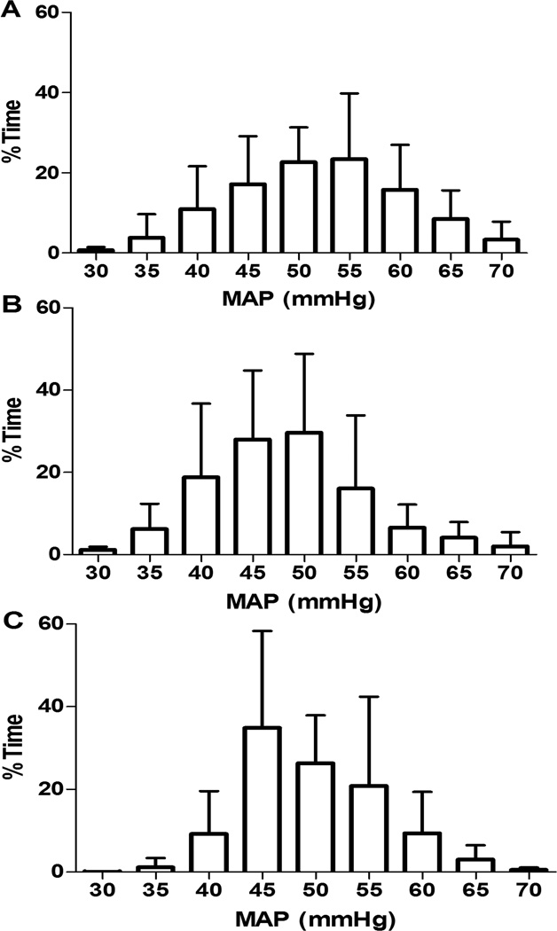
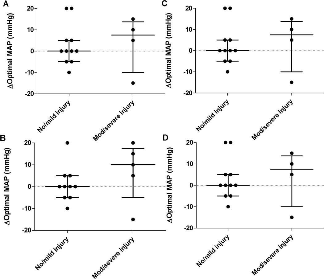
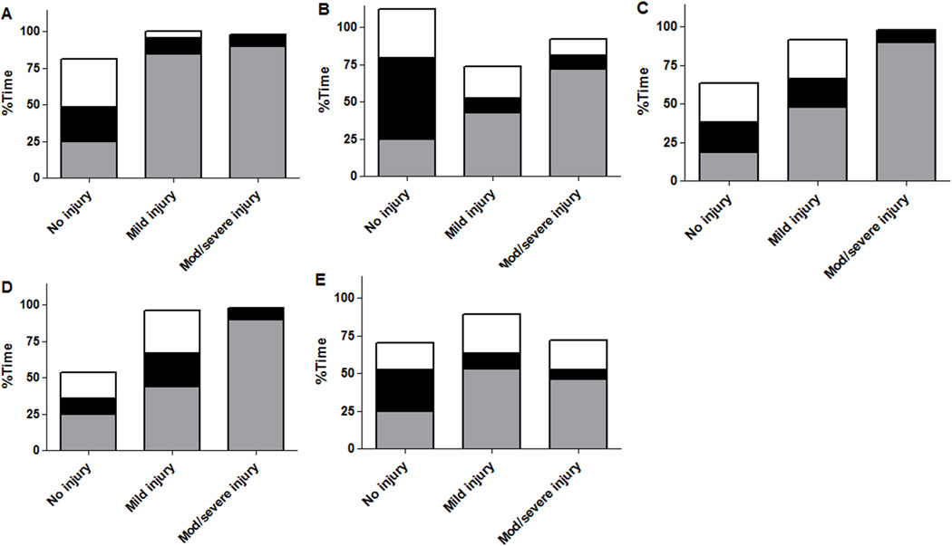
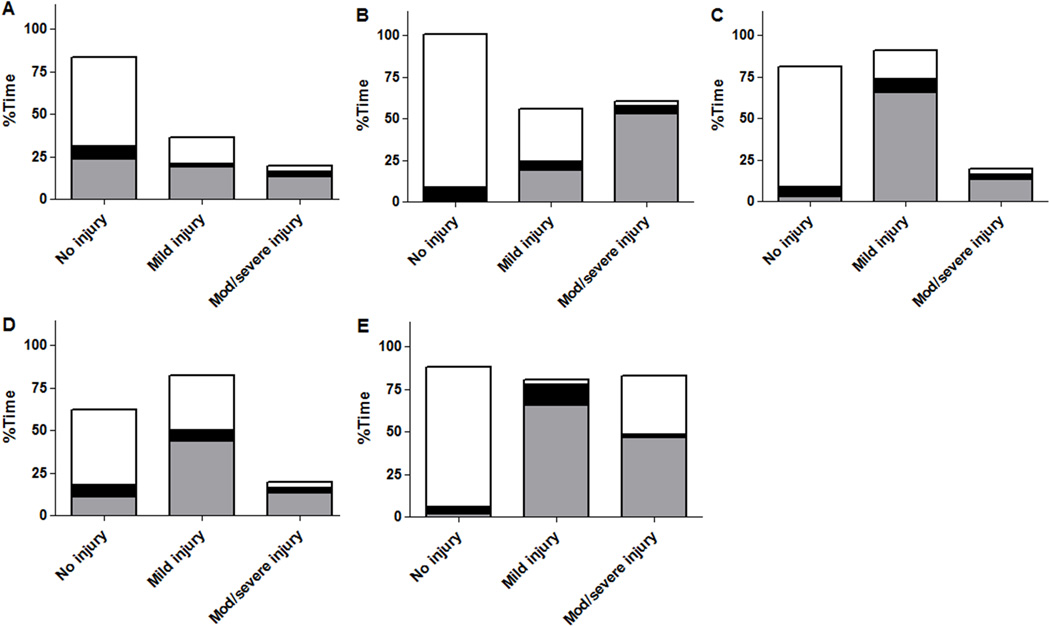
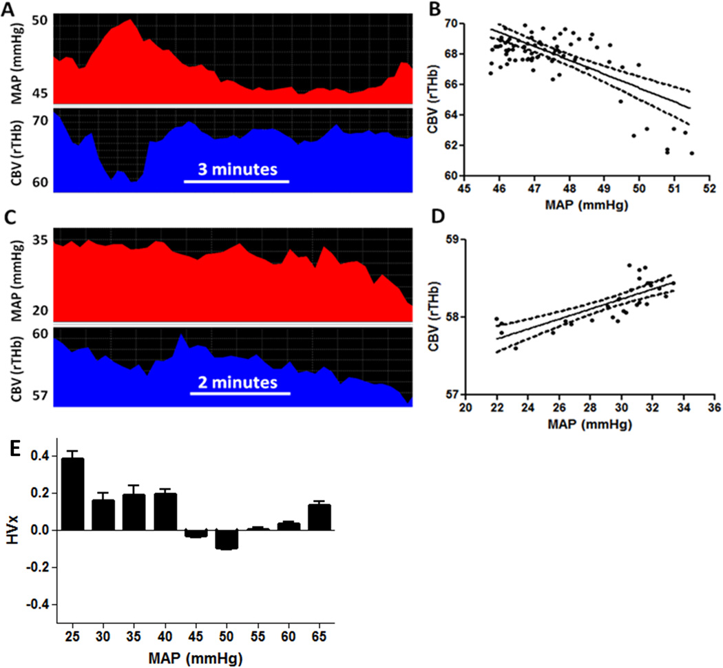
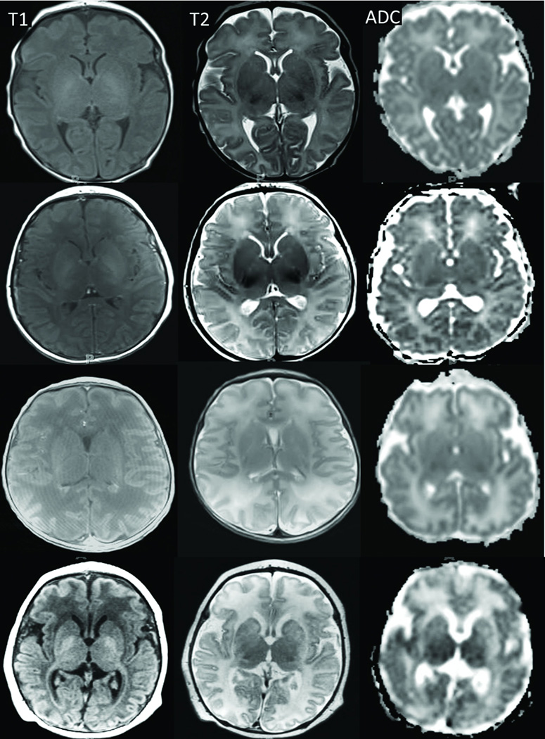
Methods: Neonates with HIE had autoregulation monitoring with the hemoglobin volume index (HVx) during therapeutic hypothermia, rewarming, and the first 6 h of normothermia. The 5-mm Hg range of mean arterial blood pressure (MAP) with best vasoreactivity (MAPOPT) was identified. The percentage of time spent with MAP below MAPOPT and deviation in MAP from MAPOPT were measured. Neonates received brain magnetic resonance imaging (MRI) 3-7 d after treatment. MRIs were coded as no, mild, or moderate/severe injury in five regions.

Results: HVx identified MAPOPT in 79% (19/24), 77% (17/22), and 86% (18/21) of the neonates during hypothermia, rewarming, and normothermia, respectively. Neonates with moderate/severe injury in paracentral gyri, white matter, basal ganglia, and thalamus spent a greater proportion of time with MAP below MAPOPT during rewarming than neonates with no or mild injury. Neonates with moderate/severe injury in paracentral gyri, basal ganglia, and thalamus had greater MAP deviation below MAPOPT during rewarming than neonates without injury.

Conclusion: Maintaining MAP within or above MAPOPT may reduce the risk of neurologic injuries in neonatal HIE.


Figures

Figure 1
Figure 1
The percentage of time during (A) hypothermia (n=24), (B) rewarming (n=22), and (C) normothermia (n=21) that neonates spent at each mean arterial blood pressure (MAP). Data are shown as means with SDs.
Figure 2
Figure 2
Fifteen neonates had an identifiable optimal mean arterial blood pressure (MAPOPT) during both hypothermia and rewarming. When progressing from hypothermia to rewarming, some individuals had a shift in MAPOPT. This shift is represented on the y-axis. For instance, a value of 5 indicates the MAPOPT increased by 5 mmHg as the patient moved from hypothermia to rewarming. A value of 0 represents no shift. Patients with no or mild injury in (A) paracentral gyri, (B) white matter, (C) basal ganglia, and (D) thalamus had no or minimal shift in MAPOPT when moving from hypothermia to rewarming. Data are shown as medians and interquartile ranges.
Figure 3
Figure 3
The percentage of time neonates (n=17) spent below, within, or above the optimal mean arterial blood pressure bin (MAPOPT) during rewarming in relation to injury in (A) paracentral gyri, (B) white matter, (C) basal ganglia, (D) thalamus, and (E) brainstem. Gray represents the percentage of time spent with blood pressure below MAPOPT. Black represents the percentage of time spent with blood pressure within MAPOPT. White represents the percentage of time spent with blood pressure above MAPOPT. Neonates with injuries in all regions spent more time with blood pressure below MAPOPT than patients without injury. The degree of injury in paracentral gyri, white matter, basal ganglia, and thalamus increased with greater time below MAPOPT. Neonates with no or mild injury spent a greater proportion of time with blood pressure within the MAPOPT bin than patients with moderate/severe injury. Data are displayed as medians.
Figure 4
Figure 4
The percentage of time neonates (n=18) spent below, within, or above the optimal mean arterial blood pressure bin (MAPOPT) during normothermia in relation to injury in (A) paracentral gyri, (B) white matter, (C) basal ganglia, (D) thalamus, and (E) brainstem. Gray represents the percentage of time spent with blood pressure below MAPOPT. Black represents the percentage of time spent with blood pressure within MAPOPT. White represents the percentage of time spent with blood pressure above MAPOPT. Patients with injury in white matter and brainstem, and patients with more severe injury in white matter spent more time with blood pressure below MAPOPT. Neonates with no or mild injury in all regions spent more time with blood pressure above MAPOPT than patients with moderate/severe injury. Injury severity was lower in paracentral gyri, white matter, basal ganglia, and thalamus with greater time spent above MAPOPT. Data are displayed as medians.
Figure 5
Figure 5
Hemoglobin volume index (HVx) calculation in a neonate with hypoxic-ischemic encephalopathy. (A, B) When mean arterial blood pressure (MAP) exceeded 45 mmHg, MAP negatively correlated with cerebral blood volume (CBV, or the relative total hemoglobin (rTHb) measured by near-infrared spectroscopy). This negative correlation yielded an HVx of −0.29, indicating pressure-reactive vasoreactivity with functional autoregulation. The linear regression line is illustrated (E(Y)=111.5−0.91X; 95% confidence interval for slope: −1.18, −0.66; p<0.0001). (C, D) When MAP was less than 35 mmHg, MAP and CBV positively correlated. This resulted in an HVx of 0.12, indicating pressure-passive vasoreactivity with impaired autoregulation. The linear regression line is illustrated (E(Y)=56.3+0.06X; 95% confidence interval for slope: 0.04, 0.08; p<0.0001). (E) Six hours of HVx monitoring. HVx was sorted into 5-mmHg bins of MAP. Optimal MAP (MAPOPT) was identified at the HVx nadir and represents the range of MAP with most robust vasoreactivity. This patient’s MAPOPT was 50 mmHg. Data in panels B and D are shown with linear regression lines and 95% confidence intervals. Data in panel E are shown as means with SDs.
Figure 6
Figure 6
Axial T1-weighted (first column) and T2-weighted (second column) images and ADC maps (third column) of four neonates with no (first row), mild (second row), moderate (third row), or severe (fourth row) injury. T1 and T2 signals increased in the cortex, basal ganglia, thalami, and posterior limb of the internal capsule with greater injury. With worsening white matter injury, the T2 signal increased and the gray-white matter differentiation became less distinct. ADC maps confirmed the injuries, particularly in the white matter, as signal increased with greater injury.

References

    1. Graham EM, Ruis KA, Hartman AL, Northington FJ, Fox HE. A systematic review of the role of intrapartum hypoxia-ischemia in the causation of neonatal encephalopathy. Am J Obstet Gynecol. 2008;199:587–595. - PubMed
    1. Shankaran S, Pappas A, McDonald SA, et al. Childhood outcomes after hypothermia for neonatal encephalopathy. N Engl J Med. 2012;366:2085–2092. - PMC - PubMed
    1. Zweifel C, Castellani G, Czosnyka M, et al. Noninvasive monitoring of cerebrovascular reactivity with near infrared spectroscopy in head-injured patients. J Neurotrauma. 2010;27:1951–1958. - PubMed
    1. Aries MJ, Czosnyka M, Budohoski KP, et al. Continuous determination of optimal cerebral perfusion pressure in traumatic brain injury. Crit Care Med. 2012;40:2456–2463. - PubMed
    1. Brady KM, Lee JK, Kibler KK, et al. The lower limit of cerebral blood flow autoregulation is increased with elevated intracranial pressure. Anesth Analg. 2009;108:1278–1283. - PubMed

Publication types