. 2013 Dec;43(12):3279-90.
doi: 10.1002/eji.201343727. Epub 2013 Sep 6.

Exosomes carrying mycobacterial antigens can protect mice against Mycobacterium tuberculosis infection

Affiliations

Exosomes carrying mycobacterial antigens can protect mice against Mycobacterium tuberculosis infection

Yong Cheng et al. Eur J Immunol. 2013 Dec.

Abstract

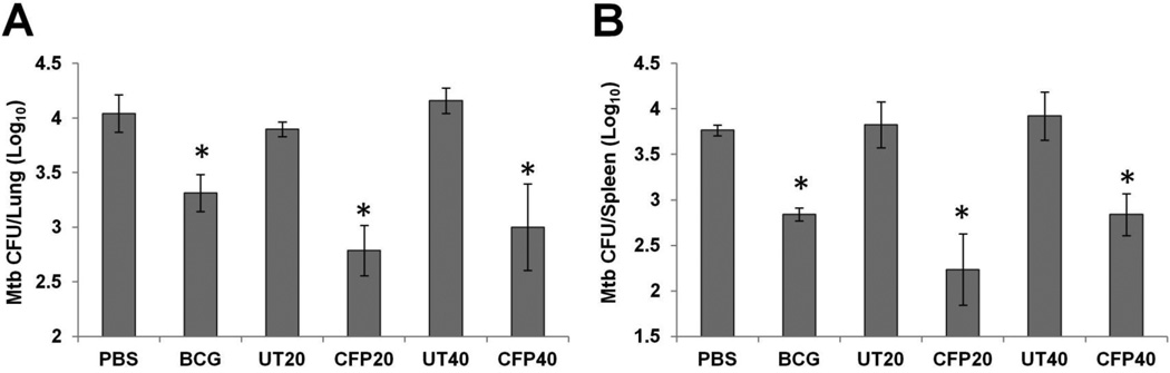
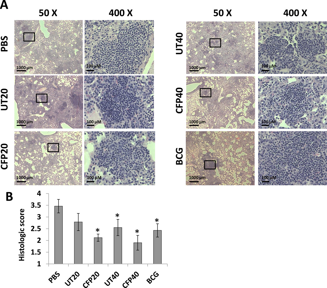
Approximately 2 billion people are infected with Mycobacterium tuberculosis, the etiological agent of tuberculosis (TB), and an estimated 1.5 million individuals die annually from TB. Presently, Mycobacterium bovis BCG remains the only licensed TB vaccine; however, previous studies suggest its protective efficacy wanes over time and fails in preventing pulmonary TB. Therefore, a safe and effective vaccine is urgently required to replace BCG or boost BCG immunizations. Our previous studies revealed that mycobacterial proteins are released via exosomes from macrophages infected with M. tuberculosis or pulsed with M. tuberculosis culture filtrate proteins (CFP). In the present study, exosomes purified from macrophages treated with M. tuberculosis CFP were found to induce antigen-specific IFN-γ and IL-2-expressing CD4(+) and CD8(+) T cells. In exosome-vaccinated mice, there was a similar TH1 immune response but a more limited TH2 response compared to BCG-vaccinated mice. Using a low-dose M. tuberculosis mouse aerosol infection model, exosomes from CFP-treated macrophages were found to both prime a protective immune response as well as boost prior BCG immunization. The protection was equal to or superior to BCG. In conclusion, our findings suggest that exosomes might serve as a novel cell-free vaccine against an M. tuberculosis infection.

Keywords: Exosomes; Mycobacterium tuberculosis; Vaccine.


Conflict of interest statement

Conflict of interest: The authors declare no financial or commercial conflict of interest.

Figures

Figure 1
Figure 1. Exosomes from CFP-treated macrophages contains both host and mycobacterial components
(A) Purified CFP analyzed by SDS-PAGE gel (15%) and stained with Coomassie. (B) Western blot analysis of CFP using a polyclonal anti-serum made against the M. tuberculosis culture filtrate proteins. (C) Exosomes released from CFP-treated (CFP exosomes) or untreated (UT exosomes) macrophages were analyzed by Western blot for the mycobacterial 19-KDa lipoprotein and the host exosomal marker LAMP1.
Figure 2
Figure 2. Antigen-specific immune response in the lungs and spleens of mice primarily vaccinated with exosomes containing mycobacterial proteins
(A–D) Flow cytometric analysis of antigen-specific cytokine – expressing CD4+ and CD8+ T cells isolated from the spleens and lungs of vaccinated or non-vaccinated mice. The frequency of INF-γ-positive CD4+ (A) or CD8+ (B) T cells as well as the frequency of IL-2 positive CD4+ (C) or CD8+ (D) T cells after ex vivo stimulation with M. tuberculosis antigens. (E and F) IFN-γ and IL-2 ELISA using spleen and lung cells isolated from vaccinated and non-vaccinated mice. The results are expressed as the cytokine concentration after ex vivo stimulation of 1×10⁶ cells with M. tuberculosis antigens. The data shown +/− standard error of pooled lung or spleen homogenate from 5 mice per given condition run in duplicate and is the representative of three independent experiments. *, p < 0.05 compared to PBS control (one-way ANOVA with Tukey post-test. UT20 and UT40, exosomes (20 or 40µg) isolated from untreated marcophages; CFP20 and CFP40, exosomes (20 or 40µg) isolated from macrophages pulsed with M. tuberculosis CFP.
Figure 3
Figure 3. BCG but not CFP-exosome vaccination induces IL-4-expressing CD4+ T cells
Splenic and lung cells were harvested 2 weeks after the final exosome vaccination and ex vivo stimulated with M. tuberculosis antigens followed by flow cytometry staining for CD4 and IL-4 expression. The data shown is the representative of three independent experiments. *, p < 0.05 (one-way ANOVA with Tukey post-test).
Figure 4
Figure 4. Exosomes from CFP-treated macrophages induce a limited TH2 immune response in mice compared to BCG vaccinated mice
Antigen-specific (A) IgG, and subtypes (B) IgG2c and (C) IgG1 endpoint titers were defined using mouse serum which was collected 2 weeks after the final vaccination with exosomes. Mean reciprocal dilutions are used as the endpoint titer (log10). The data shown +/− standard error of pooled lung or spleen homogenate from 5 mice per given condition run in duplicate and is the representative of three independent experiments. *, p < 0.05 compared to the BCG group (one-way ANOVA with Tukey post-test).
Figure 5
Figure 5. Mice vaccinated with exosomes from CFP-treated macrophages were partially protected against a low-dose aerosolized M. tuberculosis infection
Mice (n=4) were infected with virulent M. tuberculosis H37Rv by aerosol challenge 4 weeks after the final exosome vaccination. Six weeks postinfection, all mice were sacrificed and mycobacterial counts in the lungs (A) and spleens (B) were determined by plating. Results are expressed as the means ± standard errors of 4 mice per group. The data shown is the representative of three independent experiments. *, p < 0.05 compared to results with PBS control groups (one-way ANOVA with Tukey post-test).
Figure 6
Figure 6. Lung histopathology in M. tuberculosis-infected mice
H&E staining of lung sections from vaccinated and non-vaccinated mice following infection with virulent M. tuberculosis H37Rv. (A) The lung sections were analyzed under low (50X) and high (400X) magnification. (B) Histopathological scores of lung sections. PBS and BCG were used as a negative and positive control, respectively. Sections are representatives of lung sections from 4 mice per group and from one of three independent experiments. *, p<0.05 compared to the PBS group (one-way ANOVA with Tukey post-test).
Figure 7
Figure 7. Antigen-specific immune response in the lungs and spleens of mice after prime-boost vaccination
(A–D) Flow cytometric analysis of antigen-specific cytokine – expressing CD4+ and CD8+ T cells isolated from the spleens and lungs of non-vaccinated mice or mice vaccinated with BCG as the prime vaccine only or with BCG plus a booster vaccine with BCG or exosomes. The frequency of INF-γ-positive CD4+ (A) or CD8+ (B) T cells as well as the frequency of IL-2 positive CD4+ (C) or CD8+ (D) T cells after ex-vivo stimulation with M. tuberculosis antigens. (E and F) IFN-γ and IL-2 ELISA using spleen and lung cells isolated from vaccinated and non-vaccinated mice. The results are expressed as cytokine concentration after ex vivo stimulation of 1 × 10⁶ cells with M. tuberculosis antigens. The data shown +/− standard error of pooled lung or spleen homogenate from 5 mice per given condition run in duplicate and is the representative of three independent experiments. *, p < 0.05 compared to PBS control (one-way ANOVA with Tukey post-test). UT20 and UT40, exosomes (20 or 40µg) isolated from untreated macrophages; CFP20 and CFP40, exosomes (20 or 40µg) isolated from macrophages pulsed with M. tuberculosis CFP.
Figure 8
Figure 8. BCG vaccinated mice boosted with exosomes from CFP-treated macrophages were protected against a low-dose aerosolized M. tuberculosis infection
Six weeks after the final exosome vaccination mice were infected with virulent M. tuberculosis H37Rv by aerosol challenge and 6 weeks later the mycobacterial burden in the lungs (A) and spleens (B) was determined by plating the tissue homogenates on 7H10 agar plates. Results are expressed as the means ± standard error for 4 mice per group and the data shown is the representative of two independent experiments. *, p < 0.05 compared to PBS control; **, P < 0.05 compared to BCG primed/BCG boost vaccinated mice; as determined by Student paired t-test.

References

    1. WHO. Geneva: WHO; 2012. Global Tuberculosis Report 2012.
    1. Calmette A. Sur la vaccination préventive des enfants nouveau-nés contre la tuberculose par le BCG. Ann. Inst. Pasteur. 1927;41:201–232.
    1. Kaufmann SH. Envisioning future strategies for vaccination against tuberculosis. Nat. Rev. Immunol. 2006;6:699–704. - PubMed
    1. Kaufmann SH. Future vaccination strategies against tuberculosis: thinking outside the box. Immunity. 2010;33:567–577. - PubMed
    1. Trunz BB, Fine P, Dye C. Effect of BCG vaccination on childhood tuberculous meningitis and miliary tuberculosis worldwide: a meta-analysis and assessment of cost-effectiveness. Lancet. 2006;367:1173–1180. - PubMed

Publication types

MeSH terms