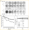
Glucocorticoids antagonize RUNX2 during osteoblast differentiation in cultures of ST2 pluripotent mesenchymal cells
- PMID: 23943595
- PMCID: PMC5771406
- DOI: 10.1002/jcb.24646
Glucocorticoids antagonize RUNX2 during osteoblast differentiation in cultures of ST2 pluripotent mesenchymal cells
Abstract
The efficacy of glucocorticoids (GCs) in treating a wide range of autoimmune and inflammatory conditions is blemished by severe side effects, including osteoporosis. The chief mechanism leading to GC-induced osteoporosis is inhibition of bone formation, but the role of RUNX2, a master regulator of osteoblast differentiation and bone formation, has not been well studied. We assessed effects of the synthetic GC dexamethasone (dex) on transcription of RUNX2-stimulated genes during the differentiation of mesenchymal pluripotent cells into osteoblasts. Dex inhibited a RUNX2 reporter gene and attenuated locus-dependently RUNX2-driven expression of several endogenous target genes. The anti-RUNX2 activity of dex was not attributable to decreased RUNX2 expression, but rather to physical interaction between RUNX2 and the GC receptor (GR), demonstrated by co-immunoprecipitation assays and co-immunofluorescence imaging. Investigation of the RUNX2/GR interaction may lead to the development of bone-sparing GC treatment modalities for the management of autoimmune and inflammatory diseases.
Keywords: GLUCOCORTICOID-INDUCED OSTEOPOROSIS; OSTEOBLAST DIFFERENTIATION; TRANSCRIPTION.
© 2013 Wiley Periodicals, Inc.
Figures


Similar articles
-
Glucocorticoids Hijack Runx2 to Stimulate Wif1 for Suppression of Osteoblast Growth and Differentiation.J Cell Physiol. 2017 Jan;232(1):145-53. doi: 10.1002/jcp.25399. Epub 2016 Apr 26. J Cell Physiol. 2017. PMID: 27061521 Free PMC article.
-
Runx2 promotes both osteoblastogenesis and novel osteoclastogenic signals in ST2 mesenchymal progenitor cells.Osteoporos Int. 2012 Apr;23(4):1399-413. doi: 10.1007/s00198-011-1728-5. Epub 2011 Sep 1. Osteoporos Int. 2012. PMID: 21881969 Free PMC article.
-
Glucocorticoids inhibit the maturation of committed osteoblasts via SOX2.J Mol Endocrinol. 2022 Apr 22;68(4):195-207. doi: 10.1530/JME-21-0213. J Mol Endocrinol. 2022. PMID: 35255002
-
Regulation of bone development and extracellular matrix protein genes by RUNX2.Cell Tissue Res. 2010 Jan;339(1):189-95. doi: 10.1007/s00441-009-0832-8. Epub 2009 Aug 1. Cell Tissue Res. 2010. PMID: 19649655 Review.
-
Distinct Glucocorticoid Receptor Actions in Bone Homeostasis and Bone Diseases.Front Endocrinol (Lausanne). 2022 Jan 10;12:815386. doi: 10.3389/fendo.2021.815386. eCollection 2021. Front Endocrinol (Lausanne). 2022. PMID: 35082759 Free PMC article. Review.
Cited by
-
Activation of cannabinoid receptor 2 alleviates glucocorticoid-induced osteonecrosis of femoral head with osteogenesis and maintenance of blood supply.Cell Death Dis. 2021 Oct 30;12(11):1035. doi: 10.1038/s41419-021-04313-3. Cell Death Dis. 2021. PMID: 34718335 Free PMC article.
-
Vitamin K2 Prevents Glucocorticoid-induced Osteonecrosis of the Femoral Head in Rats.Int J Biol Sci. 2016 Jan 28;12(4):347-58. doi: 10.7150/ijbs.13269. eCollection 2016. Int J Biol Sci. 2016. PMID: 27019620 Free PMC article.
-
Function and Regulation of Bone Marrow Adipose Tissue in Health and Disease: State of the Field and Clinical Considerations.Compr Physiol. 2024 Jun 27;14(3):5521-5579. doi: 10.1002/cphy.c230016. Compr Physiol. 2024. PMID: 39109972 Free PMC article. Review.
-
Exosomes derived from human CD34+ stem cells transfected with miR-26a prevent glucocorticoid-induced osteonecrosis of the femoral head by promoting angiogenesis and osteogenesis.Stem Cell Res Ther. 2019 Nov 15;10(1):321. doi: 10.1186/s13287-019-1426-3. Stem Cell Res Ther. 2019. PMID: 31730486 Free PMC article.
-
Dexamethasone and 1,25-dihydroxyvitamin D3 reduce oxidative stress-related DNA damage in differentiating osteoblasts.Int J Mol Sci. 2014 Sep 19;15(9):16649-64. doi: 10.3390/ijms150916649. Int J Mol Sci. 2014. PMID: 25244015 Free PMC article.
References
-
- Banerjee C, McCabe LR, Choi JY, Hiebert SW, Stein JL, Stein GS, Lian JB. Runt homology domain proteins in osteoblast differentiation: AML3/CBFA1 is a major component of a bone-specific complex. J Cell Biochem. 1997;66:1–8. - PubMed
-
- Baschant U, Lane NE, Tuckermann J. The multiple facets of glucocorticoid action in rheumatoid arthritis. Nat Rev Rheumatol. 2012;8:645–655. - PubMed
Publication types
MeSH terms
Substances
Grants and funding
LinkOut - more resources
Full Text Sources
Other Literature Sources
Medical
Molecular Biology Databases
Miscellaneous

