. 2013 Oct 4;288(40):29025-34.
doi: 10.1074/jbc.M113.501510. Epub 2013 Aug 13.

The extended transmembrane Orai1 N-terminal (ETON) region combines binding interface and gate for Orai1 activation by STIM1

Affiliations

The extended transmembrane Orai1 N-terminal (ETON) region combines binding interface and gate for Orai1 activation by STIM1

Isabella Derler et al. J Biol Chem. .

Abstract

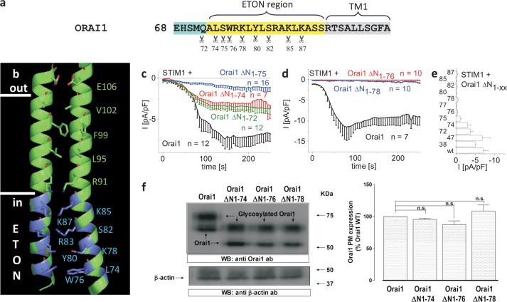
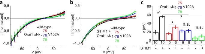
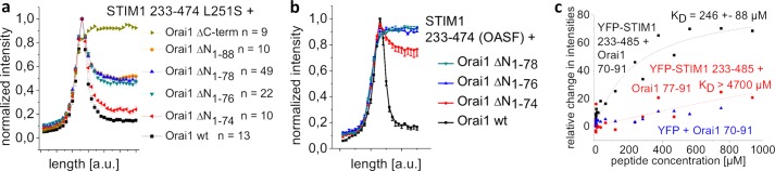
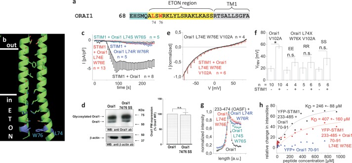
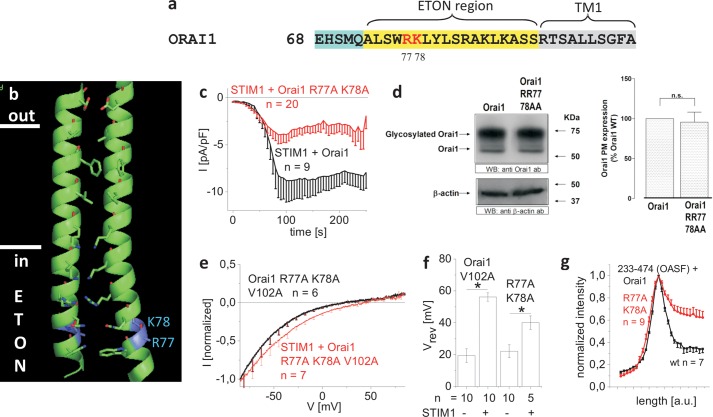
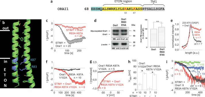
STIM1 and Orai1 represent the two molecular key components of the Ca(2+) release-activated Ca(2+) channels. Their activation involves STIM1 C terminus coupling to both the N terminus and the C terminus of Orai. Here we focused on the extended transmembrane Orai1 N-terminal (ETON, aa73-90) region, conserved among the Orai family forming an elongated helix of TM1 as recently shown by x-ray crystallography. To identify "hot spot" residues in the ETON binding interface for STIM1 interaction, numerous Orai1 constructs with N-terminal truncations or point mutations within the ETON region were generated. N-terminal truncations of the first four residues of the ETON region or beyond completely abolished STIM1-dependent Orai1 function. Loss of Orai1 function resulted from neither an impairment of plasma membrane targeting nor pore damage, but from a disruption of STIM1 interaction. In a complementary approach, we monitored STIM1-Orai interaction via Orai1 V102A by determining restored Ca(2+) selectivity as a consequence of STIM1 coupling. Orai1 N-terminal truncations that led to a loss of function consistently failed to restore Ca(2+) selectivity of Orai1 V102A in the presence of STIM1, demonstrating impairment of STIM1 binding. Hence, the major portion of the ETON region (aa76-90) is essential for STIM1 binding and Orai1 activation. Mutagenesis within the ETON region revealed several hydrophobic and basic hot spot residues that appear to control STIM1 coupling to Orai1 in a concerted manner. Moreover, we identified two basic residues, which protrude into the elongated pore to redound to Orai1 gating. We suggest that several hot spot residues in the ETON region contribute in aggregate to the binding of STIM1, which in turn is coupled to a conformational reorientation of the gate.

Keywords: Calcium Channels; Calcium Intracellular Release; Calcium Signaling; Electrophysiology; Ion Channels.


Figures

FIGURE 1.
FIGURE 1.
N-terminal truncations including Trp-76 of the ETON region or beyond abolish STIM1-dependent Orai1 function. a, amino acid sequence of Orai1 N terminus displaying those residues (indicated by scissors) in the conserved ETON region (yellow area) up to which increasing N-truncations have been performed. b, schematic representation of two TM1 domains + ETON regions highlighting those residues up to which N-truncations have been performed (blue) in the ETON region. c, time course of whole cell inward currents at −74 mV activated by passive store depletion of HEK 293 cells co-expressing CFP-STIM1 and YFP-Orai1 ΔN1–72, ΔN1–74 and ΔN1–75 in comparison with wild-type YFP-Orai1. pF, picofarads. d, time course of whole cell inward currents at −74 mV activated by passive store depletion of HEK293 cells co-expressing CFP-STIM1 and YFP-Orai1 ΔN1–76 and ΔN1–78 in comparison with wild-type YFP-Orai1. e, block diagram comparing maximal current densities of Orai1 N-truncation mutants (ΔN1–38, -47, -72, -74, -75, -76, -77, -78, -80, -82, -85, -87) with that of wild-type Orai1. Error bars indicate S.E. f, Orai1 plasma membrane expression in HEK293 cells expressing Orai1 WT, Orai1 ΔN1–74, Orai1 ΔN1–76, or Orai1 ΔN1–78 determined by biotinylation as described under “Experimental Procedures” (n = 5; *, p < 0.05). Error bars indicate S.E. n.s., not significant. WB, Western blot.; ab, antibody.
FIGURE 2.
FIGURE 2.
Nonfunctional Orai1 N-truncation mutants introduced into the Orai1 V102A core display no shift in reversal potential in the presence of STIM1. a, I/V relationships of normalized Orai1 V102A currents in comparison with those of Orai1 ΔN1–75 V102A, Orai1 ΔN1–76 V102A, and Orai1 ΔN1–78 V102A. b, analogue to panel a in the presence of STIM1. c, block diagram displaying reversal potentials of Orai1 V102A, Orai1 ΔN1–75 V102A, Orai1 ΔN1–76 V102A, and Orai1 ΔN1–78 V102A currents in the absence and presence of STIM1 (*, p < 0.05). Error bars indicate S.E. n.s., not significant.
FIGURE 3.
FIGURE 3.
Nonfunctional Orai1 N-truncation mutants display a significant reduction in STIM1 binding. a, intensity plots representing the localization of STIM1 233–474 L251S across the cell when co-expressed with Orai1 ΔN1–74, Orai1 ΔN1–76, Orai1 ΔN1–78, Orai1 ΔN1–88, and Orai1 ΔC-term when compared with wild-type Orai1. a. u., arbitrary units. b, intensity plots representing the localization of STIM1 233–474 (OASF) across the cell when co-expressed with Orai1 ΔN1–74, Orai1 ΔN1–76, and Orai1 ΔN1–78 when compared with wild-type Orai1. c, binding curve fitted to a one-site binding model for YFP-STIM1 233–485 from a HEK293 cell lysate together with Orai1 NT(aa70–91) or Orai1 NT(aa77–91) in comparison with YFP from HEK293 cell lysate together with Orai1 NT(aa70–91), obtained from thermophoresis experiments.
FIGURE 4.
FIGURE 4.
The hydrophobic residues Leu-74 and Trp-76 of the ETON region contribute in a concerted manner to the coupling with STIM1. a, amino acid sequence of Orai1 N terminus displaying Leu-74 and Trp-76 in red in the conserved ETON region (yellow area) b, schematic representation of two TM1 domains + ETON regions highlighting Leu-74 and Trp-76 (blue) in the ETON region. c, time course of whole cell inward currents at −74 mV activated by passive store depletion of HEK 293 cells co-expressing CFP-STIM1 and YFP-Orai1 L74S/W76S, YFP-Orai1 L74E/W76E, and YFP-Orai1 L74R/W76R in comparison with wild-type YFP-Orai1. pF, picofarads. d, Orai1 plasma membrane expression in HEK293 cells, expressing Orai1 WT in comparison with Orai1 L74S/W76S determined by biotinylation as described under “Experimental Procedures” (n = 5; *, p < 0.05). Error bars indicate S.E. e, I/V relationship of normalized Orai1 V102A currents in comparison with that of Orai1 L74E/W76E/V102A, shown as example. Error bars indicate S.E. n.s., not significant. WB, Western blot; ab, antibody. f, block diagram displaying reversal potentials of Orai1 V102A, Orai1 L74S/W76S/V102A (SS), Orai1 L74E/W76E/V102A (EE), and Orai1 L74R/W76R/V102A (RR) in the absence and presence of STIM1 (*, p < 0.05). Error bars indicate S.E. g, intensity plots representing the localization of STIM1 233–474 (OASF) across the cell when co-expressed with Orai1 L74S/W76S, Orai1 L74E/W76E, and Orai1 L74R/W76R when compared with wild-type Orai1. a. u., arbitrary units. h, binding curve fitted to a one-site binding model for YFP-STIM1 233–485 from a HEK293 cell lysate together with Orai1 NT(aa70–91) or Orai1 NT(aa70–91 L74E/W76E) in comparison with YFP from HEK293 cell lysate together with Orai1 NT(aa70–91), obtained from thermophoresis measurements.
FIGURE 5.
FIGURE 5.
The positively charged residues Arg-77 and Lys-78 in the ETON region also contribute to STIM1 binding. a, amino acid sequence of Orai1 N terminus displaying Arg-77/Lys-78 in red in the ETON region (yellow area). b, schematic representation of two TM1 domains + ETON regions highlighting Arg-77 and Lys-78 (blue) in the ETON region. c, time course of whole cell inward currents at −74 mV activated by passive store depletion of HEK 293 cells co-expressing CFP-STIM1 and YFP-Orai1 R77A/K78A in comparison with wild-type YFP-Orai1. pF, picofarads. d, Orai1 plasma membrane expression in HEK293 cells, expressing Orai1 in comparison with Orai1 R77A/K78A as determined by biotinylation as described under “Experimental Procedures” (n = 5; *, p < 0.05). Error bars indicate S.E. n.s., not significant. WB, Western blot; ab, antibody. e, I/V relationships of normalized Orai1 R77A/K78A/V102A currents in the absence and presence of STIM1. Error bars indicate S.E. f, block diagram displaying reversal potentials of Orai1 V102A and Orai1 R77A/K78A/V102A in the absence and presence of STIM1 (*, p < 0.05). Error bars indicate S.E. g, intensity plots representing the localization of STIM1 233–474 (OASF) across the cell when co-expressed with Orai1 R77A/K78A when compared with wild-type Orai1. Error bars indicate S.E.
FIGURE 6.
FIGURE 6.
The positively charged Arg-83 and Lys-87 residues in the ETON region contribute to both STIM1 binding and the gate. a, amino acid sequence of Orai1 N terminus displaying Arg-83/Lys-87 in red in the ETON region (yellow area). b, schematic representation of two TM1 domains + ETON regions highlighting the positively charged residues Arg-83 and Lys-87 (blue) in the ETON region. c, time course of whole cell inward currents at −74 mV activated by passive store depletion of HEK 293 cells co-expressing CFP-STIM1 and YFP-Orai1 R83A/K87A in comparison with wild-type YFP-Orai1. d, plasma membrane expression in HEK293 cells expressing Orai1 WT in comparison with Orai1 R83A/K87A determined by biotinylation as described under “Experimental Procedures” (n = 5; *, p < 0.05). Error bars indicate S.E. n.s., not significant. WB, Western blot; ab, antibody. e, intensity plots representing the localization of STIM1 233–474 (OASF) across the cell when co-expressed with Orai1 R83A/K87A when compared with wild-type Orai1. Error bars indicate S.E. f, time course of whole cell inward currents at −74 mV activated by passive store depletion of HEK 293 cells co-expressing CFP-STIM1 and YFP-Orai1 R83A/K87A/V102A in the absence and presence of STIM1. g, I/V relationships of normalized Orai1 R83A/K87A/V102A currents in the absence and presence of STIM1. h, block diagram displaying reversal potentials of Orai1 V102A and Orai1 R83A/K87A/V102A in the absence and presence of STIM1 (*, p < 0.05). Error bars indicate S.E. i, time course of whole cell inward currents at −74 mV activated by passive store depletion of HEK 293 cells co-expressing CFP-STIM1 and YFP-Orai1 ΔN1–72 R83A/K87A/V102A, YFP-Orai1 ΔN1–74 R83A/K87A/V102A, and YFP-Orai1 ΔN1–76 R83A/K87A/V102A in comparison with YFP-Orai1 R83A/K87A/V102A (ctrl).

Similar articles

Cited by

References

    1. Roos J., DiGregorio P. J., Yeromin A. V., Ohlsen K., Lioudyno M., Zhang S., Safrina O., Kozak J. A., Wagner S. L., Cahalan M. D., Veliçelebi G., Stauderman K. A. (2005) STIM1, an essential and conserved component of store-operated Ca2+ channel function. J. Cell Biol. 169, 435–445 - PMC - PubMed
    1. Zhang S. L., Yu Y., Roos J., Kozak J. A., Deerinck T. J., Ellisman M. H., Stauderman K. A., Cahalan M. D. (2005) STIM1 is a Ca2+ sensor that activates CRAC channels and migrates from the Ca2+ store to the plasma membrane. Nature 437, 902–905 - PMC - PubMed
    1. Prakriya M., Feske S., Gwack Y., Srikanth S., Rao A., Hogan P. G. (2006) Orai1 is an essential pore subunit of the CRAC channel. Nature 443, 230–233 - PubMed
    1. Vig M., Beck A., Billingsley J. M., Lis A., Parvez S., Peinelt C., Koomoa D. L., Soboloff J., Gill D. L., Fleig A., Kinet J. P., Penner R. (2006) CRACM1 multimers form the ion-selective pore of the CRAC channel. Curr. Biol. 16, 2073–2079 - PMC - PubMed
    1. Vig M., Peinelt C., Beck A., Koomoa D. L., Rabah D., Koblan-Huberson M., Kraft S., Turner H., Fleig A., Penner R., Kinet J. P. (2006) CRACM1 is a plasma membrane protein essential for store-operated Ca2+ entry. Science 312, 1220–1223 - PMC - PubMed

Publication types

MeSH terms

LinkOut - more resources