. 2013 Sep 27;288(39):28324-30.
doi: 10.1074/jbc.C113.495473. Epub 2013 Aug 13.

Adipocyte fetuin-A contributes to macrophage migration into adipose tissue and polarization of macrophages

Affiliations

Adipocyte fetuin-A contributes to macrophage migration into adipose tissue and polarization of macrophages

Priyajit Chatterjee et al. J Biol Chem. .

Abstract

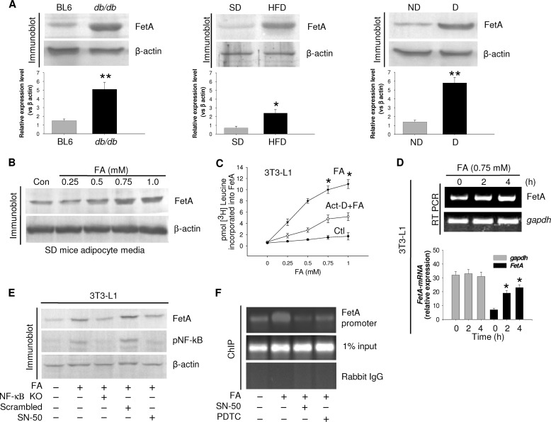
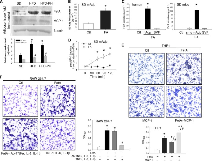
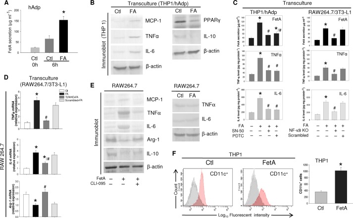
Macrophage infiltration into adipose tissue during obesity and their phenotypic conversion from anti-inflammatory M2 to proinflammatory M1 subtype significantly contributes to develop a link between inflammation and insulin resistance; signaling molecule(s) for these events, however, remains poorly understood. We demonstrate here that excess lipid in the adipose tissue environment may trigger one such signal. Adipose tissue from obese diabetic db/db mice, high fat diet-fed mice, and obese diabetic patients showed significantly elevated fetuin-A (FetA) levels in respect to their controls; partially hepatectomized high fat diet mice did not show noticeable alteration, indicating adipose tissue to be the source of this alteration. In adipocytes, fatty acid induces FetA gene and protein expressions, resulting in its copious release. We found that FetA could act as a chemoattractant for macrophages. To simulate lipid-induced inflammatory conditions when proinflammatory adipose tissue and macrophages create a niche of an altered microenvironment, we set up a transculture system of macrophages and adipocytes; the addition of fatty acid to adipocytes released FetA into the medium, which polarized M2 macrophages to M1. This was further confirmed by direct FetA addition to macrophages. Taken together, lipid-induced FetA from adipocytes is an efficient chemokine for macrophage migration and polarization. These findings open a new dimension for understanding obesity-induced inflammation.

Keywords: Adipose Tissue; Inflammation; Lipids; Macrophages; Obesity.


Figures

FIGURE 1.
FIGURE 1.
FA-induced FetA secretion from adipocytes. A, adipose tissue collected from control (Ctl) or db/db mice, SD, or HFD mice and obese diabetic (D) or non-diabetic (ND) human subjects was immunoblotted for FetA. Results are expressed as means ± S.E. (n = 5). *, p < 0.01, **, p < 0.001 (versus BL6 or SD or non-diabetic). B, SD mice adipocytes with varied concentrations of palmitate (FA) were incubated for 4 h, and medium was immunoblotted for FetA. Con, control. C, 3T3-L1 adipocytes were incubated with increasing concentrations of FA in the presence of [3H]leucine without or with actinomycin D. Media were immunoprecipitated by anti-FetA antibody and subjected for radioactive counting. *, p < 0.001 (versus Ctl). D, RNA extracted from 3T3-L1 adipocytes was incubated with FA for different periods and subjected to RT-PCR and qPCR. Data are expressed as means ± S.E. (n = 3). *, p < 0.001 (versus Ctl). E, Ctl or NF-κB siRNA (NF-κB KO) transfected or SN-50-treated 3T3-L1 cells were incubated with FA and immunoblotted with indicated antibodies. pNF-kB, phospho-NF-κB. F, human adipocytes were incubated with FA or FA+SN-50 or pyrrolidine dithiocarbamate (PDTC). NF-κB binding to the FetA promoter was determined by ChIP assay.
FIGURE 2.
FIGURE 2.
Macrophage migration due to FetA. A, adipose tissue fluid collected from SD or HFD or HFD-PH (PH = partially hepatectomized) mice was immunoblotted for FetA and MCP-1 by using the respective antibodies. B, adipocytes from SD mice (mAdp) were incubated without or with FA, and FetA release was measured by ELISA. C, FetA secretion in Ctl (media without cells) and hAdp or stromal vascular fraction (SVF) or mouse skeletal muscle cells (smc) or mouse Adp incubated with FA was measured by ELISA. D, SD mice adipocytes were incubated with FA in the presence of [3H]leucine for different time periods. On termination, medium was immunoprecipitated with anti-FetA antibody and subjected to radioactive counting. E, THP1 macrophages were added to the upper chamber of the Boyden chamber and allowed to migrate through the porous membrane into the lower chamber containing medium alone or medium with FetA (100 μg ml−1) or MCP-1(100 ng ml−1) or both. F, similar migration of RAW264.7 cells was observed in the Boyden chamber containing medium with FetA (100 μg ml−1) or FetA plus anti-TNFα, IL-6, and IL-1β antibodies (2 μg ml−1) and TNFα, IL-6, and IL-1β (2 μg ml−1). Migrated cells on the lower side of the membrane were stained and observed under a microscope. Dye was further extracted following the manufacturer's protocol, and optical density (OD) was measured at 560 nm. Data are expressed as means ± S.E. (n = 3). *, p < 0.001 (versus Ctl); #, p < 0.01 (versus FetA).
FIGURE 3.
FIGURE 3.
FetA-induced polarization of macrophage from M2 to M1 phenotype. A, hAdp were incubated without or with FA, and FetA released into the medium was estimated by ELISA. B, THP-1 and hAdp were transcultured with or without FA, and THP1 cell lysate was immunoblotted with anti-MCP-1, TNFα, IL-6, PPARγ, and IL-10 antibodies. β-Actin was used as a loading control. C, THP1 and SN-50 or pyrrolidine dithiocarbamate (PDTC)-treated hAdp or RAW264.7 and NF-κB KO 3T3-L1 adipocytes were transcultured with or without FA. FetA secretion was estimated by ELISA, whereas TNFα and IL-6 were determined in macrophage cell lysates by ELISA. D, TLR4 KO RAW264.7 and 3T3-L1 adipocytes were transcultured with or without FA, and relative mRNA expression levels of TNFα, IL-6, and Arg-1 were determined by qPCR. Results of C and D are expressed as means ± S.E. (n = 3). *, p < 0.001(versus Ctl); #, p < 0.01(versus FA). E, RAW264.7 cells were treated with or without FetA or CLI-095+FetA. Media and cell lysates were immunoblotted with anti-MCP-1, TNFα, IL-6, Arg-1, and IL-10 antibodies. RAW264.7 cells were incubated with or without FA, and media were immunoblotted against anti-TNFα and IL-6 antibodies. F, THP1 cells were incubated with or without FetA, and conversion to CD11c+ was determined by FACS analysis. Results are expressed as means ±S.E. (n = 3). *, p < 0.001 (versus Ctl).

References

    1. Kahn S. E., Hull R. L., Utzschneider K. M. (2006) Mechanisms linking obesity to insulin resistance and type 2 diabetes. Nature 444, 840–846 - PubMed
    1. Lumeng C. N., Saltiel A. R. (2011) Inflammatory links between obesity and metabolic disease. J. Clin. Invest. 121, 2111–2117 - PMC - PubMed
    1. Glass C. K., Olefsky J. M. (2012) Inflammation and lipid signaling in the etiology of insulin resistance. Cell Metab. 15, 635–645 - PMC - PubMed
    1. Gregor M. F., Hotamisligil G. S. (2011) Inflammatory mechanisms in obesity. Annu. Rev. Immunol. 29, 415–445 - PubMed
    1. Johnson A. M. F., Olefsky J. M. (2013) The origins and drivers of insulin resistance. Cell 152, 673–684 - PubMed

Publication types

LinkOut - more resources