. 2013 Oct 10;122(15):2582-90.
doi: 10.1182/blood-2012-02-407452. Epub 2013 Aug 13.

Blockade of invariant TCR-CD1d interaction specifically inhibits antibody production against blood group A carbohydrates

Affiliations

Blockade of invariant TCR-CD1d interaction specifically inhibits antibody production against blood group A carbohydrates

Hirofumi Tazawa et al. Blood. .

Abstract

Previously, we detected B cells expressing receptors for blood group A carbohydrates in the CD11b(+)CD5(+) B-1a subpopulation in mice, similar to that in blood group O or B in humans. In the present study, we demonstrate that CD1d-restricted natural killer T (NKT) cells are required to produce anti-A antibodies (Abs), probably through collaboration with B-1a cells. After immunization of wild-type (WT) mice with human blood group A red blood cells (A-RBCs), interleukin (IL)-5 exclusively and transiently increased and the anti-A Abs were elevated in sera. However, these reactions were not observed in CD1d(-/-) mice, which lack NKT cells. Administration of anti-mouse CD1d blocking monoclonal Abs (mAb) prior to immunization abolished IL-5 production by NKT cells and anti-A Ab production in WT mice. Administration of anti-IL-5 neutralizing mAb also diminished anti-A Ab production in WT mice, suggesting that IL-5 secreted from NKT cells critically regulates anti-A Ab production by B-1a cells. In nonobese diabetic/severe combined immunodeficient (NOD/SCID/γc(null)) mice, into which peripheral blood mononuclear cells from type O human volunteers were engrafted, administration of anti-human CD1d mAb prior to A-RBC immunization completely inhibited anti-A Ab production. Thus, anti-CD1d treatment might constitute a novel approach that could help in evading Ab-mediated rejection in ABO-incompatible transplant recipients.


Figures

Figure 1
Figure 1
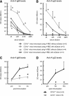
Ab production against blood group A determinants was dependent on iNKT cells but independent of T cells. (A-B) Balb/c nude mice and B6 MHC C2D mice were immunized with human group A-RBCs 2 times per week. Balb/c and B6 WT mice were used as the respective controls. The serum anti-A Ab (IgM) concentrations at 2 weeks after the second immunization were measured by ELISA. Even after immunization, the serum anti-A Ab titers were not elevated in nude mice, but Ab concentrations significantly increased in C2D mice. Balb/c nude mice, n = 3; Balb/c WT mice, n = 3; B6 C2D mice, n = 4; B6 WT mice, n = 3. (C-D) Balb/c CD1d−/− mice and B6 Jα18−/− mice were immunized with human group A-RBCs 2 times per week. Balb/c and B6 WT mice served as the respective controls. The serum anti-A Ab levels were measured 2 weeks after the second immunization. The response of anti-A Abs was completely impaired in CD1d−/− mice and partially impaired in Jα18−/− mice. Average values ± SEM for the individual groups are shown. P values were shown to compare pre- and post-levels at the bottom of each figure. Balb/c CD1d−/− mice, n = 4; Balb/c WT mice, n = 3; B6 Jα18−/− mice, n = 5; B6 WT mice, n = 4. *P < .05 compared with the respective control mice.
Figure 2
Figure 2
Effect of αGalCer administration on anti-blood group A responses in CD1d/ mice. Balb/c CD1d−/− mice and Balb/c WT (CD1d+/+) mice were immunized using blood group A-RBCs together with intraperitoneal injection of either αGalCer (4 μg/mouse) or PBS (control) 2 times per week. (A-B) The serum anti-A–specific IgM and IgG levels were determined using ELISA at 2 and 10 weeks after the last immunization, respectively. αGalCer significantly increased the blood group A-specific Ab levels in CD1d+/+ mice, but the Ab levels did not increase in CD1d−/− mice. The average values ± SEM for the individual groups are shown. *P < .05 compared with the data from CD1d+/+ mice without αGalCer. (C-D) The kinetics of the serum IgM and IgG titers against blood group A determinants in Balb/c CD1d+/+ mice and Balb/c CD1d−/− mice (25× diluted serum was used). Anti-A Ab production was elicited by intraperitoneal immunization of mice with A-RBC 2 times per week. The average values ± SEM for the individual groups are shown. Balb/c CD1d−/− mice, n = 4; WT Balb/c mice, n = 5. *P < .05 compared with the respective CD1d+/+ mice.
Figure 3
Figure 3
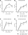
Correlation of iNKT cells with Ab production against Gal and NeuGc epitopes. B6 CD1d−/−GalT−/− and CD1d−/−CMAH−/− mice were immunized with Gal- and NeuGc-bearing rat thymocytes, and the levels of anti-Gal and anti-NeuGc Abs were then determined in their respective sera using FCM. (A-B) CD1d−/−GalT−/− mice showed increased anti-Gal Ab titer (both IgM and IgG subclasses) similar to that in CD1d+/+GalT−/− mice (n = 4 per group). (C-D) CD1d−/−CMAH−/− mice also showed increased anti-NeuGc Ab titer similar to that in CD1d+/+CMAH−/− mice (n = 4 per group). Median fluorescence intensity values were used to follow Ab levels. The average values ± SEM for the individual groups are shown.
Figure 4
Figure 4
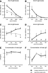
Effect of administration of anti-mouse CD1d mAb on anti-blood group A titers in mice. Balb/c WT mice received intraperitoneal injection of anti-mouse CD1d mAb (n = 6). Mice that received injections of isotype-matched Ab served as the controls (n = 5). The mice were immunized with human blood group A-RBCs (5 × 10⁸/mouse) on days 1 and 8 after mAb administration. After immunization, blood samples were obtained and the total IgM/IgG and anti-A IgM/IgG concentrations were measured using ELISA. (A-B) Treatment with anti-CD1d mAb significantly inhibited Ab production against blood group A epitopes in the mice. Anti-A IgM levels were detected 6 weeks after mAb administration, and anti-A IgG levels were detected at 8 weeks. (C-D) The kinetics of anti-A Abs in Balb/c WT mice that were injected with either anti-mouse CD1d mAb or isotype-matched Ab are shown (10× diluted serum was used). (E-F) The kinetics of the total serum immunoglobulin (IgM and IgG) levels of the Balb/c WT mice treated with anti-CD1d mAb are presented. The average values ± SEM for the individual groups are shown. *P < .05 compared with the data from WT mice treated with isotype-matched Ab.
Figure 5
Figure 5
Impact of IL-5 on anti-A Ab production after stimulation with blood group A-RBCs. (A) Balb/c CD1d−/− mice and WT CD1d+/+ mice (n = 5 in each group) were immunized with human blood group A-RBCs (5 × 10⁸/mouse). The levels of cytokines in serum were analyzed at the indicated time points using cytometric bead array flex sets (CBA). Blood group A determinants significantly increased the level of IL-5 in CD1d+/+ mice, but this cytokine level did not increase in CD1d−/− mice. In contrast, blood group A determinants did not increase the levels of IL-4, IL-9, IL-17, IL-21, and IFN-γ in either CD1d−/− or CD1d+/+ mice. (B) Balb/c WT mice received intraperitoneal injection of anti-mouse CD1d mAb (n = 5). Mice that received injections of isotype-matched Ab served as controls (n = 5). The mice were immunized with human blood group A-RBCs (5 × 10⁸/mouse) on day 1 after mAb administration. The level of IL-5 in serum was analyzed at the indicated time points using CBA. Treatment with anti-CD1d mAb significantly inhibited IL-5 production against blood group A epitopes in the mice. (C-E) Balb/c WT mice received intraperitoneal injection of anti-mouse IL-5 mAb (n = 5) 30 minutes prior to immunization with human blood group A-RBCs (5 × 10⁸/mouse). Mice that received injections of isotype-matched Ab served as controls (n = 5). The mice were immunized with human blood group A-RBCs 2 times at 1-week interval after mAb administration. (C) Anti-A IgM concentrations were measured using ELISA before immunization. (D) Anti-A IgM concentrations were measured at 2 weeks after the first immunization. (E) Anti-A IgM concentrations were measured at 3 weeks after the first immunization. Treatment with anti-IL-5 mAb significantly inhibited Ab production against blood group A epitopes in the mice. The average values ± SEM for the individual groups are shown. *P < .05 compared with the data from CD1d−/− mice and data from WT mice treated with isotype-matched Ab.
Figure 6
Figure 6
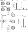
NKT cells were predominant sources of IL-5 secreted after immunization with group A-RBCs. WT CD1d+/+ Balb/c mice received intraperitoneal injection of anti-mouse CD1d mAb (n = 3). Mice that received injections of isotype-matched Ab served as controls (n = 3). The mice were immunized with human blood group A-RBCs (5 × 10⁸/mouse) on day 1 after mAb administration. The mice were sacrificed to determine the IL-5–producing cells 6 hours after immunization. (A-B) LMNCs and spleen cells were seeded. The representative pictures of ELISPOT wells are shown (A) and the frequency of IL-5 producing cells is (B). Number in each picture refers to the total cells seeded per well (×103). (C-E) Six hours after immunization with A-RBCs, the LMNCs were isolated from CD1d+/+ Balb/c mice (n = 12). The pooled cells were used in ELISPOT assay to determine the frequency of IL-5–producing cells. The LMNCs were stained with APC-conjugated anti-mouse CD1d-tetramer and PE-Cy7–conjugated anti-mouse TCRβ. NKT cells (CD1d-tetramer+, TCRβ+), T cells (CD1d-tetramer, TCRβ+), and the others (CD1d-tetramer, TCRβ) were isolated by sorting with FACS Aria. After sorting, the purities of NKT, T, and other cells were reanalyzed by FCM. (D-E) The representative pictures of ELISPOT wells are shown (D) and the frequency of IL-5–producing cells is shown (E). Number in each picture refers to the total cells seeded per well (×103). The results shown are the average ± SEM calculated from red spot number in quadruplicate wells. The results are representative of 2 similar experiments. *P < .05.
Figure 7
Figure 7
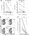
Effect of administration of anti-human CD1d mAb on anti-A Ab production in humanized mice. The same dose of PBMCs from each type O human volunteer was intraperitoneally injected into 2 NOD/SCID/γcnull mice (20 × 106 cells/mouse). Of these mice, 1 subsequently received anti-human CD1d mAb and the other received isotype-matched irrelevant control Ab at days 7 and 10 after PBMC engrafting. The humanized mice were immunized with human blood group A-RBCs 8 days after PBMC injection. (A) The serum anti-A IgM and IgG levels in the humanized mice were determined using ELISA at 14 and 21 days after engraftment. Each point represents an individual mouse. Each group contained 5 animals. (B) Three weeks after human PBMC engrafting, the humanized mice were sacrificed to determine the proportion of B cells with receptors for group A carbohydrates. Spleen cells were prepared from the humanized mice (n = 4 in each group). The pooled cells were stained with FITC-labeled A-BSA or control FITC-labeled BSA together with PE-conjugated anti-human CD19 mAb. Representative FCM results of group A-BSA–binding spleen cells. We analyzed 50 000 cells per contour plot. The percentages in the figure represent percentages of total CD19+ B cells. (C) The frequencies of A-BSA–binding B cells among the total B cell population in mice treated with either anti-human CD1d mAb or isotype-matched control Ab are shown. *P < .05 compared with the data from humanized mice treated with isotype-matched Ab.

Comment in

Similar articles

Cited by

References

    1. Kronenberg M. Toward an understanding of NKT cell biology: progress and paradoxes. Annu Rev Immunol. 2005;23:877–900. - PubMed
    1. Taniguchi M, Harada M, Kojo S, Nakayama T, Wakao H. The regulatory role of Valpha14 NKT cells in innate and acquired immune response. Annu Rev Immunol. 2003;21:483–513. - PubMed
    1. Kawano T, Cui J, Koezuka Y, et al. CD1d-restricted and TCR-mediated activation of valpha14 NKT cells by glycosylceramides. Science. 1997;278(5343):1626–1629. - PubMed
    1. Carnaud C, Lee D, Donnars O, et al. Cutting edge: Cross-talk between cells of the innate immune system: NKT cells rapidly activate NK cells. J Immunol. 1999;163(9):4647–4650. - PubMed
    1. Singh N, Hong S, Scherer DC, et al. Cutting edge: activation of NK T cells by CD1d and alpha-galactosylceramide directs conventional T cells to the acquisition of a Th2 phenotype. J Immunol. 1999;163(5):2373–2377. - PubMed

Publication types

MeSH terms